Scientists create artifical brain with 2.3 million simulated neurons

aritificial brain

Another computer is setting its wits to perform human tasks. But this computer is different. Instead of the tour de force processing of Deep Blue or Watson’s four terabytes of facts of questionable utility, Spaun attempts to play by the same rules as the human brain to figure things out. Instead of the logical elegance of a CPU, Spaun’s computations are performed by 2.3 million simulated neurons configured in networks that resemble some of the brain’s own networks. It was given a series of tasks and performed pretty well, taking a significant step toward the creation of a simulated brain.

Spaun stands for Semantic Pointer Architecture: Unified Network. It was given 6 different tasks that tested its ability to recognize digits, recall from memory, add numbers and complete patterns. Its cognitive network simulated the prefrontal cortex to handle working memory and the basal ganglia and thalamus to control movements. Like a human, Spaun can view an image and then give a motor response; that is, it is presented images that it sees through a camera and then gives a response by drawing with a robotic arm.

And its performance was similar to that of a human brain. For example, the simplest task, image recognition, Spaun was shown various numbers and asked to draw what it sees. It got 94 percent of the numbers correct. In a working memory task, however, it didn’t do as well. It was shown a series of random numbers and then asked to draw them in order. Like us with human brains, Spaun found the pattern recognition task easy, the working memory task not quite as easy.

The important thing here is not how well Spaun performed on the tasks – your average computer could find ways to perform much better than Spaun. But what’s important is that, in Spaun’s case, the task computations were carried out solely by the 2.3 million artificial neurons spiking in the way real neurons spike to carry information from one neuron to another. The visual image, for example, was processed hierarchically, with multiple levels of neurons successively extracting more complex information, just as the brain’s visual system does. Similarly, the motor response mimicked the brain’s strategy of combining many simple movements to produce an optimal, single movement while drawing.

Chris Eliasmith, from the University of Waterlook in Ontario, Canada and lead author of the study is happy with his cognitive creation. “It’s not as smart as monkeys when it comes to categorization,” he told CNN, “but it’s actually smarter than monkeys when it comes to recognizing syntactic patterns, structured patterns in the input, that monkeys won’t recognize.”

Watch Spaun work through its tasks in the following video.

One thing Spaun can’t do is perform tasks in realtime. Every second you saw Spaun performing tasks in the video actually requires 2.5 hours of numbers crunching by its artificial brain. The researchers hope to one day have it perform in realtime.

It’s important to note that Spaun isn’t actually learning anything by performing these tasks. Its neural nets are hardwired and are incapable of the modifications that real neurons undergo when we learn. But producing complex behavior from a simulated neuronal network still represents an important initial step toward building an artificial brain. Christian Machens, a neuroscientist at the Champalimaud Neuroscience Programme in Lisbon and was not involved in the study, writes in Science that the strategy for building a simulated brain is “to not simply incorporate the largest number of neurons or the greatest amount of detail, but to reproduce the largest amount of functionality and behavior.”

We’re still a long way from artificial intelligence that is sentient and self-aware. And there’s no telling if the robots of the future will have brains that look like ours or if entirely different solutions will be used to produce complex behavior. Whatever it looks like, Spaun is a noble step in the right direction.

Scientists Create Artificial Brain With 2.3 Million Simulated Neurons

How gender stereotypes warp our view of depression

74583142depressioncrop

Stereotypes about male and female roles may influence the way we perceive depressed people.

It’s a well-known fact that men and women who behave the same way in the exact same situation—whether it’s a job interview, a cocktail party, or a traffic stop—are sometimes perceived and treated differently based on their gender.

Something similar, it seems, may happen when men and women start to show signs of depression. A new study, published this week in the journal PLoS ONE, suggests that people of both sexes are less likely to view men as being depressed and in need of professional help—even if a man’s symptoms are identical to a woman’s.

“A lot of attention has been paid to depression in women, and with good reason: Depression is twice as common in women,” says Dr. James B. Potash, the editor of the study and a professor of psychiatry at the University of Iowa, in Iowa City. “There has been relatively little focus on education about depression in men. This [study] emphasizes the importance of figuring out how to get through to men that depression can be disabling and treatment is important.”

Health.com: 12 Signs of Depression in Men

In the study, researchers in the U.K. asked a group of about 600 adults to read a short description of a hypothetical depressed person. This vignette, which was designed to illustrate the diagnostic criteria for clinical depression (also known as major depression), read in part:

For the past two weeks, Kate has been feeling really down. She wakes up in the morning with a flat, heavy feeling that sticks with her all day. She isn’t enjoying things the way she normally would. In fact, nothing gives her pleasure. Even when good things happen, they don’t seem to make Kate happy.

Fifty-seven percent of the study participants recognized Kate’s symptoms—which also included difficulty concentrating, fatigue, and insomnia—as indications of a mental health disorder, and more than three-quarters of those people correctly identified the disorder as depression. Only 10% of the respondents said Kate did not have a problem.

The researchers presented the same vignette to another group of 600 people. This time, however, every mention of “Kate” was replaced by “Jack,” and all the pronouns were switched from female to male. Those minor changes had a noticeable effect: Though nearly as many people recognized Jack as having a mental health problem (52%), more than twice as many as in the Kate scenario said he did not (21%).

In addition, men themselves were less likely than women to label Jack depressed—a pattern that was not seen with Kate.

Health.com: How to Help Someone Who’s Depressed

Why the difference? Male stereotypes that emphasize traits such as toughness and strength may dissuade both women and men, and especially the latter, from identifying or acknowledging the signs of depression in men, says study author Viren Swami.

“Men are expected to be strong, deny pain and vulnerability, and conceal any emotional fragility,” says Swami, a psychologist at the University of Westminster, in London. “Because of these societal expectations, men appear to have poorer understanding of mental health and aren’t as good at detecting symptoms of depression compared with women.”

Potash says the findings also may reflect the fact that women are generally more attuned to emotions and better at articulating them. Some men might have all the outward signs of depression, and yet when asked about their mood they “may not be able to say much more than ‘I don’t know,’” he says. “A substantial minority of men just don’t describe depression.”

Health.com: 10 Careers With High Rates of Depression

On a deeper level, men’s failure to recognize the symptoms of depression in a fellow male may represent a kind of defense mechanism prompted by an “unconscious identification” with that man, says Dr. Radu Saveanu, a professor of psychiatry at the University of Miami Miller School of Medicine.

“They may think, ‘If this guy is having trouble and may need treatment, I may be in the same position someday,’” says Saveanu, who was not involved in the study. “That anxiety distorts the ability to be more objective.”

All of these dynamics may affect the likelihood of seeking or recommending treatment. In the study, men were more likely than women to recommend that Kate seek professional help, but this gap disappeared in the Jack scenario. Men also expressed less sympathy for Jack than women did.

The reluctance to seek treatment isn’t unique to men, but it does reflect an independent-minded streak that is more common among males, Potash says. Men tend to think that pulling themselves out of depression is “something they ought to be able to do,” he says. “It’s the stereotype of men who never ask for directions. They won’t admit that they can’t take care of it themselves.”

Health.com: Depressed? 12 Mental Tricks to Turn It Around

Gender, of course, isn’t the only factor that shapes how we view depression symptoms. Swami also found that respondents of either sex who held negative attitudes towards psychiatry and science felt that both Kate and Jack’s symptoms were less distressing, more difficult to treat, and less worthy of sympathy or professional help.

Swami took these trends into account, but he can’t rule out that other factors might have influenced the gender differences seen in the study. The participants’ own mental health history was unknown, for instance, though Swami says previous diagnoses do not tend to impact “mental health literacy,” or how well people understand mental health issues.

Future research will need to address these limitations, he says.

Read more: http://healthland.time.com/2012/11/15/how-gender-stereotypes-warp-our-view-of-depression/#ixzz2Es26tBvB


Pee marks the spot

sn-mice

Human beings tend to avoid places that smell of urine. But to mice, there is something positively addictive about the scent; they like to go back to a spot where they found the excretions again and again. Now, researchers have discovered that this behavior is triggered by a single protein in the urine of male mice.

Mice use scent to mark their territory, advertise their social dominance, and convey information about their health and reproductive status. But these are usually volatile pheromones that disperse quickly, and it has remained unclear what exactly stimulates a female to be attracted to a specific male.

Previous research had shown that female laboratory mice often return to a place where they have come across cage bedding soiled by males. Now, researchers at the University of Liverpool in the United Kingdom have confirmed this. Female mice spent five times as much time in a place where they had encountered a dish with male urine than at a place where they encountered water. Just 10 minutes of exposure to the urine was enough for the mice to show this place preference even after 14 days.

However, if the mice were prevented from by a mesh screen touching the urine with their nose, the place seemed to lose its attractiveness. “That suggested that the story was not as simple as everybody assumed and volatile pheromones were not responsible,” says behavioral ecologist Jane Hurst, one of the authors of the study. By separating the urine into different fractions, the scientists showed that a protein called darcin that they had identified in 2005—and which mice can only detect if their noses touch the urine—is responsible for the frequent visits. Pure darcin, produced in cell culture in the lab, elicited the same reaction, the authors report online today in Science.

“This is a really compelling story,” says Lisa Stowers, a neuroscientist at the Scripps Research Institute in San Diego, California. “Mammals were thought to be much more complex, but this study shows that a single chemical can lead [them to act] in a certain way.” The study is “very simple and elegant,” she adds. But it also raises new questions. For instance: There are many other ways a mouse could learn to return to a certain place. “So what is the benefit of evolving this [special] mechanism?”

Hurst says that what fascinates her is that the pheromones induce learning in the mice. And the animals do not only learn to be attracted to the place where they encountered the darcin. “They learn the odor cues of that specific male and are then attracted to it,” Hurst says. “Being familiar with a scent really seems to be important for whether a female is interested in a male.” The reason, Hurst suggests, is that dominant males, who make attractive mates, tend to leave the most marks in a certain territory.

The researchers showed that male mice, too, are attracted to a place if they have encountered darcin there, probably to foster a behavior called countermarking. “If males come across another male’s scent mark, they put their own, fresher urine there,” Hurst explains. This could also be the reason why some laboratory strains seem to have lost the ability to produce darcin: Because laboratory mice are usually group-housed, they have been selected to be less aggressive, and not producing darcin could help reduce tensions.

http://news.sciencemag.org/sciencenow/2012/12/pee-marks-the-spot.html?ref=em

Thanks to Dr. Rajadhyaksha for bringing this to the attention of the It’s Interesting community.

Controversial surgical treatment for addiction burns away the brain’s pleasure center


How far should doctors go in attempting to cure addiction? In China, some physicians are taking the most extreme measures. By destroying parts of the brain’s “pleasure centers” in heroin addicts and alcoholics, these neurosurgeons hope to stop drug cravings. But damaging the brain region involved in addictive desires risks permanently ending the entire spectrum of natural longings and emotions, including the ability to feel joy.

In 2004, the Ministry of Health in China banned this procedure due to lack of data on long term outcomes and growing outrage in Western media over ethical issues about whether the patients were fully aware of the risks.

However, some doctors were allowed to continue to perform it for research purposes—and recently, a Western medical journal even published a new study of the results. In 2007, The Wall Street Journal detailed the practice of a physician who claimed he performed 1000 such procedures to treat mental illnesses such as depression, schizophrenia and epilepsy, after the ban in 2004; the surgery for addiction has also since been done on at least that many people.

The November publication has generated a passionate debate in the scientific community over whether such research should be published or kept outside the pages of reputable scientific journals, where it may find undeserved legitimacy and only encourage further questionable science to flourish.

The latest study is the third published since 2003 in Stereotactic and Functional Neurosurgery, which isn’t the only journal chronicling results from the procedure, which is known as ablation of the nucleus accumbens. In October, the journal World Neurosurgery also published results from the same researchers, who are based at Tangdu Hospital in Xi’an.

The authors, led by Guodong Gao, claim that the surgery is “a feasible method for alleviating psychological dependence on opiate drugs.” At the same time, they report that more than half of the 60 patients had lasting side effects, including memory problems and loss of motivation. Within five years, 53% had relapsed and were addicted again to opiates, leaving 47% drug free.

(MORE: Addicted: Why We Get Hooked)

Conventional treatment only results in significant recovery in about 30-40% of cases, so the procedure apparently improves on that, but experts do not believe that such a small increase in benefit is worth the tremendous risk the surgery poses.  Even the most successful brain surgeries carry risk of infection, disability and death since opening the skull and cutting brain tissue for any reason is both dangerous and unpredictable. And the Chinese researchers report that 21% of the patients they studied experienced memory deficits after the surgery and 18% had “weakened motivation,” including at least one report of lack of sexual desire. The authors claim, however, that “all of these patients reported that their [adverse results] were tolerable.” In addition, 53% of patients had a change in personality, but the authors describe the majority of these changes as “mildness oriented,” presumably meaning that they became more compliant. Around 7%, however, became more impulsive.

The surgery is actually performed while patients are awake in order to minimize the chances of destroying regions necessary for sensation, consciousness or movement.  Surgeons use heat to kill cells in small sections of both sides of the brain’s nucleus accumbens.  That region is saturated with neurons containing dopamine and endogenous opioids, which are involved in pleasure and desire related both to drugs and to ordinary experiences like eating, love and sex.

(MORE: A Drug to End Drug Addiction)

In the U.S. and the U.K., reports the Wall Street Journal, around two dozen stereotactic ablations are performed each year, but only in the most intractable cases of depression and obsessive-compulsive disorder and after extensive review by institutional review boards and intensive discussions with the patient, who must acknowledge the risks. Often, a different brain region is targeted, not the nucleus accumbens. Given the unpredictable and potentially harmful consequences of the procedure, experts are united in their condemnation of using the technique to treat addictions. “To lesion this region that is thought to be involved in all types of motivation and pleasure risks crippling a human being,” says Dr. Charles O’Brien, head of the Center for Studies of Addiction at the University of Pennsylvania.

David Linden, professor of neuroscience at Johns Hopkins and author of a recent book about the brain’s pleasure systems calls the surgery “horribly misguided.”  He says “This treatment will almost certainly render the subjects unable to feel pleasure from a wide range of experiences, not just drugs of abuse.”

But some neurosurgeons see it differently. Dr. John Adler, professor emeritus of neurosurgery at Stanford University, collaborated with the Chinese researchers on the publication and is listed as a co-author.  While he does not advocate the surgery and did not perform it, he believes it can provide valuable information about how the nucleus accumbens works, and how best to attempt to manipulate it. “I do think it’s worth learning from,” he says. ” As far as I’m concerned, ablation of the nucleus accumbens makes no sense for anyone.  There’s a very high complication rate. [But] reporting it doesn’t mean endorsing it. While we should have legitimate ethical concerns about anything like this, it is a bigger travesty to put our heads in the sand and not be willing to publish it,” he says.

(MORE: Anesthesia Study Opens Window Into Consciousness)

Dr. Casey Halpern, a neurosurgery resident at the University of Pennsylvania makes a similar case. He notes that German surgeons have performed experimental surgery involving placing electrodes in the same region to treat the extreme lack of pleasure and motivation associated with otherwise intractable depression.  “That had a 60% success rate, much better than [drugs like Prozac],” he says. Along with colleagues from the University of Magdeburg in Germany, Halpern has just published a paper in the Proceedings of the New York Academy of Sciences calling for careful experimental use of DBS in the nucleus accumbens to treat addictions, which have failed repeatedly to respond to other approaches. The paper cites the Chinese surgery data and notes that addiction itself carries a high mortality risk.

DBS, however, is quite different from ablation.  Although it involves the risk of any brain surgery, the stimulation itself can be turned off if there are negative side effects, while surgical destruction of brain tissue is irreversible. That permanence—along with several other major concerns — has ethicists and addiction researchers calling for a stop to the ablation surgeries, and for journals to refuse to publish related studies.

Harriet Washington, author of Medical Apartheid:  The Dark History of Medical Experimentation on Black Americans from Colonial Times to the Present, argues that by publishing the results of unethical studies, scientists are condoning the questionable conditions under which the trials are conducted. “When medical journals publish research that violates the profession’sethical guidelines, this serves not only to sanction such abuses, but to encourage them,” she says. “In doing so, this practice encourages a relaxing of moral standards that threatens all patients and subjects, but especially  the medically vulnerable.”

(MORE: Real-Time Video: First Look at a Brain Losing Consciousness Under Anesthesia)

Shi-Min Fang, a Chinese biochemist who became a freelance journalist and recently won the journal Nature‘s Maddox prize for his exposes of widespread fraud in Chinese research, has revealed some of the subpar scientific practices behind research conducted in China, facing death threats and, as the New York Times reported, a beating with a hammer. He agrees that publishing such research only perpetuates the unethical practices. Asked by TIME to comment on the addiction surgery studies, Fang writes that publishing the research, particularly in western journals, “would encourage further unethical research, particularly in China where rewards for publication in international journals are high.”

While he doesn’t have the expertise to comment specifically on the ablation data, he says “the results of clinical research in China are very often fabricated. I suspect that the approvals by Ethics Committee mentioned in these papers were made up to meet publication requirement. I also doubt if the patients were really informed in detail about the nature of the study.” Fang also notes that two of the co-authors of the paper are advertising on the internet in Chinese, offering the surgery at a cost of 35,000 renminbi, about $5,600.  That’s more than the average annual income in China, which is about $5,000.

Given the available evidence, in fact, it’s hard to find a scientific justification for even studying the technique in people at all. Carl Hart, associate professor of psychology at Columbia University and author of the leading college textbook on psychoactive drugs, says animal studies suggest the approach may ultimately fail as an effective treatment for addiction; a 1984 experiment, for example, showed that destroying the nucleus accumbens in rats does not permanently stop them from taking opioids like heroin and later research found that it similarly doesn’t work for curbing cocaine cravings. Those results alone should discourage further work in humans. “These data are clear,” he says, “If you are going to take this drastic step, you damn well better know all of the animal literature.” [Disclosure:  Hart and I have worked on a book project together].

(MORE: Top 10 Medical Breakthroughs of 2012)

Moreover, in China, where addiction is so demonized that execution has been seen as an appropriate punishment and where the most effective known treatment for heroin addiction— methadone or buprenorphine maintenance— is illegal, it’s highly unlikely that addicted people could give genuinely informed consent for any brain surgery, let alone one that risks losing the ability to feel pleasure. And even if all of the relevant research suggested that ablating the nucleus accumbens prevented animals from seeking drugs, it would be hard to tell from rats or even primates whether the change was due to an overall reduction in motivation and pleasure or to a beneficial reduction in desiring just the drug itself.

There is no question that addiction can be difficult to treat, and in the most severe cases, where patients have suffered decades of relapses and failed all available treatments multiple times, it may make sense to consider treatments that carry significant risks, just as such dangers are accepted in fighting suicidal depression or cancer.  But in the ablation surgery studies, some of the participants were reportedly as young as 19 years old and had only been addicted for three years.  Addiction research strongly suggests that such patients are likely to recover even without treatment, making the risk-benefit ratio clearly unacceptable.

The controversy highlights the tension between the push for innovation and the reality of risk. Rules on informed consent didn’t arise from fears about theoretical abuses:  they were a response to the real scientific horrors of the Holocaust. And ethical considerations become especially important when treating a condition like addiction, which is still seen by many not as an illness but as a moral problem to be solved by punishment.  Scientific innovation is the goal, but at what price?
Read more: http://healthland.time.com/2012/12/13/controversial-surgery-for-addiction-burns-away-brains-pleasure-center/#ixzz2ExzobWQq

Thanks to Dr. Lutter for bringing this to the attention of the It’s Interesting community.


Homosexuality may start in the womb

sn-homosexuality


From a strictly Darwinian viewpoint, homosexuality shouldn’t still be around. It isn’t the best way to pass along one’s genes, and to complicate the picture further, no “gay genes” have even been identified. According to a newly released hypothesis, the explanation may not lie in DNA itself. Instead, as an embryo develops, sex-related genes are turned on and off in response to fluctuating levels of hormones in the womb, produced by both mother and child. This tug of war benefits the unborn child, keeping male or female development on a steady course even amid spikes in hormones. But if these so-called epigenetic changes persist once the child is born and has children of its own, some of those offspring may be homosexual, the study proposes.

Evolutionary geneticist William Rice of the University of California, Santa Barbara, felt there had to be a reason why homosexuality didn’t just fade away down the generations. Research estimates that about 8% of the population is gay, and homosexuality is known to run in families. If one of a set of identical twins is gay, there’s a 20% probability that the other will be, too.

Furthermore, Rice notes, “homosexuality isn’t just a human thing.” Among California gulls, which he watches from his office window, about 14% of pairs are female-female. In Australian black swans, some 6% of pairs are male-male, and 8% of male sheep are attracted exclusively to male partners.

But many genetic screens have failed to turn up genes that are responsible for sexual orientation. So to find out what makes homosexuality persist, Rice and colleagues began a comprehensive survey of the literature.

According to conventional wisdom, an embryo becomes a boy when a gene on the Y chromosome triggers the development of testes, which then begin to produce male sex hormones, including testosterone, at about the 8th week of gestation. With no Y chromosome and hence no testosterone, the embryo becomes a girl.

But testosterone doesn’t explain everything, the researchers found. For one thing, female fetuses are exposed to small amounts of the hormone from their adrenal glands, the placenta, and the mother’s endocrine system. At many key points of gestation, male and female fetuses are often exposed to similar amounts of testosterone. Levels of the hormone can even be higher than normal in females and lower than normal in males without any effect on genital or brain structure.

Rice and his co-workers were more intrigued by studies showing that male and female fetuses respond differently to the hormones that surround them, even when one hormone is temporarily higher. In their study, published online today in The Quarterly Review of Biology, the authors propose that differences in sensitivity to sex hormones result from “epigenetic” changes. These are changes that affect not the structure of a gene but when, if, and how much of it is activated—by chemically altering a gene’s promoter region or “on” switch, for example. Epigenetic changes at key points in the pathway through which testosterone exerts its effects on the fetus could blunt or enhance the hormone’s activity as needed, the authors suggest.

Although epigenetic changes are usually temporary, they involve alterations in the proteins that bind together the long strands of DNA. Thus, they can sometimes be handed down to offspring. According to the hypothesis, homosexuality may be a carry-over from one’s parents’ own prenatal resistance to the hormones of the opposite sex. The “epi-marks” that adjusted parental genes to resist excess testosterone, for example, may alter gene activation in areas of the child’s brain involved in sexual attraction and preference. “These epigenetic changes protect mom and dad during their own early development,” Rice says. The initial benefit to the parents may explain why the trait of homosexuality persists throughout evolution, he says.

“The authors have done a terrific job providing a mechanism for genetic variation, especially a variation that might not be expected to persist because it’s so tightly bound to reproduction,” says evolutionary biologist Marlene Zuk of the University of Minnesota, Twin Cities. But she adds that to go from changes in gene expression to why someone is attracted to a person of the same sex is a question for which science may never fill in all the blanks.

http://news.sciencemag.org/sciencenow/2012/12/homosexuality-may-start-in-the-w.html?ref=hp

Chinese scientists turn human urine into brain cells

NPGs_from_urine


Chinese researchers have developed a new technique for isolating kidney cells from urine and turning them into neural progenitors — –immature brain cells that can develop into various types of glial cells and neurons. Reprogramming cells has been done before, of course, but not with cells gleaned from urine and not via a method this direct. The technique could prove extremely helpful to those pursuing treatments for neurodegenerative disorders like Parkinson’s and Alzheimer’s.

The innovation here is in the source and the method. We know that embryonic stem cells offer potential treatments for neurodegenerative disorders. And we know that we can turn adult human cells–that is, non-embryonic cells gathered from adult humans–into pluripotent cells (those that can become a different type of cell) by reprogramming them, usually with genetically engineered viruses that tamper with the cells’ genetic codes.

But embryonic stem cell treatments are fraught with ethical issues and non-embryonic methods are complicated–and complexity introduces a greater chance of something going wrong (in this case that means mutations and genetic defects). The new method, which taps skin-like cells from the linings of the kidney tubes that are present in urine, converts its source cells into neurons and glia cells via a more direct route, making the process more efficient while narrowing the margin of error.

In their study, the researchers harvested kidney cells from the urine samples of three human donors and converted the cells directly to neural progenitors. Rather than using a genetically engineered virus to reprogram the cells, they used a small piece of bacterial DNA that can replicate in the cellular cytoplasm, a technique that eliminates the need to tamper directly with the chromosome (in theory, at least, this should reduce mutations) while also speeding up the entire process. After growing their progenitors into mature neurons and glial cells, the researchers transplanted the progenitors into the brains of newborn rats. A month later, the cells were still alive in the rats’ brains, though it is not yet clear that they can survive for extended periods or mesh with the brain’s wiring to become functioning parts of the neural machine.

There’s still a lot of research to be done on this method of course, but the researchers think it may provide a way to take cells gathered non-invasively and quickly and efficiently convert them into neural cells while reducing the likelihood of genetic mutations.

http://www.guardian.co.uk/science/neurophilosophy/2012/dec/09/turning-urine-into-brain-cells


Diuretic Drug Offers Latest Hope for Autism Treatment

sn-autism


A drug used for decades to treat high blood pressure and other conditions has shown promise in a small clinical trial for autism. The drug, bumetanide, reduced the overall severity of behavioral symptoms after 3 months of daily treatment. The researchers say that many parents of children who received the drug reported that their children were more “present” and engaged in social interactions after taking it. The new findings are among several recent signs that treatments to address the social deficits at the core of autism may be on the horizon.

Several lines of evidence suggest that autism interferes with the neurotransmitter GABA, which typically puts a damper on neural activity. Bumetanide may enhance the inhibitory effects of GABA, and the drug has been used safely as a diuretic to treat a wide range of heart, lung, and kidney conditions. In the new study, researchers led by Yehezkel Ben-Ari at the Mediterranean Institute of Neurobiology in Marseille, France, recruited 60 autistic children between the ages of 3 and 11 and randomly assigned them to receive either a daily pill of bumetanide or a placebo. (Neither the children’s parents nor the researchers who assessed the children knew who received the actual drug.)

As a group, those who got bumetanide improved by 5.6 points on a 60-point scale that’s often used to assess behaviors related to autism, the researchers report today in Translational Psychiatry. That was enough to nudge the group average just under the cutoff for severe autism and into the mild to medium category. The study did not look directly at whether the drug improved all symptoms equally or some more than others. “We have some indications that the symptoms particularly ameliorated with bumetanide are the genuine core symptoms of autism, namely communication and social interactions,” Ben-Ari says. More work will be needed to verify that impression. Ben-Ari says his team is now preparing for a larger, multicenter trial in Europe.

The current study already looks interesting to some. “It’s enough to make me think about trying it in a few of my autism patients who haven’t responded to other interventions,” says Randi Hagerman, a pediatrician who studies neurodevelopmental disorders at the University of California, Davis. Social interactions tend to be reinforcing, Hagerman adds, so getting an autistic child to start interacting more can have a positive effect on subsequent brain development.

Other drugs have recently shown promise for autism. In September, Hagerman and colleagues reported that arbaclofen, a drug that stimulates a type of GABA receptor, reduced social avoidance in people with fragile X syndrome, a genetic disorder that shares many features with autism. Many researchers are also hopeful about clinical trials under way with drugs that block certain receptors for glutamate, the main neurotransmitter in the brain that excites neural activity. Results from those trials should come out next year.

All of this work, including the new study, suggests that drugs that reduce neural excitation by blocking glutamate or enhance inhibition by boosting GABA may be helpful for treating autism, says Elizabeth Berry-Kravis, a pediatric neurologist at Rush University in Chicago, Illinois, and a collaborator on the recent arbaclofen study. “There seems to be this imbalance between excitation and inhibition in people with autism.”

That’s a potentially game-changing insight. Now doctors can only prescribe drugs that treat individual symptoms of autism rather than the underlying cause of the disorder, Berry-Kravis says. Doctors often prescribe antipsychotic drugs to reduce irritability, for example, but those drugs don’t address the social and communication problems at the heart of the disorder. “It’s exciting that now we’re thinking about the underlying mechanisms and treating those.”

http://news.sciencemag.org/sciencenow/2012/12/diuretic-drug-offers-latest-hope.html

Mother-Child Connection: Scientists Discover Children’s Cells Living in Mothers’ Brains, Including Male Cells Living in the Female Brain for Decades

scientists-discover-childrens-cells-living-in-mothers-brain_1


The link between a mother and child is profound, and new research suggests a physical connection even deeper than anyone thought. The profound psychological and physical bonds shared by the mother and her child begin during gestation when the mother is everything for the developing fetus, supplying warmth and sustenance, while her heartbeat provides a soothing constant rhythm.

The physical connection between mother and fetus is provided by the placenta, an organ, built of cells from both the mother and fetus, which serves as a conduit for the exchange of nutrients, gasses, and wastes. Cells may migrate through the placenta between the mother and the fetus, taking up residence in many organs of the body including the lung, thyroid muscle, liver, heart, kidney and skin. These may have a broad range of impacts, from tissue repair and cancer prevention to sparking immune disorders.

It is remarkable that it is so common for cells from one individual to integrate into the tissues of another distinct person. We are accustomed to thinking of ourselves as singular autonomous individuals, and these foreign cells seem to belie that notion, and suggest that most people carry remnants of other individuals. As remarkable as this may be, stunning results from a new study show that cells from other individuals are also found in the brain. In this study, male cells were found in the brains of women and had been living there, in some cases, for several decades. What impact they may have had is now only a guess, but this study revealed that these cells were less common in the brains of women who had Alzheimer’s disease, suggesting they may be related to the health of the brain.

We all consider our bodies to be our own unique being, so the notion that we may harbor cells from other people in our bodies seems strange. Even stranger is the thought that, although we certainly consider our actions and decisions as originating in the activity of our own individual brains, cells from other individuals are living and functioning in that complex structure. However, the mixing of cells from genetically distinct individuals is not at all uncommon. This condition is called chimerism after the fire-breathing Chimera from Greek mythology, a creature that was part serpent part lion and part goat. Naturally occurring chimeras are far less ominous though, and include such creatures as the slime mold and corals.

 Microchimerism is the persistent presence of a few genetically distinct cells in an organism. This was first noticed in humans many years ago when cells containing the male “Y” chromosome were found circulating in the blood of women after pregnancy. Since these cells are genetically male, they could not have been the women’s own, but most likely came from their babies during gestation.

In this new study, scientists observed that microchimeric cells are not only found circulating in the blood, they are also embedded in the brain. They examined the brains of deceased women for the presence of cells containing the male “Y” chromosome. They found such cells in more than 60 percent of the brains and in multiple brain regions. Since Alzheimer’s disease is more common in women who have had multiple pregnancies, they suspected that the number of fetal cells would be greater in women with AD compared to those who had no evidence for neurological disease. The results were precisely the opposite: there were fewer fetal-derived cells in women with Alzheimer’s. The reasons are unclear.

Microchimerism most commonly results from the exchange of cells across the placenta during pregnancy, however there is also evidence that cells may be transferred from mother to infant through nursing. In addition to exchange between mother and fetus, there may be exchange of cells between twins in utero, and there is also the possibility that cells from an older sibling residing in the mother may find their way back across the placenta to a younger sibling during the latter’s gestation. Women may have microchimeric cells both from their mother as well as from their own pregnancies, and there is even evidence for competition between cells from grandmother and infant within the mother.

What it is that fetal microchimeric cells do in the mother’s body is unclear, although there are some intriguing possibilities. For example, fetal microchimeric cells are similar to stem cells in that they are able to become a variety of different tissues and may aid in tissue repair. One research group investigating this possibility followed the activity of fetal microchimeric cells in a mother rat after the maternal heart was injured: they discovered that the fetal cells migrated to the maternal heart and differentiated into heart cells helping to repair the damage. In animal studies, microchimeric cells were found in maternal brains where they became nerve cells, suggesting they might be functionally integrated in the brain. It is possible that the same may true of such cells in the human brain.

These microchimeric cells may also influence the immune system. A fetal microchimeric cell from a pregnancy is recognized by the mother’s immune system partly as belonging to the mother, since the fetus is genetically half identical to the mother, but partly foreign, due to the father’s genetic contribution. This may “prime” the immune system to be alert for cells that are similar to the self, but with some genetic differences. Cancer cells which arise due to genetic mutations are just such cells, and there are studies which suggest that microchimeric cells may stimulate the immune system to stem the growth of tumors. Many more microchimeric cells are found in the blood of healthy women compared to those with breast cancer, for example, suggesting that microchimeric cells can somehow prevent tumor formation. In other circumstances, the immune system turns against the self, causing significant damage. Microchimerism is more common in patients suffering from Multiple Sclerosis than in their healthy siblings, suggesting chimeric cells may have a detrimental role in this disease, perhaps by setting off an autoimmune attack.

This is a burgeoning new field of inquiry with tremendous potential for novel findings as well as for practical applications. But it is also a reminder of our interconnectedness.

http://www.scientificamerican.com/article.cfm?id=scientists-discover-childrens-cells-living-in-mothers-brain

The Death of “Near Death” Experiences ?

near-death-experience-1


You careen headlong into a blinding light. Around you, phantasms of people and pets lost. Clouds billow and sway, giving way to a gilded and golden entrance. You feel the air, thrusted downward by delicate wings. Everything is soothing, comforting, familiar. Heaven.

It’s a paradise that some experience during an apparent demise. The surprising consistency of heavenly visions during a “near death experience” (or NDE) indicates for many that an afterlife awaits us. Religious believers interpret these similar yet varying accounts like blind men exploring an elephant—they each feel something different (the tail is a snake and the legs are tree trunks, for example); yet all touch the same underlying reality. Skeptics point to the curious tendency for Heaven to conform to human desires, or for Heaven’s fleeting visage to be so dependent on culture or time period.

Heaven, in a theological view, has some kind of entrance. When you die, this entrance is supposed to appear—a Platform 9 ¾ for those running towards the grave. Of course, the purported way to see Heaven without having to take the final run at the platform wall is the NDE. Thrust back into popular consciousness by a surgeon claiming that “Heaven is Real,” the NDE has come under both theological and scientific scrutiny for its supposed ability to preview the great gig in the sky.

But getting to see Heaven is hell—you have to die. Or do you?

This past October, neurosurgeon Dr. Eben Alexander claimed that “Heaven is Real”, making the cover of the now defunct Newsweek magazine. His account of Heaven was based on a series of visions he had while in a coma, suffering the ravages of a particularly vicious case of bacterial meningitis. Alexander claimed that because his neocortex was “inactivated” by this malady, his near death visions indicated an intellect apart from the grey matter, and therefore a part of us survives brain-death.

Alexander’s resplendent descriptions of the afterlife were intriguing and beautiful, but were also promoted as scientific proof. Because Alexander was a brain “scientist” (more accurately, a brain surgeon), his account carried apparent weight.

Scientifically, Alexander’s claims have been roundly criticized. Academic clinical neurologist Steve Novella removes the foundation of Alexander’s whole claim by noting that his assumption of cortex “inactivation” is flawed:

Alexander claims there is no scientific explanation for his experiences, but I just gave one. They occurred while his brain function was either on the way down or on the way back up, or both, not while there was little to no brain activity.

In another takedown of the popular article, neuroscientist Sam Harris (with characteristic sharpness) also points out this faulty premise, and notes that Alexander’s evidence for such inactivation is lacking:

The problem, however, is that “CT scans and neurological examinations” can’t determine neuronal inactivity—in the cortex or anywhere else. And Alexander makes no reference to functional data that might have been acquired by fMRI, PET, or EEG—nor does he seem to realize that only this sort of evidence could support his case.

Without a scientific foundation for Alexander’s claims, skeptics suggest he had a NDE later fleshed out by confirmation bias and colored by culture. Harris concludes in a follow-up post on his blog, “I am quite sure that I’ve never seen a scientist speak in a manner more suggestive of wishful thinking. If self-deception were an Olympic sport, this is how our most gifted athletes would appear when they were in peak condition.”

And these takedowns have company. Paul Raeburn in the Huffington Post, speaking of Alexander’s deathbed vision being promoted as a scientific account, wrote, “We are all demeaned, and our national conversation is demeaned, by people who promote this kind of thing as science. This is religious belief; nothing else.” We might expect this tone from skeptics, but even the faithful chime in. Greg Stier writes in the Christian post that while he fully believes in the existence of Heaven, we should not take NDE accounts like Alexander’s as proof of it.

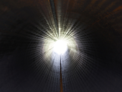
These criticisms of Alexander point out that what he saw was a classic NDE—the white light, the tunnel, the feelings of connectedness, etc. This is effective in dismantling his account of an “immaterial intellect” because, so far, most symptoms of a NDE are in fact scientifically explainable. [ another article on this site provides a thorough description of the evidence, as does this study.]

One might argue that the scientific description of NDE symptoms is merely the physical account of what happens as you cross over. A brain without oxygen may experience “tunnel vision,” but a brain without oxygen is also near death and approaching the afterlife, for example. This argument rests on the fact that you are indeed dying. But without the theological gymnastics, I think there is an overlooked yet critical aspect to the near death phenomenon, one that can render Platform 9 ¾ wholly solid. Studies have shown that you don’t have to be near death to have a near death experience.

“Dying”

In 1990, a study was published in the Lancet that looked at the medical records of people who experienced NDE-like symptoms as a result of some injury or illness. It showed that out of 58 patients who reported “unusual” experiences associated with NDEs (tunnels, light, being outside one’s own body, etc.), 30 of them were not actually in any danger of dying, although they believed they were [1]. The authors of the study concluded that this finding offered support to the physical basis of NDEs, as well as the “transcendental” basis.

Why would the brain react to death (or even imagined death) in such a way? Well, death is a scary thing. Scientific accounts of the NDE characterize it as the body’s psychological and physiological response mechanism to such fear, producing chemicals in the brain that calm the individual while inducing euphoric sensations to reduce trauma.

Imagine an alpine climber whose pick fails to catch the next icy outcropping as he or she plummets towards a craggy mountainside. If one truly believes the next experience he or she will have is an intimate acquainting with a boulder, similar NDE-like sensations may arise (i.e., “My life flashed before my eyes…”). We know this because these men and women have come back to us, emerging from a cushion of snow after their fall rather than becoming a mountain’s Jackson Pollock installation.

You do not have to be, in reality, dying to have a near-death experience. Even if you are dying (but survive), you probably won’t have one. What does this make of Heaven? It follows that if you aren’t even on your way to the afterlife, the scientifically explicable NDE symptoms point to neurology, not paradise.

This Must Be the Place

Explaining the near death experience in a purely physical way is not to say that people cannot have a transformative vision or intense mental journey. The experience is real and tells us quite a bit about the brain (while raising even more fascinating questions about consciousness). But emotional and experiential gravitas says nothing of Heaven, or the afterlife in general. A healthy imbibing of ketamine can induce the same feelings, but rarely do we consider this euphoric haze a glance of God’s paradise.

In this case, as in science, a theory can be shot through with experimentation. As Richard Feynman said, “It doesn’t matter how beautiful your theory is, it doesn’t matter how smart you are. If it doesn’t agree with experiment, it’s wrong.

The experiment is exploring an NDE under different conditions. Can the same sensations be produced when you are in fact not dying? If so, your rapping on the Pearly Gates is an illusion, even if Heaven were real. St. Peter surely can tell the difference between a dying man and a hallucinating one.

The near death experience as a foreshadowing of Heaven is a beautiful theory perhaps, but wrong.

Barring a capricious conception of “God’s plan,” one can experience a beautiful white light at the end of a tunnel while still having a firm grasp of their mortal coil. This is the death of near death. Combine explainable symptoms with a plausible, physical theory as to why we have them and you get a description of what it is like to die, not what it is like to glimpse God.

Sitting atop clouds fluffy and white, Heaven may be waiting. We can’t prove that it is not. But rather than helping to clarify, the near death experience, not dependent on death, may only point to an ever interesting and complex human brain, nothing more.

http://blogs.scientificamerican.com/guest-blog/2012/12/03/the-death-of-near-death-even-if-heaven-is-real-you-arent-seeing-it/

Sexually-deprived fruitflies drink more alcohol

drinking_drosophila

Rejection stinks. It literally hurts. But worse, it has an immediate and negative impact on our brains, producing withdrawal symptoms as if we’re quitting a serious addiction cold turkey. It’s no wonder, then, that we are tempted to turn to drugs to makeourselves feel better. But we’re not the only species that drowns our sorrows when we’re lonely – as a new study in Science reveals, rejected Drosophila do, too. Scientists have found not only will these sexually frustrated flies choose to consume more alcohol than their happily mated peers, sex and alcohol consumption activate the same neurological pathway in their brains.

Drosophila melanogaster males sure know how to woo a lady. When placed in the same container as a potential mate, a male fly will play her a delicate love song by vibrating one wing, caress her rear end, and gently nuzzle her most private of parts with his proboiscis to convince her that he is one heck of a lover. But even the most romantic fly can’t convince an already mated female Drosophila to give up the goods, so scientists were able to use the girls’ steely resolve to see how rejection affects fly drinking behavior.

“Alcohol is one of the most widely used and abused drugs in the world,” explains lead author Galit Shohat-Ophir. “The fruit fly Drosophila melanogaster is an ideal model organism to study how the social environment modulates behavior.” Previous studies have found that Drosophila melanogaster exhibit complex addiction-like behaviors. So in the controlled setting of Ulrike Heberlein’s lab at the University of California San Francisco, researchers paired male fruit flies with three types of females: 1) unmated females, which were willing and happy to mate; 2) mated females, which actively rejected the men; and 3) decapitated females, which didn’t actively reject the guys but, well, weren’t exactly willing partners either. After the flies were satisfied or frustrated, they were offered regular food and food spiked with ethanol, and the researchers measured which type they preferred to see if there was any connection between sex and drinking.

The flies that were rejected drank significantly more than their satisfied peers, but so did the ones paired with incapacitated girls, suggesting that it wasn’t the social aspect of rejection but sexual deprivation that drives male flies to increase their ethanol consumption (see the video at the end!). This alcoholic behavior was very directly related to the guy fly ever getting laid, for even after days of blue balls, if he was allowed to spend some time with a willing woman, he no longer preferred the spiked food.

What the scientists really wanted to understand, though, was why. What drives a frustrated fly to the flask? So to look at the underlying mechanism of this phenomenon, the scientists examined the flies’ brains. A body of scientific literature has connected one particular neurotransmitter, neuropeptide F (NPF), to ethanol-related behaviors in Drosophila, so it was a logical place to start. A very similar neurotransmitter in our brains, called neuropeptide Y (NPY), is linked to alcoholism.

Increased expression of NPF in mated male brains, as shown through immunochemistry.

The team found that sexual frustration caused an immediate decrease in the expression of NPF, while sex increased expression. Furthermore, when they used genetics to artificially knock down NPF levels in the satisfied flies, they drank as much as their not-so-satisfied friends. Similarly, when the researchers artificially increased NPF levels, flies stayed sober. This is the first time NPF levels have connected sexual activity to drinking. Clearly, NPF levels controlled the flies’ desire to drink, so the team further explored how NPF works in the fly’s brain.

Many animals, including ourselves, possess a neurological reward system which reinforces good behavior. Through this system, we ascribe pleasure or positive feelings to things we do that are necessary for species survival, including sex, eating, and social interaction. Drugs tap into this system, stimulating pleasure which can lead to addiction. Previous studies have shown that flies find intoxication rewarding, so the researchers hypothesized that NPF may play a role in the reward system.

Preference tests showed that artificially increasing NPF levels in the absence of sex or ethanol was rewarding to the flies, confirming the scientists’ hypothesis. This was further supported by the discovery that constantly activating NPF abolished the flies’ tendency to consider ethanol rewarding.

“NPF is a currency of reward” explains Shohat-Ophir. High NPF levels signal good behavior in Drosophila brains, thus reinforcing any activities which led to that state. This is a truly novel discovery, for while NPF and the mammal version, NPY, have been linked to alcohol consumption, no animal model has ever placed NPF/NPY in the reward system.

Understanding the role of NPF in reward-seeking behaviors may lead to better treatments for addicts. “In mammals, including humans, NPY may have a similar role [as NPF],” says Shohat-Ophir. “If so, one could argue that activating the NPY system in the proper brain regions might reverse the detrimental effects of traumatic and stressful experiences, which often lead to drug abuse.” Already, NPY and drugs that affect the function of its receptors are in clinical trials for anxiety, PTSD, mood disorders and obesity. This study suggests that perhaps they should be tested as treatment for alcoholism, too, as well as other reward-based addictions.

Research: Shohat-Ophir, G, KR Kaun & R Azanchi (2012). Sexual Deprivation Increases Ethanol Intake in Drosophila. Science 335: 1351-1355.

Click  http://blogs.scientificamerican.com/science-sushi/2012/03/15/flies-drink-upon-rejection/

to view a sequence of  three videos that show a male fly courting and successfully mating with a female fly, another male fly being rejected by a female, and a male choosing to consume an alcohol-infused solution over a non-alcohol solution. Video © Science/AAAS