Urinary biomarker of Parkinson’s disease identified

New findings indicate that phosphorylated LRRK2 (leucine-rich repeat kinase 2) protein levels in urine are elevated in patients diagnosed with idiopathic Parkinson Disease (PD), and that urinary phosphorylated LRRK2 levels correlate with the presence and severity of symptoms such as cognitive impairment in individuals with PD. Researchers affiliated with the University of Alabama at Birmingham published their findings in Neurology and in Movement Disorders (1,2).

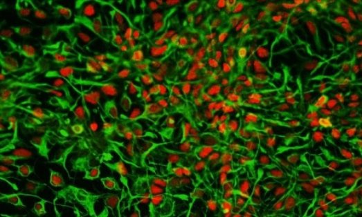
The etiology of PD is currently unknown and mechanisms of action are still not completely clarified. It is well established, however, that aging is the single most important risk factor. PD is the second most frequent age-related neurodegenerative disorder, and one of the key pathogenic features is slow and progressive neuronal death that is concomitant with cognitive dysfunction. Current therapeutic modalities are inadequate and clinical need is significant. More than 6 million individuals worldwide are diagnosed with PD.

To date, several common genetic variants, or single nucleotide polymorphisms (SNPs), have been identified that influence the risk for disease. For example, polymorphic variants in LRRK2 gene have previously been validated as genetic factors that confer susceptibility to PD.

Although the gene remains poorly characterized, five different mutations in the gene encoding LRRK2 are considered a common cause of inherited PD (3). One of the five mutations that are causal is the G2019S mutation in the LRRK2 kinase domain, a mutation that significantly increases phosphorylation activity (1,3).

“There are currently no known ways to predict which G2019S mutation carriers will develop PD,” the authors wrote in the Neurology publication. Investigators purified LRRK2 protein from urinary exosomes collected from a total of 76 men. (Exosomes are membrane vesicles of endosomal origin that are secreted by most cells in culture, and are present in most biological fluids such as urine, blood, and saliva.) Then, they compared the ratio of phosphorylated LRRK2 to total LRRK2 in urine exosomes. Results show that “elevated … phosphorylated LRRK2 predicted the risk” for onset of PD in LRRK2 G2019S mutation carriers (1).

In their follow-up study, which was published in Movement Disorders, investigators compared phosphorylated LRRK2 levels in urine samples of 79 individuals diagnosed with PD to those of 79 healthy control participants. Results show that phosphorylated LRRK2 levels were significantly elevated in patients with PD when compared to those of controls. Also, phosphorylated LRRK2 levels correlated with the severity of cognitive impairment in patients with PD (2).

“Because few viable biomarkers for PD exist … phosphorylated LRRK2 levels may be a promising candidate for further exploration,” the authors concluded in their publication.

References
1. Fraser KB, Moehle MS, Alcalay RN, et al. Urinary LRRK2 phosphorylation predicts parkinsonian phenotypes in G2019S LRRK2 carriers. Neurology. 2016;86:994-999.
2. Fraser KB, Rawlins AB, Clar RG, et al. Ser(P)-1292 LRRK2 in urinary exosomes is elevated in idiopathic Parkinson’s disease. Mov Disord. 2016. doi: 10.1002/mds.26686.
3. Greggio E, Cookson MR. Leucine-rich repeat kinase 2 mutations and Parkinson’s disease: three questions. ASN Neuro. 2009;1:e00002.

http://www.psychiatryadvisor.com/neurocognitive-disorders/urinary-biomarker-of-parkinson-disease-identified/article/508195/?DCMP=EMC-PA_Update_RD&cpn=psych_md,psych_all&hmSubId=&hmEmail=5JIkN8Id_eWz7RlW__D9F5p_RUD7HzdI0&NID=1710903786&dl=0&spMailingID=14919209&spUserID=MTQ4MTYyNjcyNzk2S0&spJobID=820575619&spReportId=ODIwNTc1NjE5S0

Cancer drug nilotinib may reverse Parkinson’s disease

by Jon Hamilton

A drug that’s already approved for treating leukemia appears to dramatically reduce symptoms in people who have Parkinson’s disease with dementia, or a related condition called Lewy body dementia.

A pilot study of 12 patients given small doses of nilotinib found that movement and mental function improved in all of the 11 people who completed the six-month trial, researchers reported Saturday at the Society for Neuroscience meeting in Chicago.

And for several patients the improvements were dramatic, says Fernando Pagan, an author of the study and director of the Movement Disorders Program at Georgetown University Medical Center. One woman regained the ability to feed herself, one man was able to stop using a walker, and three previously nonverbal patients began speaking again, Pagan says.

“After 25 years in Parkinson’s disease research, this is the most excited I’ve ever been,” Pagan says.

If the drug’s effectiveness is confirmed in larger, placebo-controlled studies, nilotinib could become the first treatment to interrupt a process that kills brain cells in Parkinson’s and other neurodegenerative diseases, including Alzheimer’s.

One of the patients in the pilot study was Alan Hoffman, 74, who lives with his wife, Nancy, in Northern Virginia.

Hoffman was diagnosed with Parkinson’s in 1997. At first, he had trouble moving his arms. Over time, walking became more difficult and his speech became slurred. And by 2007, the disease had begun to affect his thinking.

“I knew I’d dropped off in my ability to read,” Hoffman says. “People would keep giving me books and I’d have read the first chapter of about 10 of them. I had no ability to focus on it.”

“He had more and more difficulty making sense,” Nancy Hoffman says. He also became less active, less able to have conversations, and eventually stopped doing even household chores, she says.

But after a few weeks on nilotinib, Hoffman “improved in every way,” his wife says. “He began loading the dishwasher, loading the clothes in the dryer, things he had not done in a long time.”

Even more surprising, Hoffman’s scores on cognitive tests began to improve. At home, Nancy Hoffman says her husband was making sense again and regained his ability to focus. “He actually read the David McCullough book on the Wright Brothers and started reading the paper from beginning to end,” she says.

The idea of using nilotinib to treat people like Alan Hoffman came from Charbel Moussa, an assistant professor of neurology at Georgetown University and an author of the study.

Moussa knew that in people who have Parkinson’s disease with dementia or a related condition called Lewy body dementia, toxic proteins build up in certain brain cells, eventually killing them. Moussa thought nilotinib might be able to reverse this process.

His reasoning was that nilotinib activates a system in cells that works like a garbage disposal — it clears out unwanted proteins. Also, Moussa had shown that while cancer cells tend to die when exposed to nilotinib, brain cells actually become healthier.

So Moussa had his lab try the drug on brain cells in a Petri dish. “And we found that, surprisingly, with a very little amount of the drug we can clear all these proteins that are supposed to be neurotoxic,” he says.

Next, Moussa had his team give the drug to transgenic mice that were almost completely paralyzed from Parkinson’s disease. The treatment “rescued” the animals, he says, allowing them to move almost as well as healthy mice.

Moussa’s mice got the attention of Pagan from Georgetown’s Movement Disorders Program. “When Dr. Moussa showed them to me,” Pagan says, “it looked like, hey, this is type of drug that we’ve been looking for because it goes to the root of the problem.”

The pilot study was designed to determine whether nilotinib was safe for Parkinson’s patients and to determine how much drug from the capsules they were taking was reaching their brains. “But we also saw efficacy, which is really unheard of in a safety study,” Pagan says.

The study found that levels of toxic proteins in blood and spinal fluid decreased once patients began taking nilotinib. Also, tests showed that the symptoms of Parkinson’s including tremor and “freezing” decreased. And during the study patients were able to use lower doses of Parkinson’s drugs, suggesting that the brain cells that produce dopamine were working better.

But there are some caveats, Pagan says. For one thing, the study was small, not designed to measure effectiveness, and included no patients taking a placebo.

Also, nilotinib is very expensive. The cost of providing it to leukemia patients is thousands of dollars a month.

And finally, Parkinson’s and dementia patients would have to keep taking nilotinib indefinitely or their symptoms would continue to get worse.

Alan Hoffman was okay for about three weeks after the study ended and he stopped taking the drug. Since then, “There’s (been) a pretty big change,” his wife says. “He does have more problems with his speech, and he has more problems with cognition and more problems with mobility.”

The Hoffmans hope to get more nilotinib from the drug’s maker, Novartis, through a special program for people who improve during experiments like this one.

Meanwhile, the Georgetown team plans to try nilotinib in patients with another brain disease that involves toxic proteins: Alzheimer’s.

http://www.npr.org/sections/health-shots/2015/10/17/448323916/can-a-cancer-drug-reverse-parkinsons-disease-and-dementia

Eye tests may predict schizophrenia

Schizophrenia is associated with structural and functional alterations of the visual system, including specific structural changes in the eye. Tracking such changes may provide new measures of risk for, and progression of the disease, according to a literature review published online in the journal Schizophrenia Research: Cognition, authored by researchers at New York Eye and Ear Infirmary of Mount Sinai and Rutgers University.

Individuals with schizophrenia have trouble with social interactions and in recognizing what is real. Past research has suggested that, in schizophrenia, abnormalities in the way the brain processes visual information contribute to these problems by making it harder to track moving objects, perceive depth, draw contrast between light and dark or different colors, organize visual elements into shapes, and recognize facial expressions. Surprisingly though, there has been very little prior work investigating whether differences in the retina or other eye structures contribute to these disturbances.

“Our analysis of many studies suggests that measuring retinal changes may help doctors in the future to adjust schizophrenia treatment for each patient,” said study co-author Richard B. Rosen, MD, Director of Ophthalmology Research, New York Eye and Ear Infirmary of Mount Sinai, and Professor of Ophthalmology, Icahn School of Medicine at Mount Sinai. “More studies are needed to drive the understanding of the contribution of retinal and other ocular pathology to disturbances seen in these patients, and our results will help guide future research.”

The link between vision problems and schizophrenia is well established, with as many as 62 percent of adult patients with schizophrenia experience visual distortions involving form, motion, or color. One past study found that poorer visual acuity at four years of age predicted a diagnosis of schizophrenia in adulthood, and another that children who later develop schizophrenia have elevated rates of strabismus, or misalignment of the eyes, compared to the general population.

Dr. Rosen and Steven M. Silverstein, PhD, Director of the Division of Schizophrenia Research at Rutgers University Behavioral Health Care, were the lead authors of the analysis, which examined the results of approximately 170 existing studies and grouped the findings into multiple categories, including changes in the retina vs. other parts of the eye, and changes related to dopamine vs. other neurotransmitters, key brain chemicals associated with the disease.

The newly published review found multiple, replicated, indicators of eye abnormalities in schizophrenia. One of these involves widening of small blood vessels in the eyes of schizophrenia patients, and in young people at high risk for the disorder, perhaps caused by chronic low oxygen supply to the brain. This could explain several key vision changes and serve as a marker of disease risk and worsening. Also important in this regard was thinning of the retinal nerve fiber layer in schizophrenia, which is known to be related to the onset of hallucinations and visual acuity problems in patients with Parkinson’s disease. In addition, abnormal electrical responses by retinal cells exposed to light (as measured by electroretinography) suggest cellular-level differences in the eyes of schizophrenia patients, and may represents a third useful measure of disease progression, according to the authors.

In addition, the review highlighted the potentially detrimental effects of dopamine receptor-blocking medications on visual function in schizophrenia (secondary to their retinal effects), and the need for further research on effects of excessive retinal glutamate on visual disturbances in the disorder.

Interestingly, the analysis found that there are no reports of people with schizophrenia who were born blind, suggesting that congenital blindness may completely or partially protect against the development of schizophrenia. Because congenitally blind people tend to have cognitive abilities in certain domains (e.g., attention) that are superior to those of healthy individuals, understanding brain re-organization after blindness may have implications for designing cognitive remediation interventions for people with schizophrenia.

“The retina develops from the same tissue as the brain,” said Dr. Rosen. “Thus retinal changes may parallel or mirror the integrity of brain structure and function. When present in children, these changes may suggest an increased risk for schizophrenia in later life. Additional research is needed to clarify these relationships, with the goals of better predicting emergence of schizophrenia, and of predicting relapse and treatment response and people diagnosed with the condition.”

Dr. Silverstein points out that, to date, vision has been understudied in schizophrenia, and studies of the retina and other ocular structures in the disorder are in their infancy. However, he added, “because it is much faster and less expensive to obtain data on retinal structure and function, compared to brain structure and function, measures of retinal and ocular structure and function may have an important role in both future research studies and the routine clinical care of people with schizophrenia.”

http://www.eurekalert.org/pub_releases/2015-08/tmsh-rcm081715.php

Medication for Parkinson’s disease shown to lower morality and increase willingness to harm others


Healthy people who are given commonly prescribed mood-altering drugs see significant changes in the degree to which they are willing to tolerate harm against themselves and others, according to a study published Thursday. The research has implications for understanding human morality and decision-making.

A team of scientists from the University College London (UCL) and Oxford University found that healthy people who were given the serotonin-boosting antidepressant citalopram were willing to pay twice as much to prevent harm to themselves or others, compared to those given a placebo. By contrast, those who were given a dose of the dopamine-enhancing Parkinson’s drug levodopa made more selfish decisions, overcoming an existing tendency to prefer harming themselves over others.

The researchers said their findings, published in the journal Current Biology, provided clues to the neurological and chemical roots of common clinical disorders like psychopathy, which causes people to disregard the emotions of others.

The researchers compared how much pain subjects were willing to anonymously inflict on themselves or other people in exchange for money. Out of 175 subjects, 89 were given citalopram or a placebo and 86 were given levodopa or a placebo.

They were anonymously paired up into decision-makers and receivers, and all subjects were given shocks at their pain threshold. The decision-makers were then allowed to choose a different amount of money in exchange for a different amount of shocks, either to themselves or the receivers.

On average, people who were given a placebo were willing to pay about 35p per shock to prevent harm to themselves and 44p per shock to prevent harm to others. Those who were given citalopram became more averse to harm, paying an average of 60p to avoid harm to themselves and 73p per shock to avoid harm to others. This meant that citalopram users, on average, delivered 30 fewer shocks to themselves and 35 fewer shocks to others.

However, those who were given levodopa became more selfish, showing no difference in the amount they were willing to pay to prevent shocks to themselves or others. On average, they were willing to pay about 35p per shock to prevent harm to themselves or others, meaning that they delivered on average about 10 more shocks to others during the trial than those who took a placebo. They also showed less hesitation about shocking others than those given the placebo.

Similar research conducted by the same team in November found that subjects were willing to spare the stranger pain twice as often as they spared themselves, indicating that they preferred harming themselves over others for profit, a behavior known as “hyper-altruism.”

“Our findings have implications for potential lines of treatment for antisocial behavior, as they help us to understand how serotonin and dopamine affect people’s willingness to harm others for personal gain,” Molly Crockett of UCL, the study’s lead author, said in a press release. “We have shown that commonly-prescribed psychiatric drugs influence moral decisions in healthy people, raising important ethical questions about the use of such drugs.

“It is important to stress, however, that these drugs may have different effects in psychiatric patients compared to healthy people. More research is needed to determine whether these drugs affect moral decisions in people who take them for medical reasons.”

http://www.ibtimes.com/antidepressants-affect-morality-decision-making-new-study-finds-1995363

New typing test may help diagnose Parkinson’s disease

Whether it’s on a keyboard, a smartphone, or even a credit card reader, you spend a lot of your day typing. Well, researchers at MIT noticed the value of this daily habit, and are putting it to a secondary use; they’ve developed software that can gauge the speed at which a typist is tapping the keyboard to help diagnose Parkinson’s disease.

In order to type a word, your brain has to send signals down through your spinal cord to the nerves that operate your fingers. If your central nervous system is functioning perfectly, then you should be able to tap most of the keys at a fairly constant rate. But a number of conditions might slow the signal from the brain to the fingers, such as sleep deprivation (which slows all motor skills) and diseases that affect the central nervous system, including Parkinson’s.

For the first version of this study, the researchers were looking at typing patterns that indicated whether a person was sleep-deprived or well rested. They created a browser plug-in that detected the timing at which the volunteers hit they keys and found that the people who were sleepy had a much wider variation in their typing speed. They found similar results in their preliminary test with Parkinson’s patients; the 21 typists with Parkinson’s tapped the keys at much more variable rates than the 15 healthy volunteers. The researchers called it a “window into the brain.”

Right now, the algorithm they’ve developed is not refined enough to distinguish Parkinson’s patients from people who are sleep deprived, though the results might be clearer after a number of trials. The researchers plan to conduct a study with a larger group of subjects, but they hope that this type test could eventually lead to earlier diagnoses of Parkinson’s–today most people are diagnosed after they have had symptoms for 5-10 years–and to distinguish Parkinson’s from other conditions that might affect a person’s motor skills, like rheumatoid arthritis. They are currently developing a smartphone app that can test participants even more easily.

http://www.popsci.com/type-test-diagnose-parkinsons

New research shows that drug for Parkinson’s disease (benztropine) may also treat multiple sclerosis

benzotropine

A drug that treats Parkinson’s disease might also work against multiple sclerosis, or MS.

In MS patients, an aberrant immune onslaught degrades the fatty myelin sheaths that coat nerve fibers, causing blurred vision, weakness, loss of coordination and other symptoms.

Luke Lairson of the Scripps Research Institute in La Jolla, Calif., and colleagues tested a host of compounds to see which might boost regeneration of oligodendrocytes, the brain cells that make myelin and which are often lacking in MS. Using the cells’ forerunners, nascent brain cells called oligodendrocyte precursor cells, from rats and mice, the researchers found that benztropine proved adept at steering these cells to become myelin-making oligodendrocytes.

The researchers then induced in mice a disease that mimics MS and gave some of the animals benztropine, others a standard MS drug (fingolimod or interferon beta) and some no drug at all. Whether given before or after disease onset, benztropine reduced symptom severity and prevented relapses better than other MS drugs. Mice getting no drug fared the poorest, according to results appearing October 9 in Nature.

A cell count of brain tissue revealed that mice getting benztropine had substantially more mature oligodendrocytes than mice getting no drug. Further analyses suggested the animals’ symptom improvement with benztropine resulted from a rebuilding of the myelin sheaths, not from suppressing the animals’ immune systems. The researchers think the drug, if approved for use in MS, might work in concert with immune-suppressing drugs.

Old drug may have new trick

How Exercise Beefs Up the Brain

exercise

New research explains how abstract benefits of exercise—from reversing depression to fighting cognitive decline—might arise from a group of key molecules.

While our muscles pump iron, our cells pump out something else: molecules that help maintain a healthy brain. But scientists have struggled to account for the well-known mental benefits of exercise, from counteracting depression and aging to fighting Alzheimer’s and Parkinson’s disease. Now, a research team may have finally found a molecular link between a workout and a healthy brain.

Much exercise research focuses on the parts of our body that do the heavy lifting. Muscle cells ramp up production of a protein called FNDC5 during a workout. A fragment of this protein, known as irisin, gets lopped off and released into the bloodstream, where it drives the formation of brown fat cells, thought to protect against diseases such as diabetes and obesity. (White fat cells are traditionally the villains.)

While studying the effects of FNDC5 in muscles, cellular biologist Bruce Spiegelman of Harvard Medical School in Boston happened upon some startling results: Mice that did not produce a so-called co-activator of FNDC5 production, known as PGC-1α, were hyperactive and had tiny holes in certain parts of their brains. Other studies showed that FNDC5 and PGC-1α are present in the brain, not just the muscles, and that both might play a role in the development of neurons.

Spiegelman and his colleagues suspected that FNDC5 (and the irisin created from it) was responsible for exercise-induced benefits to the brain—in particular, increased levels of a crucial protein called brain-derived neurotrophic factor (BDNF), which is essential for maintaining healthy neurons and creating new ones. These functions are crucial to staving off neurological diseases, including Alzheimer’s and Parkinson’s. And the link between exercise and BDNF is widely accepted. “The phenomenon has been established over the course of, easily, the last decade,” says neuroscientist Barbara Hempstead of Weill Cornell Medical College in New York City, who was not involved in the new work. “It’s just, we didn’t understand the mechanism.”

To sort out that mechanism, Spiegelman and his colleagues performed a series of experiments in living mice and cultured mouse brain cells. First, they put mice on a 30-day endurance training regimen. They didn’t have to coerce their subjects, because running is part of a mouse’s natural foraging behavior. “It’s harder to get them to lift weights,” Spiegelman notes. The mice with access to a running wheel ran the equivalent of a 5K every night.

Aside from physical differences between wheel-trained mice and sedentary ones—“they just look a little bit more like a couch potato,” says co-author Christiane Wrann, also of Harvard Medical School, of the latter’s plumper figures—the groups also showed neurological differences. The runners had more FNDC5 in their hippocampus, an area of the brain responsible for learning and memory.

Using mouse brain cells developing in a dish, the group next showed that increasing the levels of the co-activator PGC-1α boosts FNDC5 production, which in turn drives BDNF genes to produce more of the vital neuron-forming BDNF protein. They report these results online today in Cell Metabolism. Spiegelman says it was surprising to find that the molecular process in neurons mirrors what happens in muscles as we exercise. “What was weird is the same pathway is induced in the brain,” he says, “and as you know, with exercise, the brain does not move.”

So how is the brain getting the signal to make BDNF? Some have theorized that neural activity during exercise (as we coordinate our body movements, for example) accounts for changes in the brain. But it’s also possible that factors outside the brain, like those proteins secreted from muscle cells, are the driving force. To test whether irisin created elsewhere in the body can still drive BDNF production in the brain, the group injected a virus into the mouse’s bloodstream that causes the liver to produce and secrete elevated levels of irisin. They saw the same effect as in exercise: increased BDNF levels in the hippocampus. This suggests that irisin could be capable of passing the blood-brain barrier, or that it regulates some other (unknown) molecule that crosses into the brain, Spiegelman says.

Hempstead calls the findings “very exciting,” and believes this research finally begins to explain how exercise relates to BDNF and other so-called neurotrophins that keep the brain healthy. “I think it answers the question that most of us have posed in our own heads for many years.”

The effect of liver-produced irisin on the brain is a “pretty cool and somewhat surprising finding,” says Pontus Boström, a diabetes researcher at the Karolinska Institute in Sweden. But Boström, who was among the first scientists to identify irisin in muscle tissue, says the work doesn’t answer a fundamental question: How much of exercise’s BDNF-promoting effects come from irisin reaching the brain from muscle cells via the bloodstream, and how much are from irisin created in the brain?

Though the authors point out that other important regulator proteins likely play a role in driving BDNF and other brain-nourishing factors, they are focusing on the benefits of irisin and hope to develop an injectable form of FNDC5 as a potential treatment for neurological diseases and to improve brain health with aging.

http://news.sciencemag.org/biology/2013/10/how-exercise-beefs-brain

Thanks to Dr. Rajadhyaksha for bringing this to the attention of the It’s Interesting community.

Chinese scientists turn human urine into brain cells

NPGs_from_urine


Chinese researchers have developed a new technique for isolating kidney cells from urine and turning them into neural progenitors — –immature brain cells that can develop into various types of glial cells and neurons. Reprogramming cells has been done before, of course, but not with cells gleaned from urine and not via a method this direct. The technique could prove extremely helpful to those pursuing treatments for neurodegenerative disorders like Parkinson’s and Alzheimer’s.

The innovation here is in the source and the method. We know that embryonic stem cells offer potential treatments for neurodegenerative disorders. And we know that we can turn adult human cells–that is, non-embryonic cells gathered from adult humans–into pluripotent cells (those that can become a different type of cell) by reprogramming them, usually with genetically engineered viruses that tamper with the cells’ genetic codes.

But embryonic stem cell treatments are fraught with ethical issues and non-embryonic methods are complicated–and complexity introduces a greater chance of something going wrong (in this case that means mutations and genetic defects). The new method, which taps skin-like cells from the linings of the kidney tubes that are present in urine, converts its source cells into neurons and glia cells via a more direct route, making the process more efficient while narrowing the margin of error.

In their study, the researchers harvested kidney cells from the urine samples of three human donors and converted the cells directly to neural progenitors. Rather than using a genetically engineered virus to reprogram the cells, they used a small piece of bacterial DNA that can replicate in the cellular cytoplasm, a technique that eliminates the need to tamper directly with the chromosome (in theory, at least, this should reduce mutations) while also speeding up the entire process. After growing their progenitors into mature neurons and glial cells, the researchers transplanted the progenitors into the brains of newborn rats. A month later, the cells were still alive in the rats’ brains, though it is not yet clear that they can survive for extended periods or mesh with the brain’s wiring to become functioning parts of the neural machine.

There’s still a lot of research to be done on this method of course, but the researchers think it may provide a way to take cells gathered non-invasively and quickly and efficiently convert them into neural cells while reducing the likelihood of genetic mutations.

http://www.guardian.co.uk/science/neurophilosophy/2012/dec/09/turning-urine-into-brain-cells


Wiring the Brain to Treat Depression


The procedure starts with a surgeon drilling two holes in the patient’s skull. “Every bone and tooth in my head was rattling,” says Lisa Battiloro, who was awake, but not in pain, during the eight-hour operation.

Neurologists asked her questions and issued commands as they pinpointed the exact spot in her brain for electrical stimulation. At one point, “I suddenly felt hopeful and optimistic about the future,” recalls Ms. Battiloro, who had battled severe depression for more than a decade. That’s when the doctors knew they had found Brodmann 25, an area deep in the cerebral cortex associated with negative mood. They secured the electrodes in place, then sedated Ms. Battiloro while they ran an extension wire under the skin, down the side of her head and into her chest, where they implanted a battery pack to supply her brain with a mild electrical current.

Within two months, Ms. Battiloro says, her depression had lifted considerably. Now, nearly four years later, it hasn’t returned. “My friends and family are amazed,” say Ms. Battiloro, 41, of Boynton Beach, Fla. “I’m a new and improved Lisa.”

Deep brain stimulation, sometimes called a pacemaker for the brain, has helped halt tremors in more than 100,000 patients with Parkinson’s disease and other movement disorders since 1997. Now, researchers are reporting encouraging results using the procedure for psychiatric conditions as well. Ms. Battiloro was one of 17 patients in a study published this month in the Archives of General Psychiatry. After two years of DBS, 92% reported significant relief from their major depression or bipolar disorder and more than half were in remission, with no manic side effects.

“We are seeing dramatic effects in the small numbers of subjects, and they are not just getting well, they are getting well without side effects and without relapsing,” says neurologist Helen Mayberg, who led the study at Emory University in Atlanta.

read more here:  http://online.wsj.com/article/SB10001424052970204555904577164813955136748.html#articleTabs=article


A Dearth of New Medications for Neuropsychiatric Disease on the Horizon


Neuropsychiatric diseases like schizophrenia, depression, Alzheimer’s disease, Parkinson’s disease and more cost billions per year and account for 13% of the global burden of disease (a measure of years of life lost due to premature mortality and living in a state less than full health), according to the World Health Organization.

However, pharmaceutical companies have decided that generating new drugs to treat these disorders are simply too costly to pursue, and are pulling the plug on research and development in this area.

Read more here:  http://www.scientificamerican.com/article.cfm?id=a-dearth-of-new-meds