New research shows that blood from young mice reverses aging in brain and muscle

In a trio of studies published Sunday, scientists reported that they reversed aging in the muscles and brains of old mice — simply by running the blood of young mice through their veins.

The papers, from two independent groups in Cambridge and California, used different approaches to begin to unravel the rejuvenating effects of young animals’ blood, in the hopes of eventually developing a therapy that could be tested in people.

Researchers at Harvard University administered a protein found in young blood to older mice, and found that treated mice could run longer on a treadmill and had more branching blood vessels in their brains than untreated mice. A group led by a University of California, San Francisco researcher identified a molecular switch in a memory center of the brain that appears to be turned on by blood from young mice.

“These are the tissues that are really affected by advancing age. Changes in these tissues are responsible for the changes that people worry about the most — loss of cognition and loss of independent function,” said Amy Wagers, a professor of stem cell and regenerative biology at Harvard University involved in two of the studies.

Wagers said many questions remain about the mechanism of the protein and what the best therapeutic strategy might be, but she is already working to commercialize the protein discovery. The same substance is found in human blood.

Outside scientists cautioned that the findings are limited to one strain of mice and that it is not yet clear that something so simple would have dramatic anti-aging effects in people.

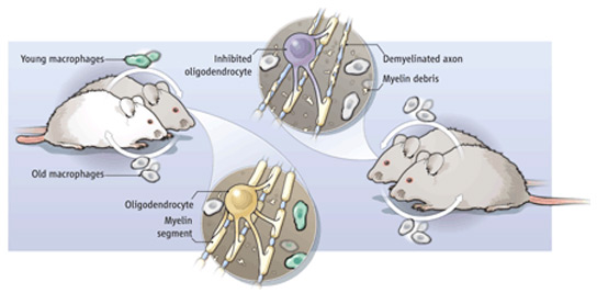
The new studies build on a decade of research that showed that young blood can have a rejuvenating effect on older mice. When scientists stitched together the circulatory systems of pairs of old and young mice, in a procedure called parabiosis, they found beneficial effects on the cells of the spinal cord, muscles, brain, and liver of the older animals. The next question was why — which of the many substances floating around in blood were responsible for the changes, and how did it work?

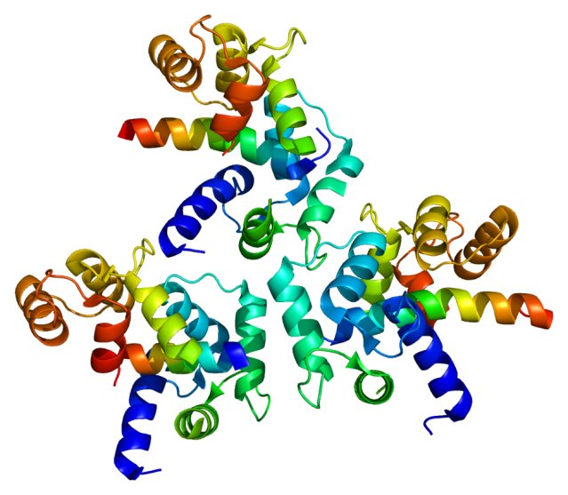
Last year, Wagers and another Harvard stem cell scientist, Dr. Richard T. Lee, found that a protein called GDF11 could cause a mouse heart thickened with age to revert to a youthful state. No one knew, however, whether the effect was specific to the heart, or would apply to aging in other tissues. Two of the new papers, published online by the journal Science, extend that work to the mouse brain and muscle.

In one study, Wagers and colleagues first connected the blood vessels of old and young mice. They measured profound changes to muscle stem cells in the older mice that made the cells appear more youthful. There were also changes to the structure of muscle. Next, they injected the protein that had been shown to rejuvenate hearts into the older mice. Although some individual mice did not change much, on average, the treated mice could run nearly twice as long on a treadmill as older mice not given the protein. The protein had no effect when injected into younger mice.

In a second study, Dr. Lee Rubin, director of translational medicine at the Harvard Stem Cell Institute, found that after parabiosis, the older mice had an increase in the branching network of blood vessels in the brain and in the rate of creation of new brain cells. Treated mice were more sensitive to changes in smell, suggesting the new neurons had an effect on their abilities. The GDF11 protein alone resulted in similar structural changes.

Wagers said that she has begun working with Atlas Venture, a venture capital firm based in Cambridge, to come up with a strategy to turn the insights about GDF11 into potential treatments that could be tested in people.

David Harrison, an aging researcher at Jackson Laboratory, a nonprofit research organization based in Bar Harbor, Maine, who was not involved in the research, said that an important caveat about the research is that it was done on a particular strain of mouse that is inbred. It will be important, he said, to test the protein’s effect in a more genetically diverse population of mice before thinking about extending the work to clinical trials.

Thomas Rando, a professor of neurology at Stanford University School of Medicine who pioneered using the parabiosis technique to study aging, said it is important to try and understand how young blood has its potent effects. But he said it seems very unlikely, given how complex aging is, that reversing it will depend on a single pathway.

“My answer always was and always will be there’s no way there’s a factor,” Rando said. “There are going to be hundreds of factors.”

In the third study published in the journal Nature Medicine, researchers from the University of California, San Francisco and Stanford used parabiosis to search for changes in gene activity in the brain that might help point to how young blood had its effects. They found changes in the activity of genes involved in the connectivity of brain cells in the hippocampus, a memory center.

Instead of using a specific protein, the researchers then gave older mice repeated transfusions of blood from young mice and found that the older animals improved on specific age-related memory tasks, such as locating an underwater platform and remembering an environment where they had experienced an unpleasant foot shock.

Saul Villeda, a UCSF faculty fellow who led the work, said that the results of the three studies reinforce one another, but they differ in their approach.

“I’m really interested to see whether GDF11 accounts for everything, or whether it’s going to be a combination of factors that together that has the full effect,” Villeda said.

All the researchers warned that people hoping to reverse aging shouldn’t get any wild ideas about infusing themselves with young blood, although they acknowledged making their share of vampire jokes.

“I am the oldest member of the team here, and I personally understand the sentiment for patients,” Rubin said. But he still wouldn’t try it.

Written by Carolyn Y. Johnson, who can be reached at cjohnson@globe.com. Follow her on Twitter @carolynyjohnson.

http://www.bostonglobe.com/news/science/2014/05/04/blood-from-young-mice-reverses-aging-brain-muscles/iepDMMf7wrLJy6WgXqpdIJ/story.html?rss_id=Top-GNP&google_editors_picks=true

Thanks to Da Brayn for bringing this to the attention of the It’s Interesting community

New study shows that anger outbursts acutely raise risk of heart attack and stroke

Getting really angry might be more dangerous than you think.

A new study found people who experienced severe anger outbursts were more at risk for cardiovascular events in the two hours following the outbursts compared to those who remained calm.

“The relative risk was similar for people who had known pre-existing heart disease and those who didn’t,” says Dr. Murray A. Mittleman, senior study author and an associate professor of medicine at Harvard Medical School.

The study was designed so that each patient was compared to his or her own baseline risk. “A person with pre-existing heart disease or cardiovascular disease, the absolute risk they are incurring is much greater than (that of) a person without cardiovascular disease or risk factors,” Mittleman says.
“If we look at somebody at higher risk for having cardiovascular events, and they get angry multiple times a day, this can lead to 650 extra heart attacks per year out of 10, 000 a year,” he says. “When we look at a person who is relatively low risk, but if they do have these episodes of anger fairly frequently, we estimate there would be about 150 extra heart attacks out of 10,000 a year.”

Smoking, high cholesterol, high blood pressure, being overweight and having diabetes are all risk factors for cardiovascular disease. An estimated 17 million people worldwide die of cardiovascular diseases, particularly heart attacks and strokes, each year, according to the Centers for Disease Control and Prevention.

The study published Monday in the European Heart Journal was a data analysis looking at nine studies where anger and cardiovascular events were self-reported over nearly two decades. The study found a 4.74 times higher risk of MI (myocardial infarction, or heart attack) or ACS (acute coronary syndrome, where the heart muscle doesn’t get enough oxygen-rich blood) following outbursts of anger.

“Anger causes our heart rate to increase through the sympathetic nervous system and causes our stress hormones to become elevated (the fight or flight mechanism),” says Dr. Mariell Jessup, president of the American Heart Association and medical director of the Penn Heart and Vascular Center at the University of Pennsylvania. “We breathe faster, all of which may trigger undesirable reactions in our blood pressure or in our arteries.”

This disruption may mean the heart or the brain doesn’t get the blood and oxygen they need resulting in a heart attack or a stroke, she says.

Researchers suggest more needs to be done to come up with effective interventions to prevent cardiovascular events triggered by anger outbursts. The American Heart Association suggests regular physical activity, finding a way to relax or talking with friends to help reduce stress and anger.

Mittleman suggests the best way to lower your risk for a heart attack or stroke during an angry outburst is to lower your overall baseline level of risk – exercise, eat healthy and don’t smoke – and then find ways to cope with stress and anger.

Angry outbursts may raise heart attack, stroke risk

Placebo Effect May Account for Half of Drug’s Efficacy

placebo-effect-one-a-day

Even when a medication works, half of its impact on a patient may be due to one aspect of the placebo effect: the positive message that a doctor provides when prescribing the treatment, according to a new study.

Researchers designed an elaborate study, in which 66 people suffering from migraine headaches were given either a placebo, or a common migraine drug called Maxalt. However, for each migraine attack the participants had during the study period, they were told something different. For example, they were told they were taking a placebo when they were actually taking Maxalt, or vice versa, and sometimes they were told the pill could be either Maxalt or a placebo.

The pain-relieving benefits of the migraine drug increased when patients were told they were taking an effective drug for the treatment of acute migraine. And when the identities of Maxalt tablets and placebo pills were switched, patients reported similar pain relief from placebo pills labeled as Maxalt as from Maxalt tablets labeled as a placebo, according to the study published January 8 in the journal Science Translational Medicine.

The results suggest that the information people have is as important as the effects of the drug in reducing pain, the researchers said.

“In many conditions, placebo effect is a big part of the effect of the drug,” said study researcher, Ted Kaptchuk, a professor of medicine at Harvard Medical School. In the new study, 50 percent of the drug’s effect could be attributed to the placebo effect, he said.

“Themore you give a positive message, the more a drug works. In this case, our message was just as important as the pharmacology of the drug,” Kaptchuk said.

In other words, patients may benefit from optimistic messages from their doctors, which may enhance the effectiveness of a good pharmaceutical, the researchers said.

“When doctors set patients’ expectations high, Maxalt [or, potentially, other migraine drugs] becomes more effective,” said study researcher Rami Burstein, a professor of anesthesia at Harvard Medical School. “Increased effectiveness means shorter migraine attacks and shorter migraine attacks mean that less medication is needed,” Burstein said.

However, physicians should be realistic when prescribing a treatment, Kaptchuk said.

“The medical community should consider what’s the positive message that is still accurate, and not an exaggeration that verges on deception,” he told LiveScience.

Migraine attacks are throbbing headaches, usually accompanied by nausea, vomiting and sensitivity to light and sound. The researchers decided to look at migraine, because it is a recurring condition, and responds well to medication, Kaptchuk said.

During the study, the participants had a total of 450 migraine attacks. Each time they were provided with one of the six available treatments: two were made with positive expectations (envelopes labeled “Maxalt”), two were made with negative expectations (envelopes labeled “placebo”), and two were made with neutral expectations (envelopes labeled “Maxalt or placebo”).

But within each of these conditions, the envelopes contained either the placebo or Maxalt. The patients then reported their pain experiences.

“When patients received Maxalt labeled as placebo, they were being treated by the medication — but without any positive expectation,” Burstein said.

For both placebo and Maxalt, patients reported great pain-relieving effects when the envelope was labeled “Maxalt.” This suggests that a positive message and a powerful medication are both important for effective clinical care, the researchers said.
The placebo effect is centered on the idea that a person’s expectations and beliefs drive changes in symptoms, even though they have received a sugar pill or a sham treatment with no effect. Knowing that they have received a placebo changes their expectations, which is expected to alter the placebo effect.

However, people in the study also reported pain relief even when they knew the pill they were receiving was a placebo, compared with no treatment at all.

This finding “contradicts the medical beliefs,” Kaptchuk said. “Because in medicine, we think you have to think it’s a real drug for placebo to work. But apparently, the body has memories, or an embodied awareness, which operates below the level of consciousness.”

One possible mechanism for this effect could be that the body is conditioned to react positively in medical situations, Kaptchuk said.

“We know from other studies that the symbols, the rituals and the words of medicine activate the brain to release neurotransmitters that change the experience of illness. It activates centers in the brain that modulate many symptoms like pain and nausea and fatigue,” he said.

http://www.livescience.com/42430-placebo-effect-half-of-drug-efficacy.html

More evidence that mindfullness meditation can help with anxiety, depression and pain

mindfulness-meditation

By Andrew M. Seaman

Mindfulness meditation may be useful in battles against anxiety, depression and pain, according to a fresh look at past research.

Using data from 47 earlier studies, researchers found moderate evidence to support the use of mindfulness meditation to treat those conditions. Meditation didn’t seem to affect mood, sleep or substance use.

“Many people have the idea that meditation means just sitting quietly and doing nothing,” wrote Dr. Madhav Goyal in an email to Reuters Health. “That is not true. It is an active training of the mind to increase awareness, and different meditation programs approach this in different ways.”

Goyal led the study at The Johns Hopkins University in Baltimore.

He and his colleagues write in JAMA Internal Medicine that meditation techniques emphasize mindfulness and concentration.

So-called mindfulness meditation is aimed at allowing the mind to pay attention to whatever thoughts enter it, such as sounds in the environment, without becoming too focused. Mantra meditation, on the other hand, involves focusing concentration on a particular word or sound.

Approximately 9 percent of people in the U.S. reported meditating in 2007, according to the National Institutes of Health. About 1 percent said they use meditation as some sort of treatment or medicine.

For the new report, the researchers searched several electronic databases that catalog medical research for trials that randomly assigned people with a certain condition – such as anxiety, pain or depression – to do meditation or another activity. These randomized controlled trials are considered the gold standard of medical research.

The researchers found 47 studies with over 3,500 participants that met their criteria.

After combining the data, Goyal said his team found between a 5 and 10 percent improvement in anxiety symptoms among people who took part in mindfulness meditation, compared to those who did another activity.

There was also about a 10 to 20 percent improvement in symptoms of depression among those who practiced mindfulness meditation, compared to the other group.

“This is similar to the effects that other studies have found for the use of antidepressants in similar populations,” Goyal said.

Mindfulness meditation was also tied to reduced pain. But Goyal said it’s hard to know what kind of pain may be most affected by meditation.

The benefits of meditation didn’t surpass what is typically associated with other treatments, such as drugs and exercise, for those conditions.

“As with many therapies, we try to get a moderate level of confidence that the therapy works before we prescribe it,” Goyal said. “If we have a high level of confidence, it is much better.”

But he noted that the researchers didn’t find anything more than moderate evidence of benefit from meditation for anxiety, depression and pain.

There was some suggestion that meditation may help improve stress and overall mental health, but the evidence supporting those findings was of low quality.

There was no clear evidence that meditation could influence positive mood, attention, substance use, eating habits, sleep or weight.

“Clinicians should be prepared to talk with their patients about the role that meditation programs could have in addressing psychological stress, particularly when symptoms are mild,” Goyal said.

Dr. Allan Goroll, who wrote an editorial accompanying the new study, told Reuters Health the analysis is an example of an area of much-needed scientific study, because many people make treatment decisions based on beliefs – not data.

“That is particularly the case with alternative and complimentary approaches to treating medical problems,” he said. “It ranges from taking vitamins to undergoing particular procedures for which the scientific evidence is very slim but people’s beliefs are very great.”

Goroll is professor at Harvard Medical School and Massachusetts General Hospital in Boston.

Goyal said people should remember that meditation was not conceived to treat any particular health problem.

“Rather, it is a path we travel on to increase our awareness and gain insight into our lives,” he wrote. “The best reason to meditate is to gain this insight. Improvements in health conditions are really a side benefit, and it’s best to think of them that way.”

SOURCE: bit.ly/WiwDtv JAMA Internal Medicine, online January 6, 2014.

How Exercise Beefs Up the Brain

exercise

New research explains how abstract benefits of exercise—from reversing depression to fighting cognitive decline—might arise from a group of key molecules.

While our muscles pump iron, our cells pump out something else: molecules that help maintain a healthy brain. But scientists have struggled to account for the well-known mental benefits of exercise, from counteracting depression and aging to fighting Alzheimer’s and Parkinson’s disease. Now, a research team may have finally found a molecular link between a workout and a healthy brain.

Much exercise research focuses on the parts of our body that do the heavy lifting. Muscle cells ramp up production of a protein called FNDC5 during a workout. A fragment of this protein, known as irisin, gets lopped off and released into the bloodstream, where it drives the formation of brown fat cells, thought to protect against diseases such as diabetes and obesity. (White fat cells are traditionally the villains.)

While studying the effects of FNDC5 in muscles, cellular biologist Bruce Spiegelman of Harvard Medical School in Boston happened upon some startling results: Mice that did not produce a so-called co-activator of FNDC5 production, known as PGC-1α, were hyperactive and had tiny holes in certain parts of their brains. Other studies showed that FNDC5 and PGC-1α are present in the brain, not just the muscles, and that both might play a role in the development of neurons.

Spiegelman and his colleagues suspected that FNDC5 (and the irisin created from it) was responsible for exercise-induced benefits to the brain—in particular, increased levels of a crucial protein called brain-derived neurotrophic factor (BDNF), which is essential for maintaining healthy neurons and creating new ones. These functions are crucial to staving off neurological diseases, including Alzheimer’s and Parkinson’s. And the link between exercise and BDNF is widely accepted. “The phenomenon has been established over the course of, easily, the last decade,” says neuroscientist Barbara Hempstead of Weill Cornell Medical College in New York City, who was not involved in the new work. “It’s just, we didn’t understand the mechanism.”

To sort out that mechanism, Spiegelman and his colleagues performed a series of experiments in living mice and cultured mouse brain cells. First, they put mice on a 30-day endurance training regimen. They didn’t have to coerce their subjects, because running is part of a mouse’s natural foraging behavior. “It’s harder to get them to lift weights,” Spiegelman notes. The mice with access to a running wheel ran the equivalent of a 5K every night.

Aside from physical differences between wheel-trained mice and sedentary ones—“they just look a little bit more like a couch potato,” says co-author Christiane Wrann, also of Harvard Medical School, of the latter’s plumper figures—the groups also showed neurological differences. The runners had more FNDC5 in their hippocampus, an area of the brain responsible for learning and memory.

Using mouse brain cells developing in a dish, the group next showed that increasing the levels of the co-activator PGC-1α boosts FNDC5 production, which in turn drives BDNF genes to produce more of the vital neuron-forming BDNF protein. They report these results online today in Cell Metabolism. Spiegelman says it was surprising to find that the molecular process in neurons mirrors what happens in muscles as we exercise. “What was weird is the same pathway is induced in the brain,” he says, “and as you know, with exercise, the brain does not move.”

So how is the brain getting the signal to make BDNF? Some have theorized that neural activity during exercise (as we coordinate our body movements, for example) accounts for changes in the brain. But it’s also possible that factors outside the brain, like those proteins secreted from muscle cells, are the driving force. To test whether irisin created elsewhere in the body can still drive BDNF production in the brain, the group injected a virus into the mouse’s bloodstream that causes the liver to produce and secrete elevated levels of irisin. They saw the same effect as in exercise: increased BDNF levels in the hippocampus. This suggests that irisin could be capable of passing the blood-brain barrier, or that it regulates some other (unknown) molecule that crosses into the brain, Spiegelman says.

Hempstead calls the findings “very exciting,” and believes this research finally begins to explain how exercise relates to BDNF and other so-called neurotrophins that keep the brain healthy. “I think it answers the question that most of us have posed in our own heads for many years.”

The effect of liver-produced irisin on the brain is a “pretty cool and somewhat surprising finding,” says Pontus Boström, a diabetes researcher at the Karolinska Institute in Sweden. But Boström, who was among the first scientists to identify irisin in muscle tissue, says the work doesn’t answer a fundamental question: How much of exercise’s BDNF-promoting effects come from irisin reaching the brain from muscle cells via the bloodstream, and how much are from irisin created in the brain?

Though the authors point out that other important regulator proteins likely play a role in driving BDNF and other brain-nourishing factors, they are focusing on the benefits of irisin and hope to develop an injectable form of FNDC5 as a potential treatment for neurological diseases and to improve brain health with aging.

http://news.sciencemag.org/biology/2013/10/how-exercise-beefs-brain

Thanks to Dr. Rajadhyaksha for bringing this to the attention of the It’s Interesting community.

New Study Ties Autism Risk to Creases in Placenta

placenta

After most pregnancies, the placenta is thrown out, having done its job of nourishing and supporting the developing baby.

But a new study raises the possibility that analyzing the placenta after birth may provide clues to a child’s risk for developing autism. The study, which analyzed placentas from 217 births, found that in families at high genetic risk for having an autistic child, placentas were significantly more likely to have abnormal folds and creases.

“It’s quite stark,” said Dr. Cheryl K. Walker, an obstetrician-gynecologist at the Mind Institute at the University of California, Davis, and a co-author of the study, published in the journal Biological Psychiatry. “Placentas from babies at risk for autism, clearly there’s something quite different about them.”

Researchers will not know until at least next year how many of the children, who are between 2 and 5, whose placentas were studied will be found to have autism. Experts said, however, that if researchers find that children with autism had more placental folds, called trophoblast inclusions, visible after birth, the condition could become an early indicator or biomarker for babies at high risk for the disorder.

“It would be really exciting to have a real biomarker and especially one that you can get at birth,” said Dr. Tara Wenger, a researcher at the Center for Autism Research at Children’s Hospital of Philadelphia, who was not involved in the study.

The research potentially marks a new frontier, not only for autism, but also for the significance of the placenta, long considered an after-birth afterthought. Now, only 10 percent to 15 percent of placentas are analyzed, usually after pregnancy complications or a newborn’s death.

Dr. Harvey J. Kliman, a research scientist at the Yale School of Medicine and lead author of the study, said the placenta had typically been given such little respect in the medical community that wanting to study it was considered equivalent to someone in the Navy wanting to scrub ships’ toilets with a toothbrush. But he became fascinated with placentas and noticed that inclusions often occurred with births involving problematic outcomes, usually genetic disorders.

He also noticed that “the more trophoblast inclusions you have, the more severe the abnormality.” In 2006, Dr. Kliman and colleagues published research involving 13 children with autism, finding that their placentas were three times as likely to have inclusions. The new study began when Dr. Kliman, looking for more placentas, contacted the Mind Institute, which is conducting an extensive study, called Marbles, examining potential causes of autism.

“This person came out of the woodwork and said, ‘I want to study trophoblastic inclusions,’ ” Dr. Walker recalled. “Now I’m fairly intelligent and have been an obstetrician for years and I had never heard of them.”

Dr. Walker said she concluded that while “this sounds like a very smart person with a very intriguing hypothesis, I don’t know him and I don’t know how much I trust him.” So she sent him Milky Way bar-size sections of 217 placentas and let him think they all came from babies considered at high risk for autism because an older sibling had the disorder. Only after Dr. Kliman had counted each placenta’s inclusions did she tell him that only 117 placentas came from at-risk babies; the other 100 came from babies with low autism risk.

She reasoned that if Dr. Kliman found that “they all show a lot of inclusions, then maybe he’s a bit overzealous” in trying to link inclusions to autism. But the results, she said, were “astonishing.” More than two-thirds of the low-risk placentas had no inclusions, and none had more than two. But 77 high-risk placentas had inclusions, 48 of them had two or more, including 16 with between 5 and 15 inclusions.

Dr. Walker said that typically between 2 percent and 7 percent of at-risk babies develop autism, and 20 percent to 25 percent have either autism or another developmental delay. She said she is seeing some autism and non-autism diagnoses among the 117 at-risk children in the study, but does not yet know how those cases match with placental inclusions.

Dr. Jonathan L. Hecht, associate professor of pathology at Harvard Medical School, said the study was intriguing and “probably true if it finds an association between these trophoblast inclusions and autism.” But he said that inclusions were the placenta’s way of responding to many kinds of stress, so they might turn out not to be specific enough to predict autism.

Dr. Kliman calls inclusions a “check-engine light, a marker of: something’s wrong, but I don’t know what it is.”

That’s how Chris Mann Sullivan sees it, too. Dr. Sullivan, a behavioral analyst in Morrisville, N.C., was not in the study, but sent her placenta to Dr. Kliman after her daughter Dania, now 3, was born. He found five inclusions. Dr. Sullivan began intensive one-on-one therapy with Dania, who has not been given a diagnosis of autism, but has some relatively mild difficulties.

“What would have happened if I did absolutely nothing, I’m not sure,” Dr. Sullivan said. “I think it’s a great way for parents to say, ‘O.K., we have some risk factors; we’re not going to ignore it.’ ”

Thanks to Dr. Rajadhyaksha for bringing this to the attention of the It’s Interesting community.

Largest psychiatric genetic study in history shows a common genetic basis that underlies 5 types of mental disorders

Protein_CACNA1C_PDB_2be6
Structure of the CACNA1C gene product, a calcium channel named Cav1.2, which is one of 4 genes that has now been found to be genetically held in common amongst schizophrenia, bipolar disorder, autism, major depression and attention deficit hyperactivity disoder. Groundbreaking work on the role of this protein on anxiety and other forms of behavior related to mental illness has previously been established in the Rajadhyaksha laboratory at Weill Cornell Medical Center.
http://weill.cornell.edu/research/arajadhyaksha/

http://www.ncbi.nlm.nih.gov/pmc/articles/PMC3481072/
http://www.ncbi.nlm.nih.gov/pmc/articles/PMC3192195/
http://www.ncbi.nlm.nih.gov/pmc/articles/PMC3077109/

From the New York Times:
The psychiatric illnesses seem very different — schizophrenia, bipolar disorder, autism, major depression and attention deficit hyperactivity disorder. Yet they share several genetic glitches that can nudge the brain along a path to mental illness, researchers report. Which disease, if any, develops is thought to depend on other genetic or environmental factors.

Their study, published online Wednesday in the Lancet, was based on an examination of genetic data from more than 60,000 people worldwide. Its authors say it is the largest genetic study yet of psychiatric disorders. The findings strengthen an emerging view of mental illness that aims to make diagnoses based on the genetic aberrations underlying diseases instead of on the disease symptoms.

Two of the aberrations discovered in the new study were in genes used in a major signaling system in the brain, giving clues to processes that might go awry and suggestions of how to treat the diseases.

“What we identified here is probably just the tip of an iceberg,” said Dr. Jordan Smoller, lead author of the paper and a professor of psychiatry at Harvard Medical School and Massachusetts General Hospital. “As these studies grow we expect to find additional genes that might overlap.”

The new study does not mean that the genetics of psychiatric disorders are simple. Researchers say there seem to be hundreds of genes involved and the gene variations discovered in the new study confer only a small risk of psychiatric disease.

Steven McCarroll, director of genetics for the Stanley Center for Psychiatric Research at the Broad Institute of Harvard and M.I.T., said it was significant that the researchers had found common genetic factors that pointed to a specific signaling system.

“It is very important that these were not just random hits on the dartboard of the genome,” said Dr. McCarroll, who was not involved in the new study.

The work began in 2007 when a large group of researchers began investigating genetic data generated by studies in 19 countries and including 33,332 people with psychiatric illnesses and 27,888 people free of the illnesses for comparison. The researchers studied scans of people’s DNA, looking for variations in any of several million places along the long stretch of genetic material containing three billion DNA letters. The question: Did people with psychiatric illnesses tend to have a distinctive DNA pattern in any of those locations?

Researchers had already seen some clues of overlapping genetic effects in identical twins. One twin might have schizophrenia while the other had bipolar disorder. About six years ago, around the time the new study began, researchers had examined the genes of a few rare families in which psychiatric disorders seemed especially prevalent. They found a few unusual disruptions of chromosomes that were linked to psychiatric illnesses. But what surprised them was that while one person with the aberration might get one disorder, a relative with the same mutation got a different one.

Jonathan Sebat, chief of the Beyster Center for Molecular Genomics of Neuropsychiatric Diseases at the University of California, San Diego, and one of the discoverers of this effect, said that work on these rare genetic aberrations had opened his eyes. “Two different diagnoses can have the same genetic risk factor,” he said.

In fact, the new paper reports, distinguishing psychiatric diseases by their symptoms has long been difficult. Autism, for example, was once called childhood schizophrenia. It was not until the 1970s that autism was distinguished as a separate disorder.

But Dr. Sebat, who did not work on the new study, said that until now it was not clear whether the rare families he and others had studied were an exception or whether they were pointing to a rule about multiple disorders arising from a single genetic glitch.

“No one had systematically looked at the common variations,” in DNA, he said. “We didn’t know if this was particularly true for rare mutations or if it would be true for all genetic risk.” The new study, he said, “shows all genetic risk is of this nature.”

The new study found four DNA regions that conferred a small risk of psychiatric disorders. For two of them, it is not clear what genes are involved or what they do, Dr. Smoller said. The other two, though, involve genes that are part of calcium channels, which are used when neurons send signals in the brain.

“The calcium channel findings suggest that perhaps — and this is a big if — treatments to affect calcium channel functioning might have effects across a range of disorders,” Dr. Smoller said.

There are drugs on the market that block calcium channels — they are used to treat high blood pressure — and researchers had already postulated that they might be useful for bipolar disorder even before the current findings.

One investigator, Dr. Roy Perlis of Massachusetts General Hospital, just completed a small study of a calcium channel blocker in 10 people with bipolar disorder and is about to expand it to a large randomized clinical trial. He also wants to study the drug in people with schizophrenia, in light of the new findings. He cautions, though, that people should not rush out to take a calcium channel blocker on their own.

“We need to be sure it is safe and we need to be sure it works,” Dr. Perlis said.

The brain’s natural valium

sn-sleep

Hitting the wall in the middle of a busy work day is nothing unusual, and a caffeine jolt is all it takes to snap most of us back into action. But people with certain sleep disorders battle a powerful urge to doze throughout the day, even after sleeping 10 hours or more at night. For them, caffeine doesn’t touch the problem, and more potent prescription stimulants aren’t much better. Now, a study with a small group of patients suggests that their condition may have a surprising source: a naturally occurring compound that works on the brain much like the key ingredients in chill pills such as Valium and Xanax.

The condition is known as primary hypersomnia, and it differs from the better known sleep disorder narcolepsy in that patients tend to have more persistent daytime sleepiness instead of sudden “sleep attacks.” The unknown cause and lack of treatment for primary hypersomnia has long frustrated David Rye, a neurologist at Emory University in Atlanta. “A third of our patients are on disability,” he says, “and these are 20- and 30-year-old people.”

Rye and colleagues began the new study with a hunch about what was going on. Several drugs used to treat insomnia promote sleep by targeting receptors for GABA, a neurotransmitter that dampens neural activity. Rye hypothesized that his hypersomnia patients might have some unknown compound in their brains that does something similar, enhancing the activity of so-called GABAA receptors. To try to find this mystery compound, he and his colleagues performed spinal taps on 32 hypersomnia patients and collected cerebrospinal fluid (CSF), the liquid that bathes and insulates the brain and spinal cord. Then they added the patients’ CSF to cells genetically engineered to produce GABAA receptors, and looked for tiny electric currents that would indicate that the receptors had been activated.

In that first pass, nothing happened. However, when the researchers added the CSF and a bit of GABA to the cells, they saw an electrical response that was nearly twice as big as that caused by GABA alone. All of this suggests that the patients’ CSF doesn’t activate GABAA receptors directly, but it does make the receptors almost twice as sensitive to GABA, the researchers report today in Science Translational Medicine. This effect is similar to that of drugs called benzodiazepines, the active ingredients in antianxiety drugs such as Valium. It did not occur when the researchers treated the cells with CSF from people with normal sleep patterns.

Follow-up experiments suggested that the soporific compound in the patients’ CSF is a peptide or small protein, presumably made by the brain, but otherwise its identity remains a mystery.

The idea that endogenous benzodiazepinelike compounds could cause hypersomnia was proposed in the early 1990s by Elio Lugaresi, a pioneering Italian sleep clinician, says Clifford Saper, a neuroscientist at Harvard Medical School in Boston. But several of Lugaresi’s patients later turned out to be taking benzodiazepines, which undermined his argument, and the idea fell out of favor. Saper says the new work makes a “pretty strong case.”

Based on these results, Rye and his colleagues designed a pilot study with seven patients using a drug called flumazenil, which counteracts benzodiazepines and is often used to treat people who overdose on those drugs. After an injection of flumazenil, the patients improved to near-normal levels on several measures of alertness and vigilance, the researchers report. Rye says these effects lasted up to a couple hours.

In hopes of longer-lasting benefits, the researchers persuaded the pharmaceutical company Hoffmann-La Roche, which makes the drug, to donate a powdered form that can be incorporated into dissolvable tablets taken under the tongue and a cream applied to the skin. One 30-something patient has been taking these formulations for 4 years and has improved dramatically, the researchers report in the paper. She has resumed her career as an attorney, from which her hypersomnia had forced her to take a leave of absence.

The findings are “certainly provocative,” Saper says, although they’ll have to be replicated in a larger, double-blind trial to be truly convincing.

Even so, says Phyllis Zee, a neurologist at Northwestern University in Evanston, Illinois: “This gives us a new window into thinking about treatments” for primary hypersomnia. “These patients don’t respond well to stimulants,” Zee says, so a better strategy may be to inhibit the sleep-promoting effects of GABA—or as Rye puts it, releasing the parking brake instead of pressing the accelerator.

The next steps are clear, Rye says: Identify the mystery compound, figure out a faster way to detect it, and conduct a larger clinical trial to test the benefits of flumazenil. However, the researchers first need someone to fund such a study. So far, Rye says, they’ve gotten no takers.

http://news.sciencemag.org/sciencenow/2012/11/putting-themselves-to-sleep.html