First-Ever LSD Microdosing Study Will Pit the Human Brain Against AI

by Daniel Oberhaus

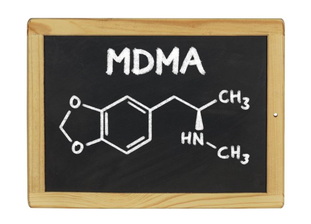
Amanda Feilding used to take lysergic acid diethylamide every day to boost creativity and productivity at work before LSD, known as acid, was made illegal in 1968. During her downtime, Feilding, who now runs the Beckley Foundation for psychedelic research, would get together with her friends to play the ancient Chinese game of Go, and came to notice something curious about her winning streaks.

“I found that if I was on LSD and my opponent wasn’t, I won more games,” Feilding told me over Skype. “For me that was a very clear indication that it improves cognitive function, particularly a kind of intuitive pattern recognition.”

An interesting observation to be sure. But was LSD actually helping Feilding in creative problem solving?

A half-century ban on psychedelic research has made answering this question in a scientific manner impossible. In recent years, however, psychedelic research has been experiencing something of a “renaissance” and now Feilding wants to put her intuition to the test by running a study in which participants will “microdose” while playing Go—a strategy game that is like chess on steroids—against an artificial intelligence.

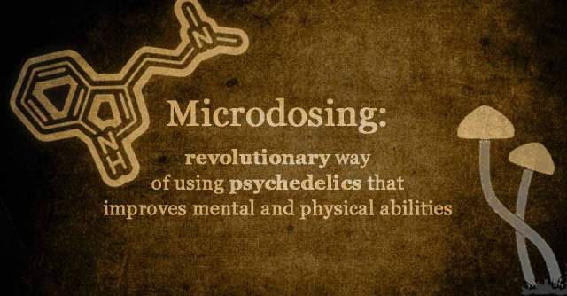
Microdosing LSD is one of the hallmarks of the so-called “Psychedelic Renaissance.” It’s a regimen that involves regularly taking doses of acid that are so low they don’t impart any of the drug’s psychedelic effects. Microdosers claim the practice results in heightened creativity, lowered depression, and even relief from chronic somatic pain.

But so far, all evidence in favor of microdosing LSD has been based on self-reports, raising the possibility that these reported positive effects could all be placebo. So the microdosing community is going to have to do some science to settle the debate. That means clinical trials with quantifiable results like the one proposed by Feilding.

As the first scientific trial to investigate the effects of microdosing, Feilding’s study will consist of 20 participants who will be given low doses—10, 20 and 50 micrograms of LSD—or a placebo on four different occasions. After taking the acid, the brains of these subjects will be imaged using MRI and MEG while they engage in a variety of cognitive tasks, such as the neuropsychology staples the Wisconsin Card Sorting test and the Tower of London test. Importantly, the participants will also be playing Go against an AI, which will assess the players’ performance during the match.

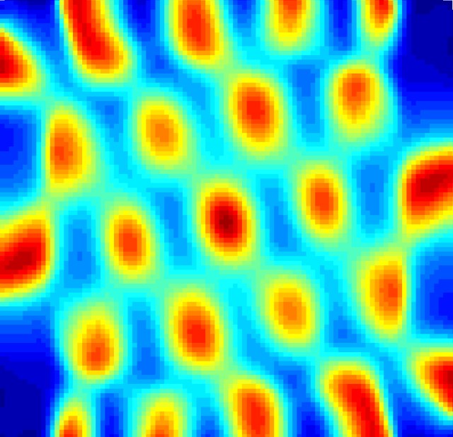
By imaging the brain while it’s under the influence of small amounts of LSD, Feilding hopes to learn how the substance changes connectivity in the brain to enhance creativity and problem solving. If the study goes forward, this will only be the second time that subjects on LSD have had their brain imaged while tripping. (That 2016 study at Imperial College London was also funded by the Beckley Foundation, which found that there was a significant uptick in neural activity in areas of the brain associated with vision during acid trips.)

Before Feilding can go ahead with her planned research, a number of obstacles remain in her way, starting with funding. She estimates she’ll need to raise about $350,000 to fund the study.

“It’s frightening how expensive this kind of research is,” Feilding said. “I’m very keen on trying to alter how drug policy categorizes these compounds because the research is much more costly simply because LSD is a controlled substance.”

To tackle this problem, Feilding has partnered with Rodrigo Niño, a New York entrepreneur who recently launched Fundamental, a platform for donations to support psychedelic research at institutions like the Beckley Foundation, Johns Hopkins University, and New York University.

The study is using smaller doses of LSD than Feilding’s previous LSD study, so she says she doesn’t anticipate problems getting ethical clearance to pursue this. A far more difficult challenge will be procuring the acid to use in her research. In 2016, she was able to use LSD that had been synthesized for research purposes by a government certified lab, but she suspects that this stash has long since been used up.

But if there’s anyone who can make the impossible possible, it would be Feilding, a psychedelic science pioneer known as much for drilling a hole in her own head (https://www.vice.com/en_us/article/drilling-a-hole-in-your-head-for-a-higher-state-of-consciousness) to explore consciousness as for the dozens of peer-reviewed scientific studies on psychedelic use she has authored in her lifetime. And according to Feilding, the potential benefits of microdosing are too great to be ignored and may even come to replace selective serotonin reuptake inhibitors, or SSRIs as a common antidepressant.

“I think the microdose is a very delicate and sensitive way of treating people,” said Feilding. “We need to continue to research it and make it available to people.”

https://motherboard.vice.com/en_us/article/first-ever-lsd-microdosing-study-will-pit-the-human-brain-against-ai

Acceleration of drug discovery by A.I.

To create a new drug, researchers have to test tens of thousands of compounds to determine how they interact. And that’s the easy part; after a substance is found to be effective against a disease, it has to perform well in three different phases of clinical trials and be approved by regulatory bodies.

It’s estimated that, on average, one new drug coming to market can take 1,000 people, 12-15 years, and up to $1.6 billion. Here is a short video on the current process.

Last week, researchers published a paper detailing an artificial intelligence system made to help discover new drugs, and significantly shorten the amount of time and money it takes to do so.

The system is called AtomNet, and it comes from San Francisco-based startup AtomWise. The technology aims to streamline the initial phase of drug discovery, which involves analyzing how different molecules interact with one another—specifically, scientists need to determine which molecules will bind together and how strongly. They use trial and error and process of elimination to analyze tens of thousands of compounds, both natural and synthetic.

AtomNet takes the legwork out of this process, using deep learning to predict how molecules will behave and how likely they are to bind together. The software teaches itself about molecular interaction by identifying patterns, similar to how AI learns to recognize images.

Remember the 3D models of atoms you made in high school, where you used pipe cleaners and foam balls to represent the connections between protons, neutrons and electrons? AtomNet uses similar digital 3D models of molecules, incorporating data about their structure to predict their bioactivity.

As AtomWise COO Alexander Levy put it, “You can take an interaction between a drug and huge biological system and you can decompose that to smaller and smaller interactive groups. If you study enough historical examples of molecules…you can then make predictions that are extremely accurate yet also extremely fast.”

“Fast” may even be an understatement; AtomNet can reportedly screen one million compounds in a day, a volume that would take months via traditional methods.

AtomNet can’t actually invent a new drug, or even say for sure whether a combination of two molecules will yield an effective drug. What it can do is predict how likely a compound is to work against a certain illness. Researchers then use those predictions to narrow thousands of options down to dozens (or less), focusing their testing where there’s more likely to be positive results.

The software has already proven itself by helping create new drugs for two diseases, Ebola and multiple sclerosis. The MS drug has been licensed to a British pharmaceutical company, and the Ebola drug is being submitted to a peer-reviewed journal for additional analysis.

Drug Discovery AI Can Do in a Day What Currently Takes Months

Thanks to Kebmodee for bringing this to the It’s Interesting community.

Compound in frog slime discovered to “blow up” the flu virus

by Katie Forster

Powerful new remedies for the flu could be created using a molecule found in frog slime after scientists discovered it destroys the virus.

Mucus from a colourful species of Indian frog contains a compound that kills influenza, according to a new study published in the scientific journal Immunity.

The frog, called hydrophylax bahuvistara, was discovered in 2015. It is a type of fungoid frog that lives in the forests of south west India and has a striking orange stripe on its upper body.

Researchers captured the frog and collected secretions from its skin after delivering a mild electric shock. They then released the amphibians back into the wild and studied the chemicals in their slime.

Joshy Jacob, a scientist at Emory University in Atlanta, who led the study, said they managed to isolate a small structure called a peptide that kills the flu virus but leaves healthy tissue intact.

“This peptide kills the viruses. It kind of blows them up,” Dr Jacob, an associate professor in microbiology, told NBC News. “There’s no collateral damage,”

Dr Jacob and his team decided to name the compound urumin – after an Indian sword called an urumi with a flexible blade that acts like a whip, used in martial arts from the southern city of Kerala.

Mice vaccinated with urumin were protected against a lethal amount of swine flu virus, also known as Influenza A of H1, which caused a pandemic in 2009.

It’s likely the frog produces the flu-fighting substance in its slime by coincidence, as one of a number of compounds that guard against harmful bacteria and fungi.

The scientists hope their discovery will lead to the development of new drugs to stop outbreaks of influenza, which is highly contagious and can be deadly, especially for the elderly and very young.

They will also continue the search for other frog slime compounds that could be used to treat other viral infections such as hepatitis, HIV and Zika.

The difficulty is finding molecules that attack flu but do not harm healthy cells as well – of the four peptides found in the hydrophylax bahuvistara mucus, only urumin did not kill red blood cells.

“In the beginning, I thought that when you do drug discovery, you have to go through thousands of drug candidates, even a million, before you get one or two hits. And here we did 32 peptides, and we had four hits,” said Dr Jacob.

Urumin is thought to target a viral surface protein called haemagluttinin – the H in H1.

“The virus needs the haemagglutinin to get inside our cells,” said Dr Jacob. “What this peptide does is it binds to the haemagglutinin and destabilises the virus. And then it kills the virus.”

http://www.independent.co.uk/news/health/frog-slime-flu-virus-compound-blows-up-kills-influenza-hydrophylax-bahuvistara-immunity-a7690141.html

Talking to a Computer May Soon Be Enough to Diagnose Illness

By Vanessa Bates Ramirez

In recent years, technology has been producing more and more novel ways to diagnose and treat illness.

Urine tests will soon be able to detect cancer: https://singularityhub.com/2016/10/14/detecting-cancer-early-with-nanosensors-and-a-urine-test/

Smartphone apps can diagnose STDs:https://singularityhub.com/2016/12/25/your-smartphones-next-big-trick-to-make-you-healthier-than-ever/

Chatbots can provide quality mental healthcare: https://singularityhub.com/2016/10/10/bridging-the-mental-healthcare-gap-with-artificial-intelligence/

Joining this list is a minimally-invasive technique that’s been getting increasing buzz across various sectors of healthcare: disease detection by voice analysis.

It’s basically what it sounds like: you talk, and a computer analyzes your voice and screens for illness. Most of the indicators that machine learning algorithms can pick up aren’t detectable to the human ear.

When we do hear irregularities in our own voices or those of others, the fact we’re noticing them at all means they’re extreme; elongating syllables, slurring, trembling, or using a tone that’s unusually flat or nasal could all be indicators of different health conditions. Even if we can hear them, though, unless someone says, “I’m having chest pain” or “I’m depressed,” we don’t know how to analyze or interpret these biomarkers.

Computers soon will, though.

Researchers from various medical centers, universities, and healthcare companies have collected voice recordings from hundreds of patients and fed them to machine learning software that compares the voices to those of healthy people, with the aim of establishing patterns clear enough to pinpoint vocal disease indicators.

In one particularly encouraging study, doctors from the Mayo Clinic worked with Israeli company Beyond Verbal to analyze voice recordings from 120 people who were scheduled for a coronary angiography. Participants used an app on their phones to record 30-second intervals of themselves reading a piece of text, describing a positive experience, then describing a negative experience. Doctors also took recordings from a control group of 25 patients who were either healthy or getting non-heart-related tests.

The doctors found 13 different voice characteristics associated with coronary artery disease. Most notably, the biggest differences between heart patients and non-heart patients’ voices occurred when they talked about a negative experience.

Heart disease isn’t the only illness that shows promise for voice diagnosis. Researchers are also making headway in the conditions below.

ADHD: German company Audioprofiling is using voice analysis to diagnose ADHD in children, achieving greater than 90 percent accuracy in identifying previously diagnosed kids based on their speech alone. The company’s founder gave speech rhythm as an example indicator for ADHD, saying children with the condition speak in syllables less equal in length.
PTSD: With the goal of decreasing the suicide rate among military service members, Boston-based Cogito partnered with the Department of Veterans Affairs to use a voice analysis app to monitor service members’ moods. Researchers at Massachusetts General Hospital are also using the app as part of a two-year study to track the health of 1,000 patients with bipolar disorder and depression.
Brain injury: In June 2016, the US Army partnered with MIT’s Lincoln Lab to develop an algorithm that uses voice to diagnose mild traumatic brain injury. Brain injury biomarkers may include elongated syllables and vowel sounds or difficulty pronouncing phrases that require complex facial muscle movements.
Parkinson’s: Parkinson’s disease has no biomarkers and can only be diagnosed via a costly in-clinic analysis with a neurologist. The Parkinson’s Voice Initiative is changing that by analyzing 30-second voice recordings with machine learning software, achieving 98.6 percent accuracy in detecting whether or not a participant suffers from the disease.
Challenges remain before vocal disease diagnosis becomes truly viable and widespread. For starters, there are privacy concerns over the personal health data identifiable in voice samples. It’s also not yet clear how well algorithms developed for English-speakers will perform with other languages.

Despite these hurdles, our voices appear to be on their way to becoming key players in our health.

https://singularityhub.com/2017/02/13/talking-to-a-computer-may-soon-be-enough-to-diagnose-illness/?utm_source=Singularity+Hub+Newsletter&utm_campaign=14105f9a16-Hub_Daily_Newsletter&utm_medium=email&utm_term=0_f0cf60cdae-14105f9a16-58158129

n Alzheimer’s, Excess Tau Protein Damages Brain’s GPS and may be responsible for wandering

Grid cell from the entorhinal cortex (EC) of the mouse brain, firing repeatedly and uniformly in a grid-like pattern. When a mouse moves through its environment, grid cells are activated, with each cell representing a specific location. This creates a triangular coordinate system that allows for spatial navigation. The accumulation of tau protein in the brain of a mouse model of Alzheimer’s disease was shown to disrupt the function of grid cells, causing problems with navigation. The findings explain why Alzheimer’s patients tend to wander and get lost. Source: Lab of Karen Duff, PhD, Columbia University Medical Center

Columbia University Medical Center (CUMC) researchers have discovered that the spatial disorientation that leads to wandering in many Alzheimer’s disease patients is caused by the accumulation of tau protein in navigational nerve cells in the brain. The findings, in mice, could lead to early diagnostic tests for Alzheimer’s and highlight novel targets for treating this common and troubling symptom.

The study was published online today in the journal Neuron.

An estimated three out of five people with Alzheimer’s disease wander and get lost, usually beginning in the early stages of the disease, leaving them vulnerable to injury. Researchers suspect that these problems originate in an area of the brain known as the entorhinal cortex (EC). The EC plays a key role in memory and navigation and is among the first brain structures affected by the buildup of neurofibrillary tangles that are largely composed of tau, a hallmark of Alzheimer’s disease. “Until now, no one has been able to show how tau pathology might lead to navigational difficulties,” said co-study leader Karen E. Duff, PhD, professor of pathology & cell biology (in psychiatry and in the Taub Institute for Research on Alzheimer’s Disease and the Aging Brain) at Columbia.

Dr. Duff and her colleagues focused their investigations on excitatory grid cells, a type of nerve cell in the EC that fires in response to movement through space, creating a grid-like internal map of a person’s environment. The researchers made electrophysiological recordings of the grid cells of older mice—including mice engineered to express tau in the EC (EC-tau mice) and normal controls—as they navigated different environments. Spatial cognitive tasks revealed that the EC-tau mice performed significantly worse compared to the controls, suggesting that tau alters grid cell function and contributes to spatial learning and memory deficits, according to co-study leader Abid Hussaini, PhD, assistant professor of neurobiology (in pathology & cell biology and the Taub Institute).

Detailed histopathological analysis of the mouse brains revealed that only the excitatory cells, but not the inhibitory cells, were killed or compromised by pathological tau, which probably resulted in the grid cells firing less. “It appears that tau pathology spared the inhibitory cells, disturbing the balance between excitatory and inhibitory cells and misaligning the animals’ grid fields,” said co-first author Hongjun Fu, PhD, associate research scientist in the Taub Institute, who led the immunohistological and behavior studies.

“This study clearly shows that tau pathology, beginning in the entorhinal cortex, can lead to deficits in grid cell firing and underlies the deterioration of spatial cognition that we see in human Alzheimer’s disease,” said Eric Kandel, MD, Nobel laureate, University Professor, and Kavli Professor of Brain Science at Columbia. “This is a classic advance in our understanding of the early stages of Alzheimer’s disease.”

“This study is the first to show a link between grid cells and Alzheimer’s disease,” said Edvard E. Moser, Nobel laureate and head of the Kavli Institute for Systems Neuroscience at Norwegian University of Science and Technology. “These findings will be crucial for future attempts to understand the development of early Alzheimer’s disease symptoms, including the tendency to wander and get lost.”

The findings raise the possibility that spatial disorientation could be treated by correcting this imbalance through transcranial stimulation, deep-brain stimulation, or light-based therapy.

“We have a lot to learn about grid cells and how they are affected by Alzheimer’s disease,” said Gustavo A. Rodriguez, PhD, a postdoctoral research scientist in the Taub Institute and a co-author of the paper. “We don’t yet know what percentage of healthy grid cells are needed for proper navigation or whether this system is rescuable once it has been compromised.”

“In the meantime,” said Dr. Duff, “our findings suggest that it may be possible to develop navigation-based cognitive tests for diagnosing Alzheimer’s disease in its initial stages. And if we can diagnose the disease early, we can start to give therapeutics earlier, when they may have a greater impact.”

The study is titled, “Tau Pathology Induces Excitatory Neuron Loss, Grid Cell Dysfunction and Spatial Memory Deficits Reminiscent of Early Alzheimer’s Disease.” The other contributors are Mathieu Herman, Sheina Emrani, Eden Nahmani, Geoffrey Barrett, Helen Y. Figueroa, and Eliana Goldberg.

The study was supported by grants from National Institutes of Health (R01NS074874 and R01AG050425) and the Alzheimer’s Association (2015-NIRG-341570).

http://newsroom.cumc.columbia.edu/blog/2017/01/19/in-alzheimers-excess-tau-protein-damages-brains-gps/

Bird flu strain may have jumped from cat to human

A veterinarian appears to have been infected with a strain of avian flu known as H7N2 that spread among more than 100 cats housed at New York City animal shelters. If confirmed, this would be the first known transmission of this bird flu strain from cat to human, officials said.

New York City health officials said the vet has recovered from a mild illness, and there’s no sign that the flu has spread to shelter workers or those who’ve adopted cats.

Still, the city’s top health official is calling for caution.

“Our investigation confirms that the risk to human health from H7N2 is low, but we are urging New Yorkers who have adopted cats from a shelter or rescue group within the past three weeks to be alert for symptoms in their pets,” city Health Commissioner Dr. Mary Bassett said in a health department news release.

“We are contacting people who may have been exposed and offering testing as appropriate,” she said.

According to officials, the outbreak of H7N2 struck cats that lived at Animal Care Centers of NYC shelters. H7N2 is a subtype of influenza A virus, also known as avian or bird flu.

More than 160 employees and volunteers were tested, and only the veterinanian, who worked with sick cats, appears to have been infected, officials said. The vet’s case has yet to be definitively confirmed.

Officials said they’ve contacted more than 80 percent of people who adopted cats from the animal shelter, and there’s no sign that any of these people have been infected.

There have only been two previous documented cases of transmission of the avian flu subtype to humans in the United States, and neither was linked to cats or to other humans, city health officials said.

For now, the city’s health department is urging people to avoid close facial contact and nuzzling with ill cats.

One cat diagnosed with the avian flu died, while the others are expected to get better. Adoptions of cats have been halted for the time being, and the sick cats will be quarantined.

Officials said no other shelter animals have been infected. For the time being, officials are urging New York City residents to not drop off cats at the Animal Care Centers of NYC shelters.

http://m.medicalxpress.com/news/2016-12-bird-flu-strain-cat-human.html

Taurine, a common additive to energy drinks, may help lessen the symptoms in first episode psychosis.

Supplementation with taurine, the additive found in many energy drinks, may improve the symptoms in young people suffering a first episode of psychosis (FEP), according to a new study presented at the International Early Psychosis Association (IEPA) meeting.

Taurine, an amino acid naturally occurring in the body, exhibits an inhibitory neuro-modulatory effect in the nervous system and also functions as a neuroprotective agent. The authors devised a study to analyze the efficacy of taurine supplementation in improving symptoms and cognition in patients with FEP.

The study included 86 individuals with FEP between the ages of 18 and 25 years. It was conducted by Dr. Colin O’Donnell, Donegal Mental Health Service, Co. Donegal, Ireland, and Professor Patrick McGorry and Dr. Kelly Allott, Orygen, The National Centre of Excellence in Youth Mental Health, Australia, and colleagues. Each participant was taking a low dose antipsychotic medication and was attending Orygen.

Forty-seven participants received 4g of taurine daily, while 39 received placebo. Symptoms were assessed Using the scoring system called BPRS (Brief Psychiatric Rating Scale) and cognition was assessed with the MCCB tool (MATRICS consensus cognitive battery).

Results showed that taurine significantly improved symptoms on the BPRS scale, in overall score and in psychosis specific analysis, however, there was no difference between the treatment and placebo group regarding cognition. Depression symptoms (rated by the Calgary Depression Scale for Schizophrenia) and general overall functioning also improved in the taurine group.

“The use of taurine warrants further investigation in larger randomised studies, particularly early in the course of psychosis,” concluded the authors, who themselves, are planning to conduct further studies into the potential benefits of taurine in the treatment of psychosis.

http://www.empr.com/news/energy-drink-additive-could-potentially-improve-psychosis-symptoms/article/567497/?DCMP=EMC-MPR_Charts_rd&cpn=&hmSubId=&NID=&c_id=&dl=0&spMailingID=16159114&spUserID=MzI5NTMwMzQ0NDIyS0&spJobID=921765029&spReportId=OTIxNzY1MDI5S0

F.D.A. Agrees to New Trials for Ecstasy as Relief for PTSD Patients

mdma

By DAVE PHILIPPS

After three tours in Iraq and Afghanistan, C. J. Hardin wound up hiding from the world in a backwoods cabin in North Carolina. Divorced, alcoholic and at times suicidal, he had tried almost all the accepted treatments for post-traumatic stress disorder: psychotherapy, group therapy and nearly a dozen different medications.

“Nothing worked for me, so I put aside the idea that I could get better,” said Mr. Hardin, 37. “I just pretty much became a hermit in my cabin and never went out.”

Then, in 2013, he joined a small drug trial testing whether PTSD could be treated with MDMA, the illegal party drug better known as Ecstasy.

“It changed my life,” he said in a recent interview in the bright, airy living room of the suburban ranch house here, where he now lives while going to college and working as an airplane mechanic. “It allowed me to see my trauma without fear or hesitation and finally process things and move forward.”

Based on promising results like Mr. Hardin’s, the Food and Drug Administration gave permission Tuesday for large-scale, Phase 3 clinical trials of the drug — a final step before the possible approval of Ecstasy as a prescription drug.

If successful, the trials could turn an illicit street substance into a potent treatment for PTSD.

Through a spokeswoman, the F.D.A. declined to comment, citing regulations that prohibit disclosing information about drugs that are being developed.

“I’m cautious but hopeful,” said Dr. Charles R. Marmar, the head of psychiatry at New York University’s Langone School of Medicine, a leading PTSD researcher who was not involved in the study. “If they can keep getting good results, it will be of great use. PTSD can be very hard to treat. Our best therapies right now don’t help 30 to 40 percent of people. So we need more options.”

But he expressed concern about the potential for abuse. “It’s a feel-good drug, and we know people are prone to abuse it,” he said. “Prolonged use can lead to serious damage to the brain.”

The Multidisciplinary Association for Psychedelic Studies, a small nonprofit created in 1985 to advocate the legal medical use of MDMA, LSD, marijuana and other banned drugs, sponsored six Phase 2 studies treating a total of 130 PTSD patients with the stimulant. It will also fund the Phase 3 research, which will include at least 230 patients.

Two trials here in Charleston focused on treating combat veterans, sexual assault victims, and police and firefighters with PTSD who had not responded to traditional prescription drugs or psychotherapy. Patients had, on average, struggled with symptoms for 17 years.

After three doses of MDMA administered under a psychiatrist’s guidance, the patients reported a 56 percent decrease of severity of symptoms on average, one study found. By the end of the study, two-thirds no longer met the criteria for having PTSD. Follow-up examinations found that improvements lasted more than a year after therapy.

“We can sometimes see this kind of remarkable improvement in traditional psychotherapy, but it can take years, if it happens at all,” said Dr. Michael C. Mithoefer, the psychiatrist who conducted the trials here. “We think it works as a catalyst that speeds the natural healing process.”

The researchers are so optimistic that they have applied for so-called breakthrough therapy status with the Food and Drug Administration, which would speed the approval process. If approved, the drug could be available by 2021.

Under the researchers’ proposal for approval, the drug would be used a limited number of times in the presence of trained psychotherapists as part of a broader course of therapy. But even in those controlled circumstances, some scientists worry that approval as a therapy could encourage more illegal recreational use.

“It sends the message that this drug will help you solve your problems, when often it just creates problems,” said Andrew Parrott, a psychologist at Swansea University in Wales who has studied the brains of chronic Ecstasy users. “This is a messy drug we know can do damage.”

Allowing doctors to administer the drug to treat a disorder, he warned, could inadvertently lead to a wave of abuse similar to the current opioid crisis.

During initial studies, patients went through 12 weeks of psychotherapy, including three eight-hour sessions in which they took MDMA. During the sessions, they lay on a futon amid candles and fresh flowers, listening to soothing music.

Dr. Mithoefer and his wife, Ann Mithoefer, and often their portly terrier mix, Flynn, sat with each patient, guiding them through traumatic memories.

“The medicine allows them to look at things from a different place and reclassify them,” said Ms. Mithoefer, a psychiatric nurse. “Honestly, we don’t have to do much. Each person has an innate ability to heal. We just create the right conditions.”

Research has shown that the drug causes the brain to release a flood of hormones and neurotransmitters that evoke feelings of trust, love and well-being, while also muting fear and negative emotional memories that can be overpowering in patients with post-traumatic stress disorder. Patients say the drug gave them heightened clarity and ability to address their problems.

For years after his combat deployments, Mr. Hardin said he was sleepless and on edge. His dreams were marked with explosions and death. The Army gave him sleeping pills and antidepressants. When they didn’t work, he turned to alcohol and began withdrawing from the world.

“I just felt hopeless and in the dark,” he said. “But the MDMA sessions showed me a light I could move toward. Now I’m out of the darkness and the world is all around me.”

Since the trial, he has gone back to school and remarried.

The chemist Alexander Shulgin first realized the euphoria-inducing traits of MDMA in the 1970s, and introduced it to psychologists he knew. Under the nickname Adam, thousands of psychologists began to use it as an aid for therapy sessions. Some researchers at the time thought the drug could be helpful for anxiety disorders, including PTSD, but before formal clinical trails could start, Adam spread to dance clubs and college campuses under the name Ecstasy, and in 1985, the Drug Enforcement Administration made it a Schedule 1 drug, barring all legal use.

Since then, the number of people seeking treatment for PTSD has exploded and psychiatry has struggled to keep pace. Two drugs approved for treating the disorder worked only mildly better than placebos in trials. Current psychotherapy approaches are often slow and many patients drop out when they don’t see results. Studies have shown combat veterans are particularly hard to treat.

In interviews, study participants said MDMA therapy had not only helped them with painful memories, but also had helped them stop abusing alcohol and other drugs and put their lives back together.

On a recent evening, Edward Thompson, a former firefighter, tucked his twin 4-year-old girls into bed, turned on their night light, then joined his wife at a backyard fire.

“If it weren’t for MDMA … ” he said.

“He’d be dead,” his wife, Laura, finished.

They both nodded.

Years of responding to gory accidents left Mr. Thompson, 30, in a near constant state of panic that he had tried to numb with alcohol and prescription opiates and benzodiazepines.

By 2015, efforts at therapy had failed, and so had several family interventions. His wife had left with their children, and he was considering jumping in front of a bus.

A member of a conservative Anglican church, Mr. Thompson had never used illegal drugs. But he was struggling with addiction from his prescription drugs, so he at first rejected a suggestion by his therapist that he enter the study. “In the end, I was out of choices,” he said.

Three sessions with the drug gave him the clarity, he said, to identify his problems and begin to work through them. He does not wish to take the drug again.

“It gave me my life back, but it wasn’t a party drug,” he said. “It was a lot of work.”

http://mobile.nytimes.com/2016/11/29/us/ptsd-mdma-ecstasy.html

Cat ownership linked to schizophrenia and other mental illness

A pair of new studies links childhood cat ownership and infection with the parasite Toxoplasma gondii (T. gondii) with later onset schizophrenia and other mental illness. Researchers published their findings in the online Schizophrenia Research and Acta Psychiatrica Scandinavica.

In the Schizophrenia Research study, investigators compared two previous studies that suggested childhood cat ownership could be a possible risk factor for schizophrenia or another serious mental illness with a third, even earlier survey on mental health to see if the finding could be replicated.

“The results were the same,” researchers reported, “suggesting that cat ownership in childhood is significantly more common in families in which the child later becomes seriously mentally ill.”

If accurate, the researchers expect the culprit to be infection with T. gondii, a parasite commonly carried by cats. At this point, though, they are urging others to conduct further studies to clarify the apparent link between cat ownership and schizophrenia.

The Acta Psychiatrica Scandinavica study was a meta-analysis of 50 previously published studies to investigate the prevalence of t. gondii infection in people diagnosed with psychiatric disorders compared with healthy controls.

In cases of schizophrenia, researchers said evidence of an association with T. gondii was “overwhelming,” CBS News reported. Specifically, people infected with T. gondii were nearly twice as likely to be diagnosed with schizophrenia as people never infected with the parasite, according to the report.

The meta-analysis also suggested associations between T. gondii infection and bipolar disorder, obsessive-compulsive disorder, and addiction. No association, however, was found for major depression.

—Jolynn Tumolo

References

1. Fuller Torrey E, Simmons W, Yolken RH. Is childhood cat ownership a risk factor for schizophrenia later in life? Schizophrenia Research. 2015 April 18. [Epub ahead of print].

2. Sutterland AL, Fond G, Kuin A, et al. Beyond the association. Toxoplasma gondii in schizophrenia, bipolar disorder, and addiction: systematic review and meta-analysis. Acta Psychiatrica Scandinavica. 2015 April 15. [Epub ahead of print].

http://www.psychcongress.com/article/studies-link-cat-ownership-schizophrenia-other-mental-illness

Pupil response to negative facial expressions predicts risk for depression relapse

Pupil dilation in reaction to negative emotional faces predicts risk for depression relapse, according to new research from Binghamton University, State University of New York.

Researchers at Binghamton University, led by PhD student Anastacia Kudinova, aimed to examine whether physiological reactivity to emotional stimuli, assessed via pupil dilation, served as a biological marker of risk for depression recurrence among individuals who are known to be at a higher risk due to having previous history of depression. Participants were 57 women with a history of major depressive disorder (MDD). The researchers recorded the change in pupil dilation in response to angry, happy, sad and neutral faces. The team found that women’s pupillary reactivity to negative (sad or angry faces) but not positive stimuli prospectively predicted MDD recurrence.

“The study focuses on trying to identify certain markers of depression risk using measures that are readily accessible, reliable and less expensive,” said Kudinova. “It is something we can put in any doctor’s office that gives us a quick and easy objective measure of risk.”

Additionally, the researchers found that both high and low reactivity to angry faces predicted risk for MDD recurrence. These findings suggest that disrupted physiological response to negative stimuli indexed via pupillary dilation could serve as a physiological marker of MDD risk, thus presenting clinicians with a convenient and inexpensive method to predict which of the at-risk women are more likely to experience depression recurrence.

“It’s a bit complicated because different patterns of findings were found for pupil reactivity to angry versus sad faces. Specifically, really high or really low pupil dilation to angry faces was associated with increased risk whereas only low dilation to sad faces was associated with risk (high dilation to sad faces was actually protective),” said Brandon Gibb, professor of psychology at Binghamton University and director of the Mood Disorders Institute and Center for Affective Science.

Other contributors to this research include Katie Burkhouse and Mary Woody, both PhD students; Max Owens, assistant professor of psychology at the University of South Florida, St. Petersburg; and Greg Siegle, associate professor of psychiatry at the University of Pittsburgh School of Medicine.
The paper, “Pupillary reactivity to negative stimuli prospectively predicts recurrence of major depressive disorder in women,” was published in Psychophysiology.

https://www.binghamton.edu/mpr/news-releases/news-release.html?id=2448