How stress can clog your arteries

By Sarah C. P. Williams

There’s a reason people say “Calm down or you’re going to have a heart attack.” Chronic stress—such as that brought on by job, money, or relationship troubles—is suspected to increase the risk of a heart attack. Now, researchers studying harried medical residents and harassed rodents have offered an explanation for how, at a physiological level, long-term stress can endanger the cardiovascular system. It revolves around immune cells that circulate in the blood, they propose.

The new finding is “surprising,” says physician and atherosclerosis researcher Alan Tall of Columbia University, who was not involved in the new study. “The idea has been out there that chronic psychosocial stress is associated with increased cardiovascular disease in humans, but what’s been lacking is a mechanism,” he notes.

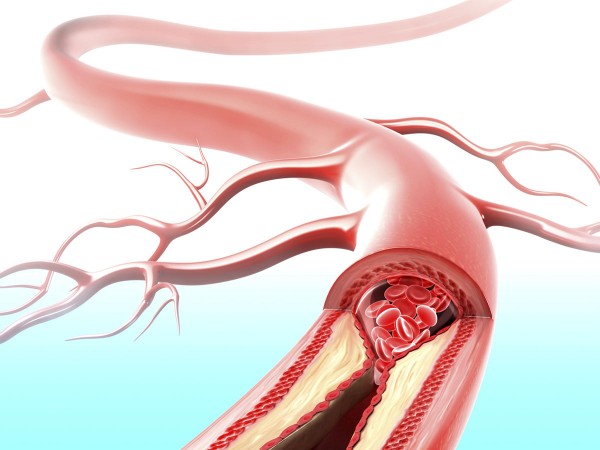
Epidemiological studies have shown that people who face many stressors—from those who survive natural disasters to those who work long hours—are more likely to develop atherosclerosis, the accumulation of fatty plaques inside blood vessels. In addition to fats and cholesterols, the plaques contain monocytes and neutrophils, immune cells that cause inflammation in the walls of blood vessels. And when the plaques break loose from the walls where they’re lodged, they can cause more extreme blockages elsewhere—leading to a stroke or heart attack.

Studying the effect of stressful intensive care unit (ICU) shifts on medical residents, biologist Matthias Nahrendorf of Harvard Medical School in Boston recently found that blood samples taken when the doctors were most stressed out had the highest levels of neutrophils and monocytes. To probe whether these white blood cells, or leukocytes, are the missing link between stress and atherosclerosis, he and his colleagues turned to experiments on mice.

Nahrendorf’s team exposed mice for up to 6 weeks to stressful situations, including tilting their cages, rapidly alternating light with darkness, or regularly switching the mice between isolation and crowded quarters. Compared with control mice, the stressed mice—like stressed doctors—had increased levels of neutrophils and monocytes in their blood.

The researchers then homed in on an explanation for the higher levels of immune cells. They already knew that chronic stress increases blood concentrations of the hormone noradrenaline; noradrenaline, Nahrendorf discovered, binds to a cell surface receptor protein called β3 on stem cells in the bone marrow. In turn, the chemical environment of the bone marrow changes and there’s an increase in the activity of the white blood cells produced by the stem cells.

“It makes sense that stress wakes up these immune cells because an enlarged production of leukocytes prepares you for danger, such as in a fight, where you might be injured,” Nahrendorf says. “But chronic stress is a different story—there’s no wound to heal and no infection.”

In mice living with chronic stress, Nahrendorf’s team reported today in Nature Medicine, atherosclerotic plaques more closely resemble plaques known to be most at risk of rupturing and causing a heart attack or stroke. When the scientists blocked the β3 receptor, though, stressed mice not only had fewer of these dangerous plaques, but also had reduced levels of the active immune cells in their plaques, pinpointing β3 as a key link between stress and atheroscelerosis.

The finding could lead to new drugs to help prevent cardiovascular disease, suggests biologist Lynn Hedrick of the La Jolla Institute for Allergy and Immunology in San Diego, California. “I think this gives us a really direct hint that the β3 receptor is important in regulating the stress-induced response by the bone marrow,” Hedrick says. “If we can develop a drug that targets the receptor, this may be very clinically relevant.”

More immediately, the new observations suggest a way that clinicians could screen patients for their risk of atherosclerosis, heart attack, and stroke, Tall says. “Rather than asking four questions about stress levels, we could use their white blood cell counts to monitor psychosocial stress,” he says.

Thanks to Dr. Rajadhyaksha for bringing this to the attention of the It’s Interesting community.

http://news.sciencemag.org/biology/2014/06/how-stress-can-clog-your-arteries

Bachelor party stumbles upon 3 million year old stegomastodon skull

Some guys have trouble remembering just what happened during their bachelor party, but a group of men on a recent stag send-off in New Mexico aren’t likely to forget their celebration very soon — since they stumbled upon a perfectly preserved three-million-year-old mastodon skull.

The party was on a hike in Elephant Butte Lake State Park near Albuquerque when they saw a bone jutting one to two inches from the sand. They started digging and uncovered the enormous skull. Their first thought was it could be the remains of a woolly mammoth so they snapped photos and sent them to the New Mexico Museum of Natural History and Science.

Scientists there made the identification — it wasn’t a woolly mammoth, but, in fact, a much more exciting find. The skull belonged to a stegomastodon — a prehistoric ancestor of the woolly mammoth — as well as today’s elephants. The massive animal stood about 9 feet tall, weighed six tons and walked the Earth during the Ice Age, according to Gary Morgan, a paleontologist at the museum who analyzed the fossil. He estimates the animal was about 50 years old when it died on a sandbar of the ancient Rio Grande River.

The family of mastodons migrated to North America around 15 million years ago and died out around 10,000 years ago.

“This is far and away the best one we’ve ever found,” said Morgan about the bachelor party’s discovery.

Scientists, following up on the party’s tip, went to the site and sealed the skull, which weighed more than 1,000 pounds, in a cast. It was transported to the museum, where it will eventually will be placed on display.

Antonia Gradillas, 33, was among the men who made the original discovery. He said, “This is the coolest thing ever. Some people with PhDs in this field might not even have this kind of opportunity. We were so lucky.”

Thanks to Dr. Rajadhyaksha for bringing this to the attention of the It’s Interesting community.

http://news.discovery.com/animals/paleontology/bachelor-party-finds-mastedon-skull-140616.htm

How male spiders keep females to themselves

Dwarf spiders don’t need to take paternity tests to know who the father is—for the most part. Right after copulation, males plug up the genital tract of females (red box in picture) to ensure that competitors can’t deposit sperm. Researchers studying the technique found that the larger the plug, the more difficult it is for subsequent males to remove. Described this month in Behavioral Ecology and Sociobiology, the “stoppers” effectively prevent 67.5% of males who show up later from breeding.

Thanks to Dr. Rajadhyaksha for bringing this to the attention of the It’s Interesting community.

http://news.sciencemag.org/sifter/2014/06/how-male-spiders-keep-females-to-themselves

Yeast from fossilized whale bones now used to make beer.

by Rebecca Cooper

Brewers have pulled yeast from pretty much everywhere to experiment with new strains — one West Coast brewery even brewed a beer using samples from the head brewer’s beard (http://www.huffingtonpost.com/2012/09/26/beard-beer-rogue-ales-yeast-john-maier_n_1917119.html) — but Lost Rhino in Ashburn may be breaking into new territory with its BoneDusters amber ale.

BoneDusters was brewed with a yeast that Lost Rhino’s Jasper Akerboom collected off a fossilized whale skeleton at the Calvert Marine Museum in Solomons, Maryland.

The collaboration came about because Akerboom, a bit of a yeast nut who handles quality assurance for Lost Rhino, is friends with Jason Osborne, a paleontologist who has donated fossilized whale skeletons to the museum.

Osborne asked Akerboom if there might be yeast present on those fossils that could be used to brew beer. Usually, yeast would not live on bone, given that it needs a sugary food source, but Akerboom decided to indulge his friend anyway.

They found a number of yeast strains on the bones, although Akerboom is pretty sure they’re more likely from the swamp where the bones were found rather than the bones themselves.

Several of the wild yeast strains flourished in Akerboom’s lab, but only one of the strains made any decent beer. The others didn’t ferment fully, making for “nasty-tasting” brews, he said.

The strain they ended up using, combined with some darker malts to create an amber ale, have yielded what Akerboom considers a tasty, well-balanced brew. The beer wasn’t made in the Belgian style, but it is “Belgian-esque,” he said, because the yeast has a slightly fruity flavor profile common in Belgian beers.

Lost Rhino plans to launch the beer June 18 at the brewery and begin distributing it to its networks after that, so it could be appearing at D.C. area bars in the next couple of weeks. A portion of the proceeds from the beer will go to Osborne’s nonprofit, Paleo Quest, which runs educational programs in the sciences.

For his part, Akerboom will keep experimenting with yeast in the lab he runs at Lost Rhino. It’s not necessarily common for a small microbrewery to have a quality assurance scientist with a Ph.D. in microbiology on staff. The Netherlands native previously isolated wild yeast from the air in Ashburn for Wild Farmwell Wheat, an “All-Virginia” beer Lost Rhino made in 2012. He now runs a yeast business on the side, and believes that focus on quality control is a big part of Lost Rhino’s consistently good beers.

“I think it adds a lot to the brewery. You have to make sure what you put in those cans is actually clean,” he said. “And you can do these kinds of projects, which keeps it fun.”

Thanks to Dr. Rajadhyaksha for bringing this to the attention of the It’s Interesting community.

http://www.bizjournals.com/washington/blog/top-shelf/2014/06/whats-the-key-ingredient-in-lost-rhino-s-newest.html?page=2

Electromagnetic helmet provides possible new treatment for depression.

A high-tech helmet has reduced symptoms of depression in two-thirds of people who’ve worn it, BBC reports. Now undergoing clinical trials, the hood works by sending electromagnetic impulses to the brain to activate the formation of new blood vessels. Patients who wear the device daily for half an hour to an hour show mood improvement in as little as week, according to the results, published in Acta Neuropsychiatrica.

Thanks to Dr. Rajadhyaksha for bringing this to the attention of the It’s Interesting community.

http://news.sciencemag.org/sifter/2014/05/watch-electromagnetic-helmet-treats-depression

Fossil Whale Offers Clues to Origins of ‘Seeing With Sound’

When you’re trying to track a fish in the murky ocean, forget about using your eyes—use your ears. Dolphins, orcas, and other toothed whales—known as odontocetes—pinpoint their prey by producing high-frequency sounds that bounce around their marine environment and reveal exactly where tricky fish are trying to hide. But when did whales evolve this sonarlike ability, known as echolocation? A newly named, 28-million-year-old whale may hold the answer.

Found in South Carolina among rocks dating back to the Oligocene epoch and christened Cotylocara macei, the fossil whale is named after Mace Brown, a curator at the College of Charleston’s Mace Brown Natural History Museum in South Carolina who acquired the specimen for his private collection about a decade ago. It was in that private accumulation of fossils that Jonathan Geisler, a paleontologist at the New York Institute of Technology College of Osteopathic Medicine in Old Westbury, first saw the skull. “I knew it was special then,” he says.

The only known specimen of the early odontocete includes a nearly complete skull and jaw, three neck vertebrae, and fragments of seven ribs. It’s the skull that makes Cotylocara so remarkable. While the whale’s soft tissue rotted away long ago, the skull bones show several features—such as a downturned snout and a slight asymmetry of the skull—that suggest Cotylocara was one of the earliest whales to use echolocation, Geisler’s team reports online today in Nature.

The strongest pieces of evidence for this hypothesis, Geisler explains, are cavities at the base of the snout and on top of the skull that probably held air sinuses. “These air sinuses are thought to have important roles in the production of high-frequency vocalizations that living odontocetes use for echolocation,” Geisler says, possibly helping direct returning sound waves or store air that can be used to make continuous sound.

“I think the authors have a good case for inferring that Cotylocara had some ability to produce some sound from its forehead, just as living toothed whales do today,” says Nicholas Pyenson, a marine mammal paleontologist at the Smithsonian Institution’s National Museum of Natural History in Washington, D.C. But even if Cotylocara made those sounds, could it have heard them? Living whales have specialized ear bones that let them hear the high-frequency sounds bouncing off their prey. The only known skull of Cotylocara doesn’t have well-preserved ear bones, and, therefore, knowing whether the whale could have actually used echolocation for hunting is unclear. “Overall, the description of Cotylocara underscores the need to investigate the inner ear of fossil Oligocene cetaceans in much more detail, because that’s where the answer will be,” Pyenson says.

Nevertheless, the whale’s probable sound-producing abilities give Cotylocara an important place in whale evolution. Whale’s biological sonar is thought to have evolved only once along the ancestral line leading to today’s toothed whales, Geisler notes. Cotylocara lies along that evolutionary stem, as do other Oligocene fossil whales that have already been found. The skull features that allowed Cotylocara to create sound, Geisler says, “can now be investigated in other fossil whales to more fully understand the evolution of echolocation.” For now, the evolutionary epic of whale echolocation is only just beginning to be heard.

http://news.sciencemag.org/paleontology/2014/03/fossil-whale-offers-clues-origins-seeing-sound

Thanks to Dr. Rajadhyaksha for bringing this to the attention of the It’s Interesting community.

New research shows that whales and dolphins can’t taste anything except salt

Tastes are a privilege. The oral sensations not only satisfy foodies, but also on a primal level, protect animals from toxic substances. Yet cetaceans—whales and dolphins—may lack this crucial ability, according to a new study. Mutations in a cetacean ancestor obliterated their basic machinery for four of the five primary tastes, making them the first group of mammals to have lost the majority of this sensory system.

The five primary tastes are sweet, bitter, umami (savory), sour, and salty. These flavors are recognized by taste receptors—proteins that coat neurons embedded in the tongue. For the most part, taste receptor genes present across all vertebrates.

Except, it seems, cetaceans. Researchers uncovered a massive loss of taste receptors in these animals by screening the genomes of 15 species. The investigation spanned the two major lineages of cetaceans: Krill-loving baleen whales—such as bowheads and minkes—were surveyed along with those with teeth, like bottlenose dolphins and sperm whales.

The taste genes weren’t gone per se, but were irreparably damaged by mutations, the team reports online this month in Genome Biology and Evolution. Genes encode proteins, which in turn execute certain functions in cells. Certain errors in the code can derail protein production—at which point the gene becomes a “pseudogene” or a lingering shell of a trait forgotten. Identical pseudogene corpses were discovered across the different cetacean species for sweet, bitter, umami, and sour taste receptors. Salty tastes were the only exception.

“The loss of bitter taste is a complete surprise, because natural toxins typically taste bitter,” says zoologist Huabin Zhao of Wuhan University in China who led the study. All whales likely descend from raccoon-esque raoellids, a group of herbivorous land mammals that transitioned to the sea where they became fish eaters. Plants range in flavors—from sugary apples to tart, poisonous rhubarb leaves—and to survive, primitive animals learned the taste cues that signal whether food is delicious or dangerous. Based on the findings, taste dissipated after this common ancestor became fully aquatic—53 million years ago—but before the group split 36 million years ago into toothed and baleen whales.

“Pseudogenes arise when a trait is no longer needed,” says evolutionary biologist Jianzhi Zhang of the University of Michigan, Ann Arbor, who was not involved in the study. “So it still raises the question as to why whales could afford to lose four of the five primary tastes.” The retention of salty taste receptors suggests that they have other vital roles, such as maintaining sodium levels and blood pressure.

But dulled taste perception might be dangerous if noxious substances spill into the water. Orcas have unwittingly migrated into oil spills, while algal toxins created by fertilizer runoff consistently seep into the fish prey of dolphins living off the Florida coast.

“When you have a sense of taste, it dictates whether you swallow or not,” says Danielle Reed, a geneticist at the Monell Chemical Senses Center in Philadelphia, Pennsylvania. She was not involved with the current work, but co-authored a 2012 paper that found the first genetic inklings that umami and sweet taste receptors were missing in cetaceans, albeit in only one species—bottlenose dolphins.

Flavors are typically released by chewing, but cetaceans tend to swallow their food whole. “The message seems clear. If you don’t chew your food and prefer swallowing food whole, then taste really becomes irrelevant,” Reed says.

http://news.sciencemag.org/biology/2014/05/whales-cant-taste-anything-salt

Thanks to Dr. Rajadhyaksha for bringing this to the attention of the It’s Interesting community.

Beaked Whales Smash Deep-Dive Record

Cuvier’s beaked whales (Ziphius cavirostris) just shattered their own deep-diving record—as well as that of elephant seals, which were the previous record-holders. The little-known species of beaked whales, cigar-shaped cetaceans with prominent snouts and which range from tropical to northern temperate seas, has long been considered one of the most extreme divers in the ocean, capable of reaching a depth of 1888 meters and staying below for 95 minutes. But a new study that tracked eight individuals off the coast of southern California via satellite tags, as in the photo above, shows they can do much more. (Male Cuvier’s beaked whales have tusks, and the scars on the back of the male in the photo are from fighting with other males.) One whale dove to 2992 meters below the surface, breaking the deep-dive record of a southern elephant seal that was tracked to 2388 meters. Another Cuvier’s beaked whale in the study remained below the surface for 2 hours and 17 minutes. Unlike elephant seals and deep-diving sperm whales, which remain at the surface for an extended period after their dives, the beaked whales headed back into the depths less than 2 minutes later, the scientists report online today in PLOS ONE. The beaked whales in the study made their deep dives about seven times a day, foraging for squid and fish; they spent more time at the surface at night. By better understanding this species’ diving behaviors, the scientists hope to solve an ongoing mystery: Why are Cuvier’s beaked whales particularly sensitive to military sonar operations? Sixty-nine percent of all recorded strandings of marine mammals that were associated with such operations involved this species. Yet these eight whales were tagged and followed on a U.S. Navy sonar training range, leading the scientists to suggest that Cuvier’s beaked whales in this area may have adapted to human noise—perhaps, in part, by becoming the most extreme of extreme divers.

http://news.sciencemag.org/plants-animals/2014/03/scienceshot-beaked-whales-smash-deep-dive-record

Thanks to Dr. Rajadhyaksha for bringing this to the attention of the It’s Interesting community.

New sweetener from the tequila plant may treat diabetes and weight loss

Could a new sugar substitute actually lower blood sugar and help you lose weight? That’s the tantalizing – but distant – promise of new research presented at the American Chemical Society (ACS) this week.

Agavins, derived from the agave plant that’s used to make tequila, were found in mouse studies to trigger insulin production and lower blood sugar, as well as help obese mice lose weight.

Unlike sucrose, glucose, and fructose, agavins aren’t absorbed by the body, so they can’t elevate blood glucose, according to research by Mercedes G. López, a researcher at the Centro de Investigación y de Estudios Avanzados, Biotechnology and Biochemistry Irapuato, in Guanajuato, Mexico.

And by boosting the level of a peptide called GLP-1 (short for glucagon-like peptide-1), which triggers the body’s production of insulin, agavins aid the body’s natural blood sugar control. Also, because agavins are type of fiber, they can make people feel fuller and reduce appetite, López’s research shows.

“We believe that agavins have a great potential as light sweeteners since they are sugars, highly soluble, have a low glycemic index, and a neutral taste, but most important, they are not metabolized by humans,” read the study abstract. “This puts agavins in a tremendous position for their consumption by obese and diabetic people.”

The caveat: The research was conducted in mice, and more study is necessary before we’ll know whether agavins are effective and safe in humans. In other words, we’re a long way from agavins appearing on grocery store shelves.

That said, with almost 26 millions of Americans living with diabetes and another 2 million diagnosed each year, a sweetener that lowered blood sugar levels rather than raised them would be quite a useful discovery. Not to mention the potential for a sugar substitute with the potential to help people lose weight.

In the study, titled “Agavins as Potential Novel Sweeteners for Obese and Diabetic People”, López added agavins to the water of mice who were fed a standard diet, weighing them and monitoring blood sugar levels every week. The majority of the mice given the agavin-supplemented water had lower blood glucose levels, ate less, and lost weight compared with other mice whose water was supplemented with glucose, sucrose, fructose, agave syrup, and aspartame.

Unlike other types of fructose, Agavins are fructans, which are long-chain fructoses that the body can’t use, so they are not absorbed into the bloodstream to raise blood sugar. And despite the similarity in the name, agavins are not to be confused with agave nectar or agave syrup, natural sweeteners that are increasingly popular sugar substitutes. In these products the fructans are broken down into fructose, which does raise blood sugar – and add calories.

López has been studying fructans for some time, and has published previous studies showing that they have protective prebiotic effects in the digestive tract and contribute to weight loss in obese mice.

A 2012 study by another team of researchers published in Plant Foods for Human Nutrition found that fructans boosted levels of the beneficial probiotics lactobacillus and bifidus. And like many types of fiber, agavins also lower levels of cholesterol and triglycerides in the blood.

But the news isn’t all good; a 2011 literature review of human studies of the relationship between fructans (not agavins specifically) and blood sugar found that of 13 randomized studies of fructans, only three documented positive results. It remains to be seen whether – as López argues – agavins are distinct from other fructans in their action.

The downside: Agavins are don’t taste as sweet as other forms of sugar such as sucrose, fructose and glucose. And not everyone can tolerate them; like other types of fiber they have the potential to cause digestive problems.

http://www.forbes.com/sites/melaniehaiken/2014/03/17/new-sweetener-from-the-tequila-plant-may-aid-diabetes-weight-loss/

Thanks to Dr. Rajadhyaksha for bringing this to the attention of the It’s Interesting community.

New Imaging Technique Speeds Removal of Non-melanoma Skin Cancers

lab to lcinic
The research team is building a smaller portable version of the laboratory’s cancer detection system. Source: Milind Rajadhyaksha, Ph. D., Memorial Sloan-Kettering Cancer Center, New York, NY.

Residual%20Cancer%20Margins

A common surgery for non-melanoma skin cancer, known as Mohs surgery, typically achieves excellent results but can be a long process, as the surgeon successively removes the area of concern until the surrounding tissue is free of cancer. To determine whether further tissue removal is necessary, the borders of the lesion must be processed in a laboratory to check for residual cancer tissue — a process that takes 20 – 45 minutes and is often repeated numerous times. Now, National Insititute of Biomedical Imaging and Bioengineering (NIBIB)-funded researchers have developed a microscopic technique to analyze removed tissue rapidly right in the clinic — dramatically reducing the length, inefficiency, and expense of this procedure.

With approximately 3.5 million new cases per year in the U.S., Mohs surgery is a fairly common procedure that many people undergo repeatedly as new skin cancers appear. It can take one to three hours, or even longer depending on the size and location of the lesion. The process is lengthy because after a section of tissue is removed, it must be frozen and stained so it can be examined to ensure the borders are clear of residual tumor. Although highly effective, the current practice is labor intensive for surgeons and assisting staff, as well as lengthy and stressful for patients. The time spent by surgical personnel and those analyzing the tissue in the lab increases the expense of the procedure, which has been estimated to cost $ 2-3 billion per year in the U.S.

NIBIB-supported researchers led by Milind Rajadhyaksha, Ph.D. at Memorial Sloan Kettering are using their expertise in optical imaging to improve this common procedure. Optical imaging is a technique that uses visible or near-infrared light to obtain detailed images of organs, tissues, and cells. The investigators developed a new pathological assessment technique called strip mosaicing confocal microscopy — a type of optical imaging — that can provide high resolution images during removal of basal cell and squamous cell carcinomas (non-melanoma skin cancers) and perhaps other tumors of the skin. The new technique uses a focused laser line that performs multiple scans of the tissue to obtain image “strips” that are then combined, like a mosaic, into a complete image of the excised tissue. The process takes only 90 seconds and eliminates the need to freeze and stain the tissue samples for analysis — a process that takes 20 to 45 minutes.

The new imaging technique was tested on 17 patients with 34 tissue samples. The overall image quality was excellent, with high resolution and contrast, providing for good visibility of the epidermis and dermis. Researchers compared the new technique against the Mohs approach with its frozen section processing. The new technique achieved a promising 94% in preliminary measures of sensitivity and specificity for detecting skin cancer margins, which is comparable to the “gold standard” Mohs procedure. These preliminary results demonstrated that the optical technique could potentially detect skin cancer margins with the same accuracy as the conventional frozen section technique.

The results of this study were obtained under laboratory conditions; a clinical trial is now being conducted to demonstrate the feasibility of using this technique in the clinical setting, the ultimate goal of the research group.

Steve Krosnick, M.D., NIBIB director for the Program for Image-Guided Interventions, explains the utility of the optical system: “The technology is particularly well-suited for Mohs-trained surgeons, who are experts at performing excisions and interpreting images of tissue samples removed during the Mohs procedure. Image quality, ability to make accurate interpretations, and time savings will be key parameters for adoption of the system in the clinical setting, and the current results are very encouraging.”

The research was conducted by a team consisting of two laboratories at Memorial Sloan-Kettering Cancer Center, New York, NY, as well as students from Bronx High School of Science, New York and Livingston High School, Livingston New Jersey. The work is published in the October 2013 issue of the British Journal of Dermatology.

http://www.nibib.nih.gov/news-events/newsroom/new-imaging-technique-speeds-removal-non-melanoma-skin-cancers

Thanks to Dr. Rajadhyaksha for bringing this to the attention of the It’s Interesting community.