Cancer drug nilotinib may reverse Parkinson’s disease

by Jon Hamilton

A drug that’s already approved for treating leukemia appears to dramatically reduce symptoms in people who have Parkinson’s disease with dementia, or a related condition called Lewy body dementia.

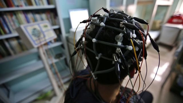
A pilot study of 12 patients given small doses of nilotinib found that movement and mental function improved in all of the 11 people who completed the six-month trial, researchers reported Saturday at the Society for Neuroscience meeting in Chicago.

And for several patients the improvements were dramatic, says Fernando Pagan, an author of the study and director of the Movement Disorders Program at Georgetown University Medical Center. One woman regained the ability to feed herself, one man was able to stop using a walker, and three previously nonverbal patients began speaking again, Pagan says.

“After 25 years in Parkinson’s disease research, this is the most excited I’ve ever been,” Pagan says.

If the drug’s effectiveness is confirmed in larger, placebo-controlled studies, nilotinib could become the first treatment to interrupt a process that kills brain cells in Parkinson’s and other neurodegenerative diseases, including Alzheimer’s.

One of the patients in the pilot study was Alan Hoffman, 74, who lives with his wife, Nancy, in Northern Virginia.

Hoffman was diagnosed with Parkinson’s in 1997. At first, he had trouble moving his arms. Over time, walking became more difficult and his speech became slurred. And by 2007, the disease had begun to affect his thinking.

“I knew I’d dropped off in my ability to read,” Hoffman says. “People would keep giving me books and I’d have read the first chapter of about 10 of them. I had no ability to focus on it.”

“He had more and more difficulty making sense,” Nancy Hoffman says. He also became less active, less able to have conversations, and eventually stopped doing even household chores, she says.

But after a few weeks on nilotinib, Hoffman “improved in every way,” his wife says. “He began loading the dishwasher, loading the clothes in the dryer, things he had not done in a long time.”

Even more surprising, Hoffman’s scores on cognitive tests began to improve. At home, Nancy Hoffman says her husband was making sense again and regained his ability to focus. “He actually read the David McCullough book on the Wright Brothers and started reading the paper from beginning to end,” she says.

The idea of using nilotinib to treat people like Alan Hoffman came from Charbel Moussa, an assistant professor of neurology at Georgetown University and an author of the study.

Moussa knew that in people who have Parkinson’s disease with dementia or a related condition called Lewy body dementia, toxic proteins build up in certain brain cells, eventually killing them. Moussa thought nilotinib might be able to reverse this process.

His reasoning was that nilotinib activates a system in cells that works like a garbage disposal — it clears out unwanted proteins. Also, Moussa had shown that while cancer cells tend to die when exposed to nilotinib, brain cells actually become healthier.

So Moussa had his lab try the drug on brain cells in a Petri dish. “And we found that, surprisingly, with a very little amount of the drug we can clear all these proteins that are supposed to be neurotoxic,” he says.

Next, Moussa had his team give the drug to transgenic mice that were almost completely paralyzed from Parkinson’s disease. The treatment “rescued” the animals, he says, allowing them to move almost as well as healthy mice.

Moussa’s mice got the attention of Pagan from Georgetown’s Movement Disorders Program. “When Dr. Moussa showed them to me,” Pagan says, “it looked like, hey, this is type of drug that we’ve been looking for because it goes to the root of the problem.”

The pilot study was designed to determine whether nilotinib was safe for Parkinson’s patients and to determine how much drug from the capsules they were taking was reaching their brains. “But we also saw efficacy, which is really unheard of in a safety study,” Pagan says.

The study found that levels of toxic proteins in blood and spinal fluid decreased once patients began taking nilotinib. Also, tests showed that the symptoms of Parkinson’s including tremor and “freezing” decreased. And during the study patients were able to use lower doses of Parkinson’s drugs, suggesting that the brain cells that produce dopamine were working better.

But there are some caveats, Pagan says. For one thing, the study was small, not designed to measure effectiveness, and included no patients taking a placebo.

Also, nilotinib is very expensive. The cost of providing it to leukemia patients is thousands of dollars a month.

And finally, Parkinson’s and dementia patients would have to keep taking nilotinib indefinitely or their symptoms would continue to get worse.

Alan Hoffman was okay for about three weeks after the study ended and he stopped taking the drug. Since then, “There’s (been) a pretty big change,” his wife says. “He does have more problems with his speech, and he has more problems with cognition and more problems with mobility.”

The Hoffmans hope to get more nilotinib from the drug’s maker, Novartis, through a special program for people who improve during experiments like this one.

Meanwhile, the Georgetown team plans to try nilotinib in patients with another brain disease that involves toxic proteins: Alzheimer’s.

http://www.npr.org/sections/health-shots/2015/10/17/448323916/can-a-cancer-drug-reverse-parkinsons-disease-and-dementia

New research shows that infants need to be able to freely move their tongues in order to distinguish sounds.

A team of researchers led by Dr Alison Bruderer, a postdoctoral fellow at the University of British Columbia, has discovered a direct link between tongue movements of infants and their ability to distinguish speech sounds.

“Until now, research in speech perception development and language acquisition has primarily used the auditory experience as the driving factor. Researchers should actually be looking at babies’ oral-motor movements as well,” said Dr Bruderer, who is the lead author on a study published in the Proceedings of the National Academy of Sciences on October 12, 2015.

In the study, teething toys were placed in the mouths of six-month-old English-learning infants while they listened to speech sounds – two different Hindi ‘d’ sounds that infants at this age can readily distinguish.

When the teethers restricted movements of the tip of the tongue, the infants were unable to distinguish between the two sounds.

But when their tongues were free to move, the babies were able to make the distinction.

“Before infants are able to speak, their articulatory configurations affect the way they perceive speech, suggesting that the speech production system shapes speech perception from early in life,” the scientists said.

“These findings implicate oral-motor movements as more significant to speech perception development and language acquisition than current theories would assume and point to the need for more research.”

http://www.sci-news.com/othersciences/psychology/science-infants-tongue-movement-speech-sounds-03336.html?utm_source=feedburner&utm_medium=feed&utm_campaign=Feed%3A+BreakingScienceNews+%28Breaking+Science+News%29

Scientists encode memories in a way that bypasses damaged brain tissue

Researchers at University of South Carolina (USC) and Wake Forest Baptist Medical Center have developed a brain prosthesis that is designed to help individuals suffering from memory loss.

The prosthesis, which includes a small array of electrodes implanted into the brain, has performed well in laboratory testing in animals and is currently being evaluated in human patients.

Designed originally at USC and tested at Wake Forest Baptist, the device builds on decades of research by Ted Berger and relies on a new algorithm created by Dong Song, both of the USC Viterbi School of Engineering. The development also builds on more than a decade of collaboration with Sam Deadwyler and Robert Hampson of the Department of Physiology & Pharmacology of Wake Forest Baptist who have collected the neural data used to construct the models and algorithms.

When your brain receives the sensory input, it creates a memory in the form of a complex electrical signal that travels through multiple regions of the hippocampus, the memory center of the brain. At each region, the signal is re-encoded until it reaches the final region as a wholly different signal that is sent off for long-term storage.

If there’s damage at any region that prevents this translation, then there is the possibility that long-term memory will not be formed. That’s why an individual with hippocampal damage (for example, due to Alzheimer’s disease) can recall events from a long time ago – things that were already translated into long-term memories before the brain damage occurred – but have difficulty forming new long-term memories.

Song and Berger found a way to accurately mimic how a memory is translated from short-term memory into long-term memory, using data obtained by Deadwyler and Hampson, first from animals, and then from humans. Their prosthesis is designed to bypass a damaged hippocampal section and provide the next region with the correctly translated memory.

That’s despite the fact that there is currently no way of “reading” a memory just by looking at its electrical signal.

“It’s like being able to translate from Spanish to French without being able to understand either language,” Berger said.

Their research was presented at the 37th Annual International Conference of the IEEE Engineering in Medicine and Biology Society in Milan on August 27, 2015.

The effectiveness of the model was tested by the USC and Wake Forest Baptist teams. With the permission of patients who had electrodes implanted in their hippocampi to treat chronic seizures, Hampson and Deadwyler read the electrical signals created during memory formation at two regions of the hippocampus, then sent that information to Song and Berger to construct the model. The team then fed those signals into the model and read how the signals generated from the first region of the hippocampus were translated into signals generated by the second region of the hippocampus.

In hundreds of trials conducted with nine patients, the algorithm accurately predicted how the signals would be translated with about 90 percent accuracy.

“Being able to predict neural signals with the USC model suggests that it can be used to design a device to support or replace the function of a damaged part of the brain,” Hampson said.
Next, the team will attempt to send the translated signal back into the brain of a patient with damage at one of the regions in order to try to bypass the damage and enable the formation of an accurate long-term memory.

http://medicalxpress.com/news/2015-09-scientists-bypass-brain-re-encoding-memories.html#nRlv

The human brain is particularly vulnerable to trauma at two distinct ages

Our brain’s ability to process information and adapt effectively is dependent on a number of factors, including genes, nutrition, and life experiences. These life experiences wield particular influence over the brain during a few sensitive periods when our most important muscle is most likely to undergo physical, chemical, and functional remodeling.

According to Tara Swart, a neuroscientist and senior lecturer at MIT, your “terrible twos” and those turbulent teen years are when the brain’s wiring is most malleable. As a result, traumatic experiences that occur during these time periods can alter brain activity and ultimately change gene expressions—sometimes for good.

Throughout the first two years of life, the brain develops at a rapid pace. However, around the second year, something important happens—babies begin to speak.

“We start to understand speech first, then we start to articulate speech ourselves and that’s a really complex thing that goes on in the brain,” Swart, who conducts ongoing research on the brain and how it affects how we become leaders, told Quartz. “Additionally, children start to walk—so from a physical point of view, that’s also a huge achievement for the brain.

Learning and understanding a new language forces your brain to work in new ways, connecting neurons and forming new pathways. This is a mentally taxing process, which is why learning a new language or musical instrument often feels exhausting.

With so many important changes happening to the brain in such a short period of time, physical or emotional trauma can cause potentially momentous interruptions to neurological development. Even though you won’t have any memories of the interruptions (most people can’t remember much before age five), any kind of traumatic event—whether it’s abuse, neglect, ill health, or separation from your loved ones—can lead to lasting behavioral and cognitive deficits later in life, warns Swart.

To make her point, Swart points to numerous studies on orphans in Romania during the 1980s and 1990s. After the nation’s communist regime collapsed, an economic decline swept throughout the region and 100,000 children found themselves in harsh, overcrowded government institutions.

“[The children] were perfectly well fed, clothed, washed, but for several reasons—one being that people didn’t want to spread germs—they were never cuddled or played with,” explains Swart. “There was a lot of evidence that these children grew up with some mental health problems and difficulty holding down jobs and staying in relationships.”

Swart continues: “When brain scanning became possible, they scanned the brains of these children who had grown up into adults and showed that they had issues in the limbic system, the part of the brain [that controls basic emotions].”

In short, your ability to maintain proper social skills and develop a sense of empathy is largely dependent on the physical affection, eye contact, and playtime of those early years. Even something as simple as observing facial expressions and understanding what those expressions mean is tied to your wellbeing as a toddler.

The research also found that the brains of the Romanian orphans had lower observable brain activity and were physically smaller than average. As a result, researchers concluded that children adopted into loving homes by age two have a much better chance of recovering from severe emotional trauma or disturbances.

The teenage years

By the time you hit your teenage years, the brain has typically reached its adult weight of about three pounds. Around this same time, the brain is starting to eliminate, or “prune” fragile connections and unused neural pathways. The process is similar to how one would prune a garden—cutting back the deadwood allows other plants to thrive.

During this period, the brain’s frontal lobes, especially the prefrontal cortex, experience increased activity and, for the first time, the brain is capable of comparing and analyzing several complex concepts at once. Similar to a baby learning how to speak, this period in an adolescent’s life is marked by a need for increasingly advanced communication skills and emotional maturity.

“At that age, they’re starting to become more understanding of social relationships and politics. It’s really sophisticated,” Swart noted. All of this brain activity is also a major reason why teenagers need so much sleep.

Swart’s research dovetails with the efforts of many other scientists who have spent decades attempting to understand how the brain develops, and when. The advent of MRIs and other brain-scanning technology has helped speed along this research, but scientists are still working to figure out what exactly the different parts of the brain do.

What is becoming more certain, however, is the importance of stability and safety in human development, and that such stability is tied to cognitive function. At any point in time, a single major interruption has the ability to throw off the intricate workings of our brain. We may not really understand how these events affect our lives until much later.

http://qz.com/470751/your-brain-is-particularly-vulnerable-to-trauma-at-two-distinct-ages/

New evidence that sleeping on your side may be better for the brain

Removal of waste, including soluble amyloid β (Aβ), from the brain may be most efficient in the lateral vs. the prone position, according to an experimental study published in the August 5 issue of the Journal of Neuroscience.

Hedok Lee, PhD, from Stony Brook University in New York, and colleagues examined whether body posture impacts cerebrospinal fluid (CSF)-interstitial fluid (ISF) exchange efficiency. They quantified CSF-ISF exchange rates using dynamic-contrast-enhanced magnetic resonance imaging (MRI) and kinetic modeling in the brains of rodents in supine, prone, or lateral positions. Fluorescence microscopy and radioactive tracers were used to validate the MRI data and assess the influence of body posture on clearance of Aβ.

The researchers found that glymphatic transport was most efficient in the lateral vs. the supine or prone positions. Transport was characterized by “retention” of the tracer, slower clearance, and more CSF efflux along larger caliber cervical vessels in the prone position, in which the rat’s head was in the most upright position (mimicking posture during the awake state). Glymphatic transport and Aβ clearance were superior in the lateral and supine positions in optical imaging and radiotracer studies.

“We propose that the most popular sleep posture (lateral) has evolved to optimize waste removal during sleep and that posture must be considered in diagnostic imaging procedures developed in the future to assess CSF-ISF transport in humans,” the authors write.

Psychiatry’s Identity Crisis

psych

By Richard A Friedman, a professor of clinical psychiatry at Weill Cornell Medical College

American psychiatry is facing a quandary: Despite a vast investment in basic neuroscience research and its rich intellectual promise, we have little to show for it on the treatment front.

With few exceptions, every major class of current psychotropic drugs — antidepressants, antipsychotics, anti-anxiety medications — basically targets the same receptors and neurotransmitters in the brain as did their precursors, which were developed in the 1950s and 1960s.

Sure, the newer drugs are generally safer and more tolerable than the older ones, but they are no more effective.

Even the new brain stimulatory treatments like repetitive transcranial magnetic stimulation don’t come close to the efficacy of electroconvulsive treatment, developed in the 1940s. (Deep brain stimulation is promising as a treatment for intractable depression, but it is an invasive treatment and little is known about its long-term safety or efficacy.)

At the same time, judging from research funding priorities, it seems that leaders in my field are turning their backs on psychotherapy and psychotherapy research. In 2015, 10 percent of the overall National Institute of Mental Health research funding has been allocated to clinical trials research, of which slightly more than half — a mere 5.4 percent of the whole research allotment — goes to psychotherapy clinical trials research.

As a psychiatrist and psychopharmacologist who loves neuroscience, I find this trend very disturbing. First, psychotherapy has been shown in scores of well-controlled clinical trials to be as effective as psychotropic medication for very common psychiatric illnesses like major depression and anxiety disorders; second, a majority of Americans clearly prefer psychotherapy to taking medication. For example, in a meta-analysis of 34 studies, Dr. R. Kathryn McHugh at McLean Hospital found that patients were three times more likely to want psychotherapy than psychotropic drugs.

Finally, many of our patients have histories of trauma, sexual abuse, the stress of poverty or deprivation. There is obviously no quick biological fix for these complex problems.

Still, there has been a steady decline in the number of Americans receiving psychotherapy along with a concomitant increase in the use of psychotropic medication in those who are treated in the outpatient setting. These trends are most likely driven by many factors, including cost and the limited availability that most Americans have to mental health practitioners. It is clearly cheaper and faster to give a pill than deliver psychotherapy.

The doubling down on basic neuroscience research seems to reflect the premise that if we can unravel the function of the brain, we will have a definitive understanding of the mind and the causes of major psychiatric disorders. Indeed, an editorial in May in one of the most respected journals in our field, JAMA Psychiatry, echoed this view: “The diseases that we treat are diseases of the brain,” the authors wrote.

Even if this premise were true — and many would consider it reductionist and simplistic — an undertaking as ambitious as unraveling the function of the brain would most likely take many years. Moreover, a complete understanding of neurobiology is unlikely to elucidate the complex interactions between genes and the environment that lie at the heart of many mental disorders. Anyone who thinks otherwise should remember the Decade of the Brain, which ended 15 years ago without yielding a significant clue about the underlying causes of psychiatric illnesses.

Sure, we now have astounding new techniques for studying the brain, like optogenetics, in which neurons can be controlled by light, allowing researchers to understand how neurons work alone and in networks. But no one thinks breakthrough biological treatments are just around the corner.

More fundamentally, the fact that all feelings, thoughts and behavior require brain activity to happen does not mean that the only or best way to change — or understand — them is with medicine. We know, for instance, that not all psychiatric disorders can be adequately treated with biological therapy. Personality disorders, like borderline and narcissistic personality disorders, which are common and can cause impairment and suffering comparable to that of severe depression, are generally poorly responsive to psychotropic drugs, but are very treatable with various types of psychotherapy.

There is often no substitute for the self-understanding that comes with therapy. Sure, as a psychiatrist, I can quell a patient’s anxiety, improve mood and clear psychosis with the right medication. But there is no pill — and probably never will be — for any number of painful and disruptive emotional problems we are heir to, like narcissistic rage and paralyzing ambivalence, to name just two.

This requires patients to re-experience the circumstances of their traumatic event, which is meant to desensitize them and teach them that their belief that they are in danger is no longer true.

But we know that many patients with PTSD do not respond to exposure, and many of them find the process emotionally upsetting or intolerable.

Dr. John C. Markowitz, a professor of clinical psychiatry at Columbia University, recently showed for the first time that PTSD is treatable with a psychotherapy that does not involve exposure. Dr. Markowitz and his colleagues randomly assigned a group of patients with PTSD to one of three treatments: prolonged exposure, relaxation therapy and interpersonal psychotherapy, which focuses on patients’ emotional responses to interpersonal relationships and helps them to solve problems and improve these relationships. His federally funded study, published in May’s American Journal of Psychiatry, reported that the response rate to interpersonal therapy (63 percent) was comparable to that of exposure therapy (47 percent).

PTSD is a serious public mental health problem, particularly given the rates of PTSD in our veterans returning from war. This study now gives clinicians a powerful new therapy for this difficult-to-treat disorder. Imagine how many more studies like Dr. Markowitz’s might be possible if the federal funding of psychotherapy research were not so stingy.

The brain is notoriously hard to study and won’t give up its secrets easily. In contrast, psychotherapy research can yield relatively quick and powerful results. Given the critically important value — and popularity — of therapy, psychotherapy research deserves a much larger share of research dollars than it currently receives.

Don’t get me wrong. I’m all for cutting-edge neuroscience research — and lots of it. But we are more than a brain in a jar. Just ask anyone who has benefited from psychotherapy.

Can a smell test be used to diagnose early autism?


An image depicting the measurement of nasal airflow while a child is presented with pleasant and unpleasant odors. Throughout the 10-minute study the children were seated comfortably in front of a computer monitor while viewing a cartoon. The nasal airflow measurement and the presentation of odorants were done using a modified pediatric nasal cannula and a custom built olfactometer.

Imagine the way you might smell a rose. You’d take a nice big sniff to breathe in the sweet but subtle floral scent. Upon walking into a public restroom, you’d likely do just the opposite–abruptly limiting the flow of air through your nose. Now, researchers reporting in the Cell Press journal Current Biology on July 2 have found that people with autism spectrum disorder (ASD) don’t make this natural adjustment like other people do. Autistic children go right on sniffing in the same way, no matter how pleasant or awful the scent.

The findings suggest that non-verbal tests related to smell might serve as useful early indicators of ASD, the researchers say.

“The difference in sniffing pattern between the typically developing children and children with autism was simply overwhelming,” says Noam Sobel of the Weizmann Institute of Science in Israel.

Earlier evidence had indicated that people with autism have impairments in “internal action models,” the brain templates we rely on to seamlessly coordinate our senses and actions. It wasn’t clear if this impairment would show up in a test of the sniff response, however.

To find out, Sobel, along with Liron Rozenkrantz and their colleagues, presented 18 children with ASD and 18 normally developing children (17 boys and 1 girl in each group) with pleasant and unpleasant odors and measured their sniff responses. The average age of children in the study was 7. While typical children adjusted their sniffing within 305 milliseconds of smelling an odor, the researchers report, children on the autism spectrum showed no such response.

That difference in sniff response between the two groups of kids was enough to correctly classify them as children with or without a diagnosis of ASD 81% of the time. Moreover, the researchers report that increasingly aberrant sniffing was associated with increasingly severe autism symptoms, based on social but not motor impairments.

The findings suggest that a sniff test could be quite useful in the clinic, although the researchers emphasize that their test is in no way ready for that yet.

“We can identify autism and its severity with meaningful accuracy within less than 10 minutes using a test that is completely non-verbal and entails no task to follow,” Sobel says. “This raises the hope that these findings could form the base for development of a diagnostic tool that can be applied very early on, such as in toddlers only a few months old. Such early diagnosis would allow for more effective intervention.”

The researchers now plan to test whether the sniff-response pattern they’ve observed is specific to autism or whether it might show up also in people with other neurodevelopmental conditions. They also want to find out how early in life such a test might be used. But the most immediate question for Sobel is “whether an olfactory impairment is at the heart of the social impairment in autism.”

Current Biology, Rozenkrantz et al.: “A Mechanistic Link between Olfaction and Autism Spectrum Disorder” http://dx.​doi.​org/​10.​1016/​j.​cub.​2015.​05.​048

Hip-hop music can improve mental health

by John Haltiwanger

As a musical genre, hip-hop is often denigrated for seemingly condoning misogyny, materialism, violence and crime. But this is an unfair characterization and an overgeneralization.

Yes, there are some rap artists who write songs containing nothing of substance. More often than not, however, hip-hop offers many of us an insightful view into a dark world we’re unfamiliar with: the impoverished inner city.

In this sense, hip-hop has the potential to educate and foster empathy.

To borrow from Jay Z:

I think that hip-hop has done more for racial relations than most cultural icons. Save Martin Luther King, because his dream speech we realized when President Obama got elected.

[Hip-hop] music didn’t only influence kids from urban areas. People listen to this music all around the world, and [they] took to this music.

Once you have people partying, dancing and singing along to the same music, then conversations naturally happen after that.

We all realize that we’re more alike than we’re separate.

Indeed, hip-hop breaches ostensibly impenetrable cultural divides, breeding solidarity among people with disparate backgrounds.

This is precisely why recent albums like Kendrick Lamar’s To Pimp a Butterfly have been widely celebrated and even used by high school teachers to teach lessons about race and oppression.

Beyond enlightening people on race, poverty, the War on Drugs and the inner city, it also appears hip-hop has a hidden benefit as a powerful tool against mental illness.

A study from Cambridge University found that hip-hop is extremely effective in combatting depression, bipolar disorder and addiction.

When you think about the themes hip-hop encompasses, this makes a lot of sense. Many artists rap about overcoming numerous obstacles in the ghetto, from gang violence and poverty to drugs and police brutality.

The overall narrative of hip-hop is one of progress. Artists tell dynamic stories of advancing from deeply oppressive environments to living out their wildest dreams.

Fundamentally, the message of hip-hop is one of hope.

Thus, hip-hop has the effect of “positive visual imagery,” helping people see the light when the whole world feels dark.

In other words, during bipolar episodes or periods of depression, listening to hip-hop can help people visualize or imagine a more positive place and where they’d like to be in the future. In turn, they arrive at a more secure mental state.

The study was conducted by neuroscientist Dr. Becky Inkster and psychiatrist Dr. Akeem Sule.

As Dr. Sule puts it:

Much of hip-hop comes from areas of great socioeconomic deprivation, so it’s inevitable that its lyrics will reflect the issues faced by people brought up in these areas, including poverty, marginalization, crime and drugs.

We can see in the lyrics many of the key risk factors for mental illness, from which it can be difficult to escape.

Hip-hop artists use their skills and talents not only to describe the world they see, but also as a means of breaking free.

We believe that hip-hop, with its rich, visual narrative style, can be used to make therapies that are more effective for specific populations and can help patients with depression to create more positive images of themselves, their situations and their future.

One of the prime examples utilized in the study is that of the Notorious B.I.G.’s “Juicy,” a hip-hop classic.

In the song, Biggie details his rise from deprivation on the harsh streets of Brooklyn to the covers of magazines and a life of affluence. It’s a song about making it against impossible odds.

There are so many other examples like this within the world of hip-hop. From Jay Z’s “On To The Next One” to the more recent Kendrick Lamar track, “i.”

Interestingly enough, not long ago, Lamar stated he penned the song as a form of encouragement and inspiration for prison inmates and suicidal teenagers:

I wrote a record for the homies that’s in the penitentiary right now, and I also wrote a record for these kids that come up to my shows with these slashes on they wrists, saying they don’t want to live no more.

Accordingly, it’s apparent some hip-hop artists are already deliberately attempting to help people with mental illness.

Regardless of the criticism it receives, hip-hop is a form of artistic expression with limitless educative and therapeutic potential.

The rapper Killer Mike has noted there is a commonly held view that hip-hop poses a threat or danger to society, but as he explains:

The kids spending hours per day writing rap songs aren’t a threat to society; they are often trying to escape the threats from society.

People with recurrent depression have significantly smaller hippocampus than healthy individuals

The brains of people with recurrent depression have a significantly smaller hippocampus – the part of the brain most associated with forming new memories – than healthy individuals, a new global study of nearly 9,000 people reveals.

Published in Molecular Psychiatry, the ENIGMA study is co-authored by University of Sydney scholars at the Brain and Mind Research Institute.

The research is the largest international study to compare brain volumes in people with and without major depression. It highlights the need to identify and treat depression effectively when it first occurs, particularly among teenagers and young adults.

Using magnetic resonance imaged (MRI) brain scans, and clinical data from 1,728 people with major depression and 7,199 healthy individuals, the study combined 15 datasets from Europe, the USA and Australia.

Major depression is a common condition affecting at least one in six people during their lifetime. It is a serious clinical mood disorder in which feelings of sadness, frustration, loss, or anger interfere with a person’s everyday life for weeks, months or years at a time.

The key finding that people with major depression have a smaller hippocampus confirms earlier clinical work conducted at the BMRI. In this study, the key finding was largely explained by subjects with recurrent depression.

People with recurrent depression represented 65 per cent of study subjects with major depression.

People with an early age of onset of major depression (before the age of 21 years) also had a smaller hippocampus than healthy individuals, consistent with the notion that many of these young people go on to have recurrent disorders.

However, people who had a first episode of major depression (34 per cent of study subjects with major depression) did not have a small hippocampus than healthy individuals, indicating that the changes are due to the adverse effects of depressive illness on the brain.

“These findings shed new light on brain structures and possible mechanisms responsible for depression,” says Associate Professor Jim Lagopoulos of the University of Sydney’s Brain and Mind Research Institute.

“Despite intensive research aimed at identifying brain structures linked to depression in recent decades, our understanding of what causes depression is still rudimentary.

“One reason for this has been the lack of sufficiently large studies, variability in the disease and treatments provided, and the complex interactions between clinical characteristics and brain structure.”

Commenting on the clinical significance of the findings, Co-Director of the Brain and Mind Research Institute, Professor Ian Hickie says: “This large study confirms the need to treat first episodes of depression effectively, particularly in teenagers and young adults, to prevent the brain changes that accompany recurrent depression.

“This is another reason that we need to ensure that young people receive effective treatments for depression – a key goal of our Centre of Research Excellence in Optimising Early Interventions for Young People with Emerging Mood Disorder.

“This new finding of smaller hippocampal volume in people with major depression may offer some support to the neurotrophic hypothesis of depression,” adds Jim Lagopoulos.

“This hypothesis argues that a range of neurobiological processes such as elevated glucocorticoid levels in those with chronic depression may induce brain shrinkage.

“Clearly, there’s a need for longitudinal studies that can track changes in hippocampal volume among people with depression over time, to better clarify whether hippocampal abnormalities result from prolonged duration of chronic stress, or represent a vulnerability factor for depression, or both,” he said.
http://www.news-medical.net/news/20150630/People-with-recurrent-depression-have-significantly-smaller-hippocampus-than-healthy-individuals.aspx

New genetic evidence for the link between creativity and bipolar disorder / schizophrenia


Results imply creative people are 25% more likely to carry genes that raise risk of bipolar disorder and schizophrenia. But others argue the evidence is flimsy.

The ancient Greeks were first to make the point. Shakespeare raised the prospect too. But Lord Byron was, perhaps, the most direct of them all: “We of the craft are all crazy,” he told the Countess of Blessington, casting a wary eye over his fellow poets.

The notion of the tortured artist is a stubborn meme. Creativity, it states, is fuelled by the demons that artists wrestle in their darkest hours. The idea is fanciful to many scientists. But a new study claims the link may be well-founded after all, and written into the twisted molecules of our DNA.

In a large study published on Monday, scientists in Iceland report that genetic factors that raise the risk of bipolar disorder and schizophrenia are found more often in people in creative professions. Painters, musicians, writers and dancers were, on average, 25% more likely to carry the gene variants than professions the scientists judged to be less creative, among which were farmers, manual labourers and salespeople.

Kari Stefansson, founder and CEO of deCODE, a genetics company based in Reykjavik, said the findings, described in the journal Nature Neuroscience, point to a common biology for some mental disorders and creativity. “To be creative, you have to think differently,” he told the Guardian. “And when we are different, we have a tendency to be labelled strange, crazy and even insane.”

The scientists drew on genetic and medical information from 86,000 Icelanders to find genetic variants that doubled the average risk of schizophrenia, and raised the risk of bipolar disorder by more than a third. When they looked at how common these variants were in members of national arts societies, they found a 17% increase compared with non-members.

The researchers went on to check their findings in large medical databases held in the Netherlands and Sweden. Among these 35,000 people, those deemed to be creative (by profession or through answers to a questionnaire) were nearly 25% more likely to carry the mental disorder variants.

Stefansson believes that scores of genes increase the risk of schizophrenia and bipolar disorder. These may alter the ways in which many people think, but in most people do nothing very harmful. But for 1% of the population, genetic factors, life experiences and other influences can culminate in problems, and a diagnosis of mental illness.

“Often, when people are creating something new, they end up straddling between sanity and insanity,” said Stefansson. “I think these results support the old concept of the mad genius. Creativity is a quality that has given us Mozart, Bach, Van Gogh. It’s a quality that is very important for our society. But it comes at a risk to the individual, and 1% of the population pays the price for it.”

Stefansson concedes that his study found only a weak link between the genetic variants for mental illness and creativity. And it is this that other scientists pick up on. The genetic factors that raise the risk of mental problems explained only about 0.25% of the variation in peoples’ artistic ability, the study found. David Cutler, a geneticist at Emory University in Atlanta, puts that number in perspective: “If the distance between me, the least artistic person you are going to meet, and an actual artist is one mile, these variants appear to collectively explain 13 feet of the distance,” he said.

Most of the artist’s creative flair, then, is down to different genetic factors, or to other influences altogether, such as life experiences, that set them on their creative journey.

For Stefansson, even a small overlap between the biology of mental illness and creativity is fascinating. “It means that a lot of the good things we get in life, through creativity, come at a price. It tells me that when it comes to our biology, we have to understand that everything is in some way good and in some way bad,” he said.

But Albert Rothenberg, professor of psychiatry at Harvard University is not convinced. He believes that there is no good evidence for a link between mental illness and creativity. “It’s the romantic notion of the 19th century, that the artist is the struggler, aberrant from society, and wrestling with inner demons,” he said. “But take Van Gogh. He just happened to be mentally ill as well as creative. For me, the reverse is more interesting: creative people are generally not mentally ill, but they use thought processes that are of course creative and different.”

If Van Gogh’s illness was a blessing, the artist certainly failed to see it that way. In one of his last letters, he voiced his dismay at the disorder he fought for so much of his life: “Oh, if I could have worked without this accursed disease – what things I might have done.”

In 2014, Rothernberg published a book, “Flight of Wonder: an investigation of scientific creativity”, in which he interviewed 45 science Nobel laureates about their creative strategies. He found no evidence of mental illness in any of them. He suspects that studies which find links between creativity and mental illness might be picking up on something rather different.

“The problem is that the criteria for being creative is never anything very creative. Belonging to an artistic society, or working in art or literature, does not prove a person is creative. But the fact is that many people who have mental illness do try to work in jobs that have to do with art and literature, not because they are good at it, but because they’re attracted to it. And that can skew the data,” he said. “Nearly all mental hospitals use art therapy, and so when patients come out, many are attracted to artistic positions and artistic pursuits.”

http://www.theguardian.com/science/2015/jun/08/new-study-claims-to-find-genetic-link-between-creativity-and-mental-illness