14th-century plague skeletons unearthed at London station

68218_Farringon-Charterhouse-Square-archaeology_-March-2013-thumb-600x460-174532

If you find yourself walking in central London, think about this: not far beneath your feet there may well be human remains. On the edge of Charterhouse Square in the district of Farringdon, engineers were digging an access tunnel for the new Crossrail underground railway when they uncovered 12 skeletons.

“We suspected there might be bodies there,” says Crossrail’s chief archaeologist, Jay Carver. “When the excavation machine uncovered the first bones, we went in and excavated by hand.”

Historical documents suggest the then-lord mayor of London ordered an emergency burial ground to be prepared in Farringdon, in response to the Black Death sweeping Europe in the 14th century.

Relatively few people died in the early stages of the plague and so they were buried in an orderly, east-west orientation. In later years there were more dead, and in their graves bodies are essentially heaped on top of one another. The newly discovered Farringdon bodies, just 2.5 metres below the surface, are neatly oriented and were probably wrapped in shrouds and interred: the Crossrail team have found shroud pins but no fabric remains and no sign of coffins. Pottery found at the same depth as the bodies has been dated to before 1350.

The skeletons will now be removed to the Museum of London Archaeology, where radiocarbon dating will determine the approximate age of the bodies. Skeletons discovered in a plague pit in nearby Smithfield yielded DNA markers identifying the plague bacterium Yersinia pestis.

“Our evidence suggests these are burials associated with that period and therefore that these are people buried during the emergency black death period,” says Carver. “If we can find a signature of that bacterium it will provide some interesting new data about this important historical event.”

The Crossrail team have a licence from the Ministry of Justice allowing them to exhume the remains, and at some point the archaeologists will make a decision about curation. Will the skeletons be reburied?

“They may be placed in a charnel store in a crypt, in case future generations want to study them,” says Carver. “It’s an academic and legal decision.”

http://www.newscientist.com/blogs/shortsharpscience/2013/03/crossrail-plague-skeleton.html?cmpid=RSS|NSNS|2012-GLOBAL|online-news

Astrobiologists Find Ancient Fossils in Fireball Fragments

Polonnaruwa%20meteorite

On 29 December 2012, a fireball lit up the early evening skies over the Sri Lankan province of Polonnaruwa. Hot, sparkling fragments of the fireball rained down across the countryside and witnesses reported the strong odour of tar or asphalt.

Over the next few days, the local police gathered numerous examples of these stones and sent them to the Sri Lankan Medical Research Institute of the Ministry of Health in Colombo. After noticing curious features inside these stones, officials forwarded the samples to a team of astrobiologists at Cardiff University in the UK for further analysis.

The results of these tests, which the Cardiff team reveal today, are extraordinary. They say the stones contain fossilised biological structures fused into the rock matrix and that their tests clearly rule out the possibility of terrestrial contamination.

In total, Jamie Wallis at Cardiff University and a few buddies received 628 stone fragments collected from rice fields in the region. However, they were able to clearly identify only three as possible meteorites.

The general properties of these three stones immediately mark them out as unusual. One stone, for example, had a density of less than 1 gram per cubic centimetre, less than all known carbonaceous meteorites. It had a partially fused crust, good evidence of atmospheric heating, a carbon content of up to 4 per cent and contained an abundance of organic compounds with a high molecular weight, which is not unknown in meteorites. On this evidence, Wallis and co think the fireball was probably a small comet.

The most startling claims, however, are based on electron microscope images of structures within the stones (see above). Wallis and co say that one image shows a complex, thick-walled, carbon-rich microfossil about 100 micrometres across that bares similarities with a group of largely extinct marine dinoflagellate algae.

They say another image shows well-preserved flagella that are 2 micrometres in diameter and 100 micrometres long. By terrestrial standards, that’s extremely long and thin, which Wallis and co interpret as evidence of formation in a low-gravity, low-pressure environment.

Wallis and co also measured the abundance of various elements in the samples to determine their origin. They say that low levels of nitrogen in particular rule out the possibility of contamination by modern organisms which would have a much higher nitrogen content. The fact that these samples are also buried within the rock matrix is further evidence, they say.

Wallis and co are convinced that the lines of evidence they have gathered are powerful and persuasive. “This provides clear and convincing evidence that these obviously ancient remains of extinct marine algae found embedded in the Polonnaruwa meteorite are indigenous to the stones and not the result of post-arrival microbial contaminants,” they conclude.

There’s no question that a claim of this kind is likely to generate controversy. Critics have already pointed out that the stones could have been formed by lightning strikes on Earth although Wallis and co counter by saying there was no evidence of lightning at the time of the fireball and that in any case, the stones do not bear the usual characteristics of this kind of strike. What’s more, the temperatures generated by lightning would have destroyed any biological content.

Nevertheless, extraordinary claims require extraordinary evidence and Wallis and co will need to make their samples and evidence available to the scientific community for further study before the claims will be taken seriously.

If the paper is taken at face value, one obvious question that arises is where these samples came from. Wallis and co have their own ideas: “The presence of fossilized biological structures provides compelling evidence in support of the theory of cometary panspermia first proposed over thirty years ago,” they say.

This is an idea put forward by Fred Hoyle and Chandra Wickramasinghe, the latter being a member of the team who has carried out this analysis.

There are other explanations, of course. One is that the fireball was of terrestrial origin, a remnant of one of the many asteroid impacts in Earth’s history that that have ejected billions of tonnes of rock and water into space, presumably with biological material inside. Another is that the structures are not biological and have a different explanation.

Either way, considerably more work will have to be done before the claims from this team can be broadly accepted. Exciting times ahead!

http://www.technologyreview.com/view/512381/astrobiologists-find-ancient-fossils-in-fireball-fragments/

Thanks to Kebmodee for bringing this to the attention of the It’s Interesting community.

Largest psychiatric genetic study in history shows a common genetic basis that underlies 5 types of mental disorders

Protein_CACNA1C_PDB_2be6
Structure of the CACNA1C gene product, a calcium channel named Cav1.2, which is one of 4 genes that has now been found to be genetically held in common amongst schizophrenia, bipolar disorder, autism, major depression and attention deficit hyperactivity disoder. Groundbreaking work on the role of this protein on anxiety and other forms of behavior related to mental illness has previously been established in the Rajadhyaksha laboratory at Weill Cornell Medical Center.
http://weill.cornell.edu/research/arajadhyaksha/

http://www.ncbi.nlm.nih.gov/pmc/articles/PMC3481072/
http://www.ncbi.nlm.nih.gov/pmc/articles/PMC3192195/
http://www.ncbi.nlm.nih.gov/pmc/articles/PMC3077109/

From the New York Times:
The psychiatric illnesses seem very different — schizophrenia, bipolar disorder, autism, major depression and attention deficit hyperactivity disorder. Yet they share several genetic glitches that can nudge the brain along a path to mental illness, researchers report. Which disease, if any, develops is thought to depend on other genetic or environmental factors.

Their study, published online Wednesday in the Lancet, was based on an examination of genetic data from more than 60,000 people worldwide. Its authors say it is the largest genetic study yet of psychiatric disorders. The findings strengthen an emerging view of mental illness that aims to make diagnoses based on the genetic aberrations underlying diseases instead of on the disease symptoms.

Two of the aberrations discovered in the new study were in genes used in a major signaling system in the brain, giving clues to processes that might go awry and suggestions of how to treat the diseases.

“What we identified here is probably just the tip of an iceberg,” said Dr. Jordan Smoller, lead author of the paper and a professor of psychiatry at Harvard Medical School and Massachusetts General Hospital. “As these studies grow we expect to find additional genes that might overlap.”

The new study does not mean that the genetics of psychiatric disorders are simple. Researchers say there seem to be hundreds of genes involved and the gene variations discovered in the new study confer only a small risk of psychiatric disease.

Steven McCarroll, director of genetics for the Stanley Center for Psychiatric Research at the Broad Institute of Harvard and M.I.T., said it was significant that the researchers had found common genetic factors that pointed to a specific signaling system.

“It is very important that these were not just random hits on the dartboard of the genome,” said Dr. McCarroll, who was not involved in the new study.

The work began in 2007 when a large group of researchers began investigating genetic data generated by studies in 19 countries and including 33,332 people with psychiatric illnesses and 27,888 people free of the illnesses for comparison. The researchers studied scans of people’s DNA, looking for variations in any of several million places along the long stretch of genetic material containing three billion DNA letters. The question: Did people with psychiatric illnesses tend to have a distinctive DNA pattern in any of those locations?

Researchers had already seen some clues of overlapping genetic effects in identical twins. One twin might have schizophrenia while the other had bipolar disorder. About six years ago, around the time the new study began, researchers had examined the genes of a few rare families in which psychiatric disorders seemed especially prevalent. They found a few unusual disruptions of chromosomes that were linked to psychiatric illnesses. But what surprised them was that while one person with the aberration might get one disorder, a relative with the same mutation got a different one.

Jonathan Sebat, chief of the Beyster Center for Molecular Genomics of Neuropsychiatric Diseases at the University of California, San Diego, and one of the discoverers of this effect, said that work on these rare genetic aberrations had opened his eyes. “Two different diagnoses can have the same genetic risk factor,” he said.

In fact, the new paper reports, distinguishing psychiatric diseases by their symptoms has long been difficult. Autism, for example, was once called childhood schizophrenia. It was not until the 1970s that autism was distinguished as a separate disorder.

But Dr. Sebat, who did not work on the new study, said that until now it was not clear whether the rare families he and others had studied were an exception or whether they were pointing to a rule about multiple disorders arising from a single genetic glitch.

“No one had systematically looked at the common variations,” in DNA, he said. “We didn’t know if this was particularly true for rare mutations or if it would be true for all genetic risk.” The new study, he said, “shows all genetic risk is of this nature.”

The new study found four DNA regions that conferred a small risk of psychiatric disorders. For two of them, it is not clear what genes are involved or what they do, Dr. Smoller said. The other two, though, involve genes that are part of calcium channels, which are used when neurons send signals in the brain.

“The calcium channel findings suggest that perhaps — and this is a big if — treatments to affect calcium channel functioning might have effects across a range of disorders,” Dr. Smoller said.

There are drugs on the market that block calcium channels — they are used to treat high blood pressure — and researchers had already postulated that they might be useful for bipolar disorder even before the current findings.

One investigator, Dr. Roy Perlis of Massachusetts General Hospital, just completed a small study of a calcium channel blocker in 10 people with bipolar disorder and is about to expand it to a large randomized clinical trial. He also wants to study the drug in people with schizophrenia, in light of the new findings. He cautions, though, that people should not rush out to take a calcium channel blocker on their own.

“We need to be sure it is safe and we need to be sure it works,” Dr. Perlis said.

Microbes discovered to be thriving high in the atmosphere

sn-atmosphere

Each year, hundreds of millions of metric tons of dust, water, and humanmade pollutants make their way into the atmosphere, often traveling between continents on jet streams. Now a new study confirms that some microbes make the trip with them, seeding the skies with billions of bacteria and other organisms—and potentially affecting the weather. What’s more, some of these high-flying organisms may actually be able to feed while traveling through the clouds, forming an active ecosystem high above the surface of the Earth.

The discovery came about when a team of scientists based at the Georgia Institute of Technology in Atlanta hitched a ride on nine NASA airplane flights aimed at studying hurricanes. Previous studies carried out at the tops of mountains hinted that researchers were likely to find microorganisms at high altitudes, but no one had ever attempted to catalog the microscopic life floating above the oceans—let alone during raging tropical storms. After all, it isn’t easy to take air samples while your plane is flying through a hurricane.

Despite the technical challenges, the researchers managed to collect thousands upon thousands of airborne microorganisms floating in the troposphere about 10 kilometers over the Caribbean, as well as the continental United States and the coast of California. Studying their genes back on Earth, the scientists counted an average of 5100 bacterial cells per cubic meter of air, they report in the Proceedings of the National Academy of Sciences. Although the researchers also captured various types of fungal cells, the bacteria were over two orders of magnitude more abundant in their samples. Well over 60% of all the microbes collected were still alive.

The researchers cataloged a total of 314 different families of bacteria in their samples. Because the type of genetic analysis they used didn’t allow them to identify precise species, it’s not clear if any of the bugs they found are pathogens. Still, the scientists offer the somewhat reassuring news that bacteria associated with human and animal feces only showed up in the air samples taken after Hurricanes Karl and Earl. In fact, these storms seemed to kick up a wide variety of microbes, especially from populated areas, that don’t normally make it to the troposphere.

This uptick in aerial microbial diversity after hurricanes supports the idea that the storms “serve as an atmospheric escalator,” plucking dirt, dust, seawater, and, now, microbes off Earth’s surface and carrying them high into the sky, says Dale Griffin, an environmental and public health microbiologist with the U.S. Geological Survey in St. Petersburg, Florida, who was not involved in the study.

Although many of the organisms borne aloft are likely occasional visitors to the upper troposphere, 17 types of bacteria turned up in every sample. Researchers like environmental microbiologist and co-author Kostas Konstantinidis suspect that these microbes may have evolved to survive for weeks in the sky, perhaps as a way to travel from place to place and spread their genes across the globe. “Not everybody makes it up there,” he says. “It’s only a few that have something unique about their cells” that allows them survive the trip.

The scientists point out that two of the 17 most common families of bacteria in the upper troposphere feed on oxalic acid, one of the most abundant chemical compounds in the sky. This observation raises the question of whether the traveling bacteria might be eating, growing, and perhaps even reproducing 10 kilometers above the surface of Earth. “That’s a big question in the field right now,” Griffin says. “Can you view [the atmosphere] as an ecosystem?”

David Smith, a microbiologist at NASA’s Kennedy Space Center in Florida, warns against jumping to such dramatic conclusions. He also observed a wide variety of microbes in the air above Oregon’s Mount Bachelor in a separate study, but he believes they must hibernate for the duration of their long, cold trips between far-flung terrestrial ecosystems. “While it’s really exciting to think about microorganisms in the atmosphere that are potentially making a living, there’s no evidence of that so far.”

Even if microbes spend their atmospheric travels in dormancy, that doesn’t mean they don’t have a job to do up there. Many microbial cells are the perfect size and texture to cause water vapor to condense or even form ice around them, meaning that they may be able to seed clouds. If these microorganisms are causing clouds to form, they could be having a substantial impact on the weather. By continuing to study the sky’s microbiome, Konstantinidis and his team hope to soon be able to incorporate its effects into atmospheric models.

http://news.sciencemag.org/sciencenow/2013/01/microbes-survive-and-maybe-thriv.html

DNA pioneer James Watson takes aim at “cancer establishments”

File photo of Watson receiving data encompassing his personal genome sequence in Houston

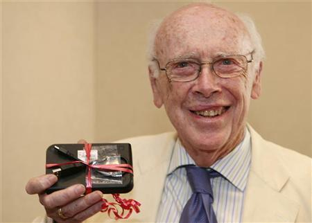
James Watson, co-discoverer of the double helix structure of DNA, lit into targets large and small. On government officials who oversee cancer research, he wrote in a paper published on Tuesday in the journal Open Biology, “We now have no general of influence, much less power … leading our country’s War on Cancer.”

On the $100 million U.S. project to determine the DNA changes that drive nine forms of cancer: It is “not likely to produce the truly breakthrough drugs that we now so desperately need,” Watson argued. On the idea that antioxidants such as those in colorful berries fight cancer: “The time has come to seriously ask whether antioxidant use much more likely causes than prevents cancer.”

That Watson’s impassioned plea came on the heels of the annual cancer report was coincidental. He worked on the paper for months, and it represents the culmination of decades of thinking about the subject. Watson, 84, taught a course on cancer at Harvard University in 1959, three years before he shared the Nobel Prize in medicine for his role in discovering the double helix, which opened the door to understanding the role of genetics in disease.

Other cancer luminaries gave Watson’s paper mixed reviews.

“There are a lot of interesting ideas in it, some of them sustainable by existing evidence, others that simply conflict with well-documented findings,” said one eminent cancer biologist who asked not to be identified so as not to offend Watson. “As is often the case, he’s stirring the pot, most likely in a very productive way.”

There is wide agreement, however, that current approaches are not yielding the progress they promised. Much of the decline in cancer mortality in the United States, for instance, reflects the fact that fewer people are smoking, not the benefits of clever new therapies.

“The great hope of the modern targeted approach was that with DNA sequencing we would be able to find what specific genes, when mutated, caused each cancer,” said molecular biologist Mark Ptashne of Memorial Sloan-Kettering Cancer Center in New York. The next step was to design a drug to block the runaway proliferation the mutation caused.

But almost none of the resulting treatments cures cancer. “These new therapies work for just a few months,” Watson told Reuters in a rare interview. “And we have nothing for major cancers such as the lung, colon and breast that have become metastatic.”

The main reason drugs that target genetic glitches are not cures is that cancer cells have a work-around. If one biochemical pathway to growth and proliferation is blocked by a drug such as AstraZeneca’s Iressa or Genentech’s Tarceva for non-small-cell lung cancer, said cancer biologist Robert Weinberg of MIT, the cancer cells activate a different, equally effective pathway.

That is why Watson advocates a different approach: targeting features that all cancer cells, especially those in metastatic cancers, have in common.

One such commonality is oxygen radicals. Those forms of oxygen rip apart other components of cells, such as DNA. That is why antioxidants, which have become near-ubiquitous additives in grocery foods from snack bars to soda, are thought to be healthful: they mop up damaging oxygen radicals.

That simple picture becomes more complicated, however, once cancer is present. Radiation therapy and many chemotherapies kill cancer cells by generating oxygen radicals, which trigger cell suicide. If a cancer patient is binging on berries and other antioxidants, it can actually keep therapies from working, Watson proposed.

“Everyone thought antioxidants were great,” he said. “But I’m saying they can prevent us from killing cancer cells.”

Research backs him up. A number of studies have shown that taking antioxidants such as vitamin E do not reduce the risk of cancer but can actually increase it, and can even shorten life. But drugs that block antioxidants – “anti-antioxidants” – might make even existing cancer drugs more effective.

Anything that keeps cancer cells full of oxygen radicals “is likely an important component of any effective treatment,” said cancer biologist Robert Benezra of Sloan-Kettering.

Watson’s anti-antioxidant stance includes one historical irony. The first high-profile proponent of eating lots of antioxidants (specifically, vitamin C) was biochemist Linus Pauling, who died in 1994 at age 93. Watson and his lab mate, Francis Crick, famously beat Pauling to the discovery of the double helix in 1953.

One elusive but promising target, Watson said, is a protein in cells called Myc. It controls more than 1,000 other molecules inside cells, including many involved in cancer. Studies suggest that turning off Myc causes cancer cells to self-destruct in a process called apoptosis.

“The notion that targeting Myc will cure cancer has been around for a long time,” said cancer biologist Hans-Guido Wendel of Sloan-Kettering. “Blocking production of Myc is an interesting line of investigation. I think there’s promise in that.”

Targeting Myc, however, has been a backwater of drug development. “Personalized medicine” that targets a patient’s specific cancer-causing mutation attracts the lion’s share of research dollars.

“The biggest obstacle” to a true war against cancer, Watson wrote, may be “the inherently conservative nature of today’s cancer research establishments.” As long as that’s so, “curing cancer will always be 10 or 20 years away.”

http://www.reuters.com/article/2013/01/09/us-usa-cancer-watson-idUSBRE90805N20130109

‘Scarecrow’ Gene: Key to Efficient Crops, Could Lead to Staple Crops With Much Higher Yields

scarecrow gene
Cross section of a mature maize leaf showing Kranz (German for wreath) anatomy around a large vein. The bundle sheath cells (lighter red) encircle the vascular core (light blue). Mesophyll cells (dark red) encircle the bundle sheath cells. The interaction and cooperation between the mesophyll and bundle sheath is essential for the C4 photosynthetic mechanism. (Credit: Thomas Slewinski)

With projections of 9.5 billion people by 2050, humankind faces the challenge of feeding modern diets to additional mouths while using the same amounts of water, fertilizer and arable land as today.

Cornell researchers have taken a leap toward meeting those needs by discovering a gene that could lead to new varieties of staple crops with 50 percent higher yields.

The gene, called Scarecrow, is the first discovered to control a special leaf structure, known as Kranz anatomy, which leads to more efficient photosynthesis. Plants photosynthesize using one of two methods: C3, a less efficient, ancient method found in most plants, including wheat and rice; and C4, a more efficient adaptation employed by grasses, maize, sorghum and sugarcane that is better suited to drought, intense sunlight, heat and low nitrogen.

“Researchers have been trying to find the underlying genetics of Kranz anatomy so we can engineer it into C3 crops,” said Thomas Slewinski, lead author of a paper that appeared online in November in the journal Plant and Cell Physiology. Slewinski is a postdoctoral researcher in the lab of senior author Robert Turgeon, professor of plant biology in the College of Arts and Sciences.

The finding “provides a clue as to how this whole anatomical key is regulated,” said Turgeon. “There’s still a lot to be learned, but now the barn door is open and you are going to see people working on this Scarecrow pathway.” The promise of transferring C4 mechanisms into C3 plants has been fervently pursued and funded on a global scale for decades, he added.

If C4 photosynthesis is successfully transferred to C3 plants through genetic engineering, farmers could grow wheat and rice in hotter, dryer environments with less fertilizer, while possibly increasing yields by half, the researchers said.

C3 photosynthesis originated at a time in Earth’s history when the atmosphere had a high proportion of carbon dioxide. C4 plants have independently evolved from C3 plants some 60 times at different times and places. The C4 adaptation involves Kranz anatomy in the leaves, which includes a layer of special bundle sheath cells surrounding the veins and an outer layer of cells called mesophyll. Bundle sheath cells and mesophyll cells cooperate in a two-step version of photosynthesis, using different kinds of chloroplasts.

By looking closely at plant evolution and anatomy, Slewinski recognized that the bundle sheath cells in leaves of C4 plants were similar to endodermal cells that surrounded vascular tissue in roots and stems.

Slewinski suspected that if C4 leaves shared endodermal genes with roots and stems, the genetics that controlled those cell types may also be shared. Slewinski looked for experimental maize lines with mutant Scarecrow genes, which he knew governed endodermal cells in roots. When the researchers grew those plants, they first identified problems in the roots, then checked for abnormalities in the bundle sheath. They found that the leaves of Scarecrow mutants had abnormal and proliferated bundle sheath cells and irregular veins.

In all plants, an enzyme called RuBisCo facilitates a reaction that captures carbon dioxide from the air, the first step in producing sucrose, the energy-rich product of photosynthesis that powers the plant. But in C3 plants RuBisCo also facilitates a competing reaction with oxygen, creating a byproduct that has to be degraded, at a cost of about 30-40 percent overall efficiency. In C4 plants, carbon dioxide fixation takes place in two stages. The first step occurs in the mesophyll, and the product of this reaction is shuttled to the bundle sheath for the RuBisCo step. The RuBisCo step is very efficient because in the bundle sheath cells, the oxygen concentration is low and the carbon dioxide concentration is high. This eliminates the problem of the competing oxygen reaction, making the plant far more efficient.

The study was funded by the National Science Foundation and the U.S. Department of Agriculture.

http://www.sciencedaily.com/releases/2013/01/130124134051.htm

Dog’s dinner was key to domestication

dog

Dogs now have an excuse for waiting under the dinner table: domestication may have adapted them to thrive on the starch-filled foods that their owners eat.

A study published in Nature found that dogs possess genes for digesting starches, setting them apart from their carnivore cousins — wolves.

The authors say the results support the contentious idea that dogs became domesticated by lingering around human settlements. “While it’s possible that humans might have gone out to take wolf pups and domesticated them, it may have been more attractive for dogs to start eating from the scrap heaps as modern agriculture started,” says Kerstin Lindblad-Toh, a geneticist at Uppsala University in Sweden, who led the work.

Canine-domestication researchers agree that all dogs, from beagles to border collies, are the smaller, more sociable and less aggressive descendants of wolves. But neither the time nor the location of the first domestication is known: fossils place the earliest dogs anywhere from 33,000 years ago in Siberia to 11,000 years ago in Israel, whereas DNA studies of modern dogs put domestication at least 10,000 years ago, and in either Southeast Asia or the Middle East. Many researchers believe that dogs were domesticated more than once, and that even after domestication, they occasionally interbred with wild wolves.

Lindblad-Toh and her team catalogued the genetic changes involved in domestication by looking for differences between the genomes of 12 wolves and 60 dogs from 14 different breeds. Their search identified 36 regions of the genome that set dogs apart from wolves — but are not responsible for variation between dog breeds.

Nineteen of those regions contained genes with a role in brain development or function. These genes, says Lindblad-Toh, may explain why dogs are so much more friendly than wolves. Surprisingly, the team also found ten genes that help dogs to digest starches and break down fats. Lab work suggested that changes in three of those genes make dogs better than meat-eating wolves at splitting starches into sugars and then absorbing those sugars.

Most humans have also evolved to more easily digest starches. Lindblad-Toh suggests that the rise of farming, beginning around 10,000 years ago in the Middle East, led to the adaptations in both species. “This is a striking sign of parallel evolution,” she says. “It really shows how dogs and humans have evolved together to be able to eat starch.”

However, Greger Larson, an evolutionary archaeologist at Durham University, UK, very much doubts that genes involved in digesting starches catalysed domestication, pointing out that the earliest dog fossils predate the dawn of agriculture. His team plans to analyse DNA preserved in dog fossils, to discover when the genetic variations involved in domestication first emerged.

Robert Wayne, a geneticist at the University of California, Los Angeles, who is also studying ancient dog genomes, says that starch metabolism could have been an important adaptation for dogs. However, he thinks that such traits probably developed after behavioural changes that emerged when humans first took dogs in, back when most of our forebears still hunted large game.

Nevertheless, the study adds to evidence that dogs should not eat the same food as wolves, says Wayne, who points out that dog food is rich in carbohydrates and low in protein compared with plain meat. “Every day I get an email from a dog owner who asks, should they feed their dog like a wolf,” says Wayne. “I think this paper answers that question: no.”

http://www.nature.com/news/dog-s-dinner-was-key-to-domestication-1.12280

Thanks to Dr. Lutter for bringing this to the attention of the It’s Interesting community.

Smoking Smothers Your Genes

sn-epigenetic

Cigarettes leave you with more than a smoky scent on your clothes and fingernails. A new study has found strong evidence that tobacco use can chemically modify and affect the activity of genes known to increase the risk of developing cancer. The finding may give researchers a new tool to assess cancer risk among people who smoke.

DNA isn’t destiny. Chemical compounds that affect the functioning of genes can bind to our genetic material, turning certain genes on or off. These so-called epigenetic modifications can influence a variety of traits, such as obesity and sexual preference. Scientists have even identified specific epigenetic patterns on the genes of people who smoke. None of the modified genes has a direct link to cancer, however, making it unclear whether these chemical alterations increase the risk of developing the disease.

In the new study, published in Human Molecular Genetics, researchers analyzed epigenetic signatures in blood cells from 374 individuals enrolled in the European Prospective Investigation into Cancer and Nutrition. EPIC, as it’s known, is a massive study aimed at linking diet, lifestyle, and environmental factors to the incidence of cancer and other chronic diseases. Half of the group consisted of people who went on to develop colon or breast cancer 5 to 7 years after first joining the study, whereas the other half remained healthy.

The team, led by James Flanagan, a human geneticist at Imperial College London, discovered a distinct “epigenetic footprint” in study subjects who were smokers. Compared with people who had never smoked, these individuals had fewer chemical tags known as methyl groups—a common type of epigenetic change—on 20 different regions of their DNA. When the researchers extended the analysis to a separate group of patients and mice that had been exposed to tobacco smoke, they narrowed down the epigenetic modifications to several sites located in four genes that have been weakly linked to cancer before. All of these changes should increase the activity of these genes, Flanagan says. It’s unclear why increasing the activity of the genes would cause cancer, he says, but individuals who don’t have cancer tend not to have these modifications.

The study is the first to establish a close link between epigenetic modifications on a cancer gene and the risk of developing the disease, says Robert Philibert, a behavioral geneticist at the University of Iowa in Iowa City. “To the best of my knowledge, no previous genome-wide epigenetics study has taken such efforts from initial discovery to replication to experimental validation,” adds Lutz Breitling, an epidemiologist at the German Cancer Research Center in Heidelberg, Germany.

The work may lead to new ways to asses cancer risks from smoking. “Previous research into smoking has often asked people to fill out questionnaires, … which have their obvious drawbacks and inaccuracies,” Flanagan says. The new study, he says, may make it possible for doctors to quantify a person’s cancer risk simply through an epigenetic analysis of their DNA.

http://news.sciencemag.org/sciencenow/2012/12/smoking-smothers-your-genes.html

Thanks to Dr. Rajadhyaksha for bringing this to the attention of the It’s Interesting community.

Bullying by Childhood Peers Leaves a Trace That Can Change the Expression of a Gene Linked to Mood

bull

A recent study by a researcher at the Centre for Studies on Human Stress (CSHS) at the Hôpital Louis-H. Lafontaine and professor at the Université de Montréal suggests that bullying by peers changes the structure surrounding a gene involved in regulating mood, making victims more vulnerable to mental health problems as they age.

The study published in the journal Psychological Medicine seeks to better understand the mechanisms that explain how difficult experiences disrupt our response to stressful situations. “Many people think that our genes are immutable; however this study suggests that environment, even the social environment, can affect their functioning. This is particularly the case for victimization experiences in childhood, which change not only our stress response but also the functioning of genes involved in mood regulation,” says Isabelle Ouellet-Morin, lead author of the study.

A previous study by Ouellet-Morin, conducted at the Institute of Psychiatry in London (UK), showed that bullied children secrete less cortisol — the stress hormone — but had more problems with social interaction and aggressive behaviour. The present study indicates that the reduction of cortisol, which occurs around the age of 12, is preceded two years earlier by a change in the structure surrounding a gene (SERT) that regulates serotonin, a neurotransmitter involved in mood regulation and depression.

To achieve these results, 28 pairs of identical twins with a mean age of 10 years were analyzed separately according to their experiences of bullying by peers: one twin had been bullied at school while the other had not. “Since they were identical twins living in the same conditions, changes in the chemical structure surrounding the gene cannot be explained by genetics or family environment. Our results suggest that victimization experiences are the source of these changes,” says Ouellet-Morin. According to the author, it would now be worthwhile to evaluate the possibility of reversing these psychological effects, in particular, through interventions at school and support for victims.

Journal Reference:

1.I. Ouellet-Morin, C. C. Y. Wong, A. Danese, C. M. Pariante, A. S. Papadopoulos, J. Mill, L. Arseneault. Increased serotonin transporter gene (SERT) DNA methylation is associated with bullying victimization and blunted cortisol response to stress in childhood: a longitudinal study of discordant monozygotic twins. Psychological Medicine, 2012; DOI: 10.1017/S0033291712002784

http://www.sciencedaily.com/releases/2012/12/121218081615.htm

Mother-Child Connection: Scientists Discover Children’s Cells Living in Mothers’ Brains, Including Male Cells Living in the Female Brain for Decades

scientists-discover-childrens-cells-living-in-mothers-brain_1


The link between a mother and child is profound, and new research suggests a physical connection even deeper than anyone thought. The profound psychological and physical bonds shared by the mother and her child begin during gestation when the mother is everything for the developing fetus, supplying warmth and sustenance, while her heartbeat provides a soothing constant rhythm.

The physical connection between mother and fetus is provided by the placenta, an organ, built of cells from both the mother and fetus, which serves as a conduit for the exchange of nutrients, gasses, and wastes. Cells may migrate through the placenta between the mother and the fetus, taking up residence in many organs of the body including the lung, thyroid muscle, liver, heart, kidney and skin. These may have a broad range of impacts, from tissue repair and cancer prevention to sparking immune disorders.

It is remarkable that it is so common for cells from one individual to integrate into the tissues of another distinct person. We are accustomed to thinking of ourselves as singular autonomous individuals, and these foreign cells seem to belie that notion, and suggest that most people carry remnants of other individuals. As remarkable as this may be, stunning results from a new study show that cells from other individuals are also found in the brain. In this study, male cells were found in the brains of women and had been living there, in some cases, for several decades. What impact they may have had is now only a guess, but this study revealed that these cells were less common in the brains of women who had Alzheimer’s disease, suggesting they may be related to the health of the brain.

We all consider our bodies to be our own unique being, so the notion that we may harbor cells from other people in our bodies seems strange. Even stranger is the thought that, although we certainly consider our actions and decisions as originating in the activity of our own individual brains, cells from other individuals are living and functioning in that complex structure. However, the mixing of cells from genetically distinct individuals is not at all uncommon. This condition is called chimerism after the fire-breathing Chimera from Greek mythology, a creature that was part serpent part lion and part goat. Naturally occurring chimeras are far less ominous though, and include such creatures as the slime mold and corals.

 Microchimerism is the persistent presence of a few genetically distinct cells in an organism. This was first noticed in humans many years ago when cells containing the male “Y” chromosome were found circulating in the blood of women after pregnancy. Since these cells are genetically male, they could not have been the women’s own, but most likely came from their babies during gestation.

In this new study, scientists observed that microchimeric cells are not only found circulating in the blood, they are also embedded in the brain. They examined the brains of deceased women for the presence of cells containing the male “Y” chromosome. They found such cells in more than 60 percent of the brains and in multiple brain regions. Since Alzheimer’s disease is more common in women who have had multiple pregnancies, they suspected that the number of fetal cells would be greater in women with AD compared to those who had no evidence for neurological disease. The results were precisely the opposite: there were fewer fetal-derived cells in women with Alzheimer’s. The reasons are unclear.

Microchimerism most commonly results from the exchange of cells across the placenta during pregnancy, however there is also evidence that cells may be transferred from mother to infant through nursing. In addition to exchange between mother and fetus, there may be exchange of cells between twins in utero, and there is also the possibility that cells from an older sibling residing in the mother may find their way back across the placenta to a younger sibling during the latter’s gestation. Women may have microchimeric cells both from their mother as well as from their own pregnancies, and there is even evidence for competition between cells from grandmother and infant within the mother.

What it is that fetal microchimeric cells do in the mother’s body is unclear, although there are some intriguing possibilities. For example, fetal microchimeric cells are similar to stem cells in that they are able to become a variety of different tissues and may aid in tissue repair. One research group investigating this possibility followed the activity of fetal microchimeric cells in a mother rat after the maternal heart was injured: they discovered that the fetal cells migrated to the maternal heart and differentiated into heart cells helping to repair the damage. In animal studies, microchimeric cells were found in maternal brains where they became nerve cells, suggesting they might be functionally integrated in the brain. It is possible that the same may true of such cells in the human brain.

These microchimeric cells may also influence the immune system. A fetal microchimeric cell from a pregnancy is recognized by the mother’s immune system partly as belonging to the mother, since the fetus is genetically half identical to the mother, but partly foreign, due to the father’s genetic contribution. This may “prime” the immune system to be alert for cells that are similar to the self, but with some genetic differences. Cancer cells which arise due to genetic mutations are just such cells, and there are studies which suggest that microchimeric cells may stimulate the immune system to stem the growth of tumors. Many more microchimeric cells are found in the blood of healthy women compared to those with breast cancer, for example, suggesting that microchimeric cells can somehow prevent tumor formation. In other circumstances, the immune system turns against the self, causing significant damage. Microchimerism is more common in patients suffering from Multiple Sclerosis than in their healthy siblings, suggesting chimeric cells may have a detrimental role in this disease, perhaps by setting off an autoimmune attack.

This is a burgeoning new field of inquiry with tremendous potential for novel findings as well as for practical applications. But it is also a reminder of our interconnectedness.

http://www.scientificamerican.com/article.cfm?id=scientists-discover-childrens-cells-living-in-mothers-brain