New research may help explain why curiosity promotes better memory

Everyone knows it’s easier to learn about a topic you’re curious about. Now, a new study reveals what’s going on in the brain during that process, revealing that such curiosity may give a person a memory boost.

When participants in the study were feeling curious, they were better at remembering information even about unrelated topics, and brain scans showed activity in areas linked to reward and memory.

The results, detailed October 2 in the journal Neuron, hint at ways to improve learning and memory in both healthy people and those with neurological disorders, the researchers said.

“Curiosity may put the brain in a state that allows it to learn and retain any kind of information, like a vortex that sucks in what you are motivated to learn, and also everything around it,” Matthias Gruber, a memory researcher at the University of California, Davis, said in a statement. “These findings suggest ways to enhance learning in the classroom and other settings.”

Gruber and his colleagues put people in a magnetic resonance imaging (MRI) scanner and showed them a series of trivia questions, asking them to rate their curiosity about the answers to those questions. Later, the participants were shown selected trivia questions, then a picture of a neutral face during a 14-second delay, followed by the answer. Afterward, the participants were given a surprise memory test of the faces, and then a memory test of the trivia answers.

Not surprisingly, the study researchers found that people remembered more information about the trivia when they were curious about the trivia answers. But unexpectedly, when the participants were curious, they were also better at remembering the faces, an entirely unrelated task. Participants who were curious were also more likley than others to remember both the trivia information and unrelated faces a day later, the researchers found.

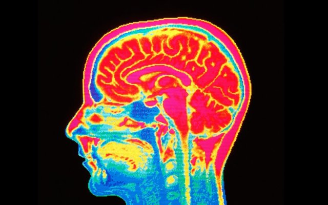
The brain scans showed that, compared with when their curiosity wasn’t piqued, when people were curious, they showed more activation of brain circuits in the nucleus accumbens, an area involved in reward. These same circuits, mediated by the neurochemical messenger dopamine, are involved in forms of external motivation, such as food, sex or drug addiction.

Finally, being curious while learning seemed to produce a spike of activity in the hippocampus, an area involved in forming new memories, and strengthened the link between memory and reward brain circuits.

The study’s findings not only highlight the importance of curiosity for learning in healthy people, but could also give insight into neurological conditions. For example, as people age, their dopamine circuits tend to deteriorate, so understanding how curiosity affects these circuits could help scientists develop treatments for patients with memory disorders, the researchers said.

http://www.livescience.com/48121-curiosity-boosts-memory-learning.html

Study Finds Pedophiles’ Brains Wired to Find Children Attractive

Pedophiles’ brains are “abnormally tuned” to find young children attractive, according to a new study published this week. The research, led by Jorge Ponseti at Germany’s University of Kiel, means that it may be possible to diagnose pedophiles in the future before they are able to offend.

The findings, published in scientific journal Biology Letters, discovered that pedophiles have the same neurological reaction to images of those they find attractive as those of people with ordinary sexual predilections, but that all the relevant cerebral areas become engaged when they see children, as opposed to fellow adults. The occipital areas, prefrontal cortex, putamen, and nucleus caudatus become engaged whenever a person finds another attractive, but the subject of this desire is inverted for pedophiles.

While studies into the cognitive wiring of sex offenders have long been a source of debate, this latest research offers some fairly conclusive proof that there is a neural pattern behind their behavior.

The paper explains: “The human brain contains networks that are tuned to face processing, and these networks appear to activate different processing streams of the reproductive domain selectively: nurturing processing in the case of child faces and sexual processing in the case of sexually preferred adult faces. This implies that the brain extracts age-related face cues of the preferred sex that inform appropriate response selection in the reproductive domains: nurturing in the case of child faces and mating in the case of adult faces.”

Usually children’s faces elicit feelings of caregiving from both sexes, whereas those of adults provide stimuli in choosing a mate. But among pedophiles, this trend is skewed, with sexual, as opposed to nurturing, emotions burgeoning.

The study analyzed the MRI scans of 56 male participants, a group that included 13 homosexual pedophiles and 11 heterosexual pedophiles, exposing them to “high arousing” images of men, women, boys, and girls. Participants then ranked each photo for attractiveness, leading researchers to their conclusion that the brain network of pedophiles is activated by sexual immaturity.

The critical new finding is that face processing is also tuned to face cues revealing the developmental stage that is sexually preferred,” the paper reads.

Dr. James Cantor, associate professor at the University of Toronto’s Faculty of Medicine, said he was “delighted” by the study’s results. “I have previously described pedophilia as a ‘cross-wiring’ of sexual and nurturing instincts, and this data neatly verifies that interpretation.”

Cantor has undertaken extensive research into the area, previously finding that pedophiles are more likely to be left-handed, 2.3 cm shorter than the average male, and 10 to 15 IQ points lower than the norm.

He continued: “This [new] study is definitely a step in the right direction, and I hope other researchers repeat this kind of work. There still exist many contradictions among scientists’ observations, especially in identifying exactly which areas of the brain are the most central to pedophilia. Because financial support for these kinds of studies is quite small, these studies have been quite small, permitting them to achieve only incremental progress. Truly definitive studies about what in the brain causes pedophilia, what might detect it, and what might prevent it require much more significant support.”

Ponseti said that he hoped to investigate this area further by examining whether findings could be emulated when images of children’s faces are the sole ones used. This could lead to gauging a person’s predisposition to pedophilia far more simply than any means currently in place. “We could start to look at the onset of pedophilia, which is probably in puberty at about 12 or 14 years [old],” he told The Independent.

While Cantor is correct in citing the less than abundant size of the study, the research is certainly significant in providing scope for future practicable testing that could reduce the number of pedophilic crimes committed. By being able to run these tests and examine a person’s tendency toward being sexually attracted to underage children, rehabilitative care and necessary precautions could be taken to safeguard children and ensure that those at risk of committing a crime of this ilk would not be able to do so.

http://www.thedailybeast.com/articles/2014/05/23/study-finds-pedophiles-brains-wired-to-find-children-attractive.html#

Scientists Construct First Detailed Map of How the Brain Organizes Everything We See

121219142257-large

Our eyes may be our window to the world, but how do we make sense of the thousands of images that flood our retinas each day? Scientists at the University of California, Berkeley, have found that the brain is wired to put in order all the categories of objects and actions that we see. They have created the first interactive map of how the brain organizes these groupings.

The result — achieved through computational models of brain imaging data collected while the subjects watched hours of movie clips — is what researchers call “a continuous semantic space.”

“Our methods open a door that will quickly lead to a more complete and detailed understanding of how the brain is organized. Already, our online brain viewer appears to provide the most detailed look ever at the visual function and organization of a single human brain,” said Alexander Huth, a doctoral student in neuroscience at UC Berkeley and lead author of the study published Dec. 19 in the journal Neuron.

A clearer understanding of how the brain organizes visual input can help with the medical diagnosis and treatment of brain disorders. These findings may also be used to create brain-machine interfaces, particularly for facial and other image recognition systems. Among other things, they could improve a grocery store self-checkout system’s ability to recognize different kinds of merchandise.

“Our discovery suggests that brain scans could soon be used to label an image that someone is seeing, and may also help teach computers how to better recognize images,” said Huth.

It has long been thought that each category of object or action humans see — people, animals, vehicles, household appliances and movements — is represented in a separate region of the visual cortex. In this latest study, UC Berkeley researchers found that these categories are actually represented in highly organized, overlapping maps that cover as much as 20 percent of the brain, including the somatosensory and frontal cortices.

To conduct the experiment, the brain activity of five researchers was recorded via functional Magnetic Resonance Imaging (fMRI) as they each watched two hours of movie clips. The brain scans simultaneously measured blood flow in thousands of locations across the brain.

Researchers then used regularized linear regression analysis, which finds correlations in data, to build a model showing how each of the roughly 30,000 locations in the cortex responded to each of the 1,700 categories of objects and actions seen in the movie clips. Next, they used principal components analysis, a statistical method that can summarize large data sets, to find the “semantic space” that was common to all the study subjects.

The results are presented in multicolored, multidimensional maps showing the more than 1,700 visual categories and their relationships to one another. Categories that activate the same brain areas have similar colors. For example, humans are green, animals are yellow, vehicles are pink and violet and buildings are blue.

“Using the semantic space as a visualization tool, we immediately saw that categories are represented in these incredibly intricate maps that cover much more of the brain than we expected,” Huth said.

Other co-authors of the study are UC Berkeley neuroscientists Shinji Nishimoto, An T. Vu and Jack Gallant.

Journal Reference:

1.Alexander G. Huth, Shinji Nishimoto, An T. Vu, Jack L. Gallant. A Continuous Semantic Space Describes the Representation of Thousands of Object and Action Categories across the Human Brain. Neuron, 2012; 76 (6): 1210 DOI: 10.1016/j.neuron.2012.10.014

http://www.sciencedaily.com/releases/2012/12/121219142257.htm

Parents-to-be in Japan can buy a 3D printed model of their fetus

EXPECTANT parents in Japan who can’t wait to show the world what their baby will look like can now buy a 3D model of the fetus to share with their friends.

Based on an MRI scan, a 3D printer can create a 9-centimetre resin model of the white fetus, encased in a transparent block in the shape of the mother’s body.

“As it is only once in a lifetime that you are pregnant with that child, we received requests for these kind of models from pregnant women who… do not want to forget the feelings and experience of that time,” said Tomohiro Kinoshita of FASOTEC, the company offering the service.

The 3D model is called Shape of an Angel and costs 100,000 yen ($A1171).

It also comes with a miniature version that could be a nice adornment to a mobile phone, he added. Many young women in Japan add decorations to their phone strap.

The company said the ideal time for a scan is about eight or nine months into the pregnancy.

For those who would like a less pricey version, the company will offer a 3D model of the face of the fetus for 50,000 yen in December.

It will use ultrasound images taken at a medical clinic in Tokyo that is working with the company.

FASOTEC, originally a supplier of devices including 3D printers, uses a layering technique to build up three-dimensional structures.

The technique has been touted as a solution to localised manufacture on a small scale.

http://www.theaustralian.com.au/australian-it/parents-to-be-in-japan-can-buy-3d-printed-model-of-fetus/story-e6frgakx-1226525367570

Thanks to Kebmodee for bringing this to the attention of the It’s Interesting community.