How Exercise Beefs Up the Brain

exercise

New research explains how abstract benefits of exercise—from reversing depression to fighting cognitive decline—might arise from a group of key molecules.

While our muscles pump iron, our cells pump out something else: molecules that help maintain a healthy brain. But scientists have struggled to account for the well-known mental benefits of exercise, from counteracting depression and aging to fighting Alzheimer’s and Parkinson’s disease. Now, a research team may have finally found a molecular link between a workout and a healthy brain.

Much exercise research focuses on the parts of our body that do the heavy lifting. Muscle cells ramp up production of a protein called FNDC5 during a workout. A fragment of this protein, known as irisin, gets lopped off and released into the bloodstream, where it drives the formation of brown fat cells, thought to protect against diseases such as diabetes and obesity. (White fat cells are traditionally the villains.)

While studying the effects of FNDC5 in muscles, cellular biologist Bruce Spiegelman of Harvard Medical School in Boston happened upon some startling results: Mice that did not produce a so-called co-activator of FNDC5 production, known as PGC-1α, were hyperactive and had tiny holes in certain parts of their brains. Other studies showed that FNDC5 and PGC-1α are present in the brain, not just the muscles, and that both might play a role in the development of neurons.

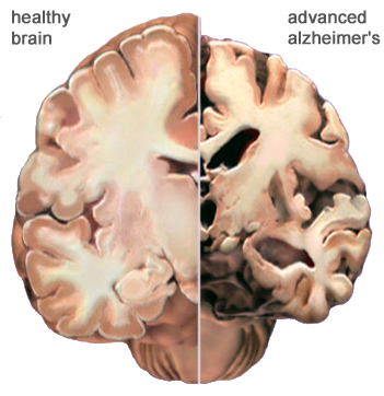
Spiegelman and his colleagues suspected that FNDC5 (and the irisin created from it) was responsible for exercise-induced benefits to the brain—in particular, increased levels of a crucial protein called brain-derived neurotrophic factor (BDNF), which is essential for maintaining healthy neurons and creating new ones. These functions are crucial to staving off neurological diseases, including Alzheimer’s and Parkinson’s. And the link between exercise and BDNF is widely accepted. “The phenomenon has been established over the course of, easily, the last decade,” says neuroscientist Barbara Hempstead of Weill Cornell Medical College in New York City, who was not involved in the new work. “It’s just, we didn’t understand the mechanism.”

To sort out that mechanism, Spiegelman and his colleagues performed a series of experiments in living mice and cultured mouse brain cells. First, they put mice on a 30-day endurance training regimen. They didn’t have to coerce their subjects, because running is part of a mouse’s natural foraging behavior. “It’s harder to get them to lift weights,” Spiegelman notes. The mice with access to a running wheel ran the equivalent of a 5K every night.

Aside from physical differences between wheel-trained mice and sedentary ones—“they just look a little bit more like a couch potato,” says co-author Christiane Wrann, also of Harvard Medical School, of the latter’s plumper figures—the groups also showed neurological differences. The runners had more FNDC5 in their hippocampus, an area of the brain responsible for learning and memory.

Using mouse brain cells developing in a dish, the group next showed that increasing the levels of the co-activator PGC-1α boosts FNDC5 production, which in turn drives BDNF genes to produce more of the vital neuron-forming BDNF protein. They report these results online today in Cell Metabolism. Spiegelman says it was surprising to find that the molecular process in neurons mirrors what happens in muscles as we exercise. “What was weird is the same pathway is induced in the brain,” he says, “and as you know, with exercise, the brain does not move.”

So how is the brain getting the signal to make BDNF? Some have theorized that neural activity during exercise (as we coordinate our body movements, for example) accounts for changes in the brain. But it’s also possible that factors outside the brain, like those proteins secreted from muscle cells, are the driving force. To test whether irisin created elsewhere in the body can still drive BDNF production in the brain, the group injected a virus into the mouse’s bloodstream that causes the liver to produce and secrete elevated levels of irisin. They saw the same effect as in exercise: increased BDNF levels in the hippocampus. This suggests that irisin could be capable of passing the blood-brain barrier, or that it regulates some other (unknown) molecule that crosses into the brain, Spiegelman says.

Hempstead calls the findings “very exciting,” and believes this research finally begins to explain how exercise relates to BDNF and other so-called neurotrophins that keep the brain healthy. “I think it answers the question that most of us have posed in our own heads for many years.”

The effect of liver-produced irisin on the brain is a “pretty cool and somewhat surprising finding,” says Pontus Boström, a diabetes researcher at the Karolinska Institute in Sweden. But Boström, who was among the first scientists to identify irisin in muscle tissue, says the work doesn’t answer a fundamental question: How much of exercise’s BDNF-promoting effects come from irisin reaching the brain from muscle cells via the bloodstream, and how much are from irisin created in the brain?

Though the authors point out that other important regulator proteins likely play a role in driving BDNF and other brain-nourishing factors, they are focusing on the benefits of irisin and hope to develop an injectable form of FNDC5 as a potential treatment for neurological diseases and to improve brain health with aging.

http://news.sciencemag.org/biology/2013/10/how-exercise-beefs-brain

Thanks to Dr. Rajadhyaksha for bringing this to the attention of the It’s Interesting community.

Chinese scientists turn human urine into brain cells

NPGs_from_urine


Chinese researchers have developed a new technique for isolating kidney cells from urine and turning them into neural progenitors — –immature brain cells that can develop into various types of glial cells and neurons. Reprogramming cells has been done before, of course, but not with cells gleaned from urine and not via a method this direct. The technique could prove extremely helpful to those pursuing treatments for neurodegenerative disorders like Parkinson’s and Alzheimer’s.

The innovation here is in the source and the method. We know that embryonic stem cells offer potential treatments for neurodegenerative disorders. And we know that we can turn adult human cells–that is, non-embryonic cells gathered from adult humans–into pluripotent cells (those that can become a different type of cell) by reprogramming them, usually with genetically engineered viruses that tamper with the cells’ genetic codes.

But embryonic stem cell treatments are fraught with ethical issues and non-embryonic methods are complicated–and complexity introduces a greater chance of something going wrong (in this case that means mutations and genetic defects). The new method, which taps skin-like cells from the linings of the kidney tubes that are present in urine, converts its source cells into neurons and glia cells via a more direct route, making the process more efficient while narrowing the margin of error.

In their study, the researchers harvested kidney cells from the urine samples of three human donors and converted the cells directly to neural progenitors. Rather than using a genetically engineered virus to reprogram the cells, they used a small piece of bacterial DNA that can replicate in the cellular cytoplasm, a technique that eliminates the need to tamper directly with the chromosome (in theory, at least, this should reduce mutations) while also speeding up the entire process. After growing their progenitors into mature neurons and glial cells, the researchers transplanted the progenitors into the brains of newborn rats. A month later, the cells were still alive in the rats’ brains, though it is not yet clear that they can survive for extended periods or mesh with the brain’s wiring to become functioning parts of the neural machine.

There’s still a lot of research to be done on this method of course, but the researchers think it may provide a way to take cells gathered non-invasively and quickly and efficiently convert them into neural cells while reducing the likelihood of genetic mutations.

http://www.guardian.co.uk/science/neurophilosophy/2012/dec/09/turning-urine-into-brain-cells


Mother-Child Connection: Scientists Discover Children’s Cells Living in Mothers’ Brains, Including Male Cells Living in the Female Brain for Decades

scientists-discover-childrens-cells-living-in-mothers-brain_1


The link between a mother and child is profound, and new research suggests a physical connection even deeper than anyone thought. The profound psychological and physical bonds shared by the mother and her child begin during gestation when the mother is everything for the developing fetus, supplying warmth and sustenance, while her heartbeat provides a soothing constant rhythm.

The physical connection between mother and fetus is provided by the placenta, an organ, built of cells from both the mother and fetus, which serves as a conduit for the exchange of nutrients, gasses, and wastes. Cells may migrate through the placenta between the mother and the fetus, taking up residence in many organs of the body including the lung, thyroid muscle, liver, heart, kidney and skin. These may have a broad range of impacts, from tissue repair and cancer prevention to sparking immune disorders.

It is remarkable that it is so common for cells from one individual to integrate into the tissues of another distinct person. We are accustomed to thinking of ourselves as singular autonomous individuals, and these foreign cells seem to belie that notion, and suggest that most people carry remnants of other individuals. As remarkable as this may be, stunning results from a new study show that cells from other individuals are also found in the brain. In this study, male cells were found in the brains of women and had been living there, in some cases, for several decades. What impact they may have had is now only a guess, but this study revealed that these cells were less common in the brains of women who had Alzheimer’s disease, suggesting they may be related to the health of the brain.

We all consider our bodies to be our own unique being, so the notion that we may harbor cells from other people in our bodies seems strange. Even stranger is the thought that, although we certainly consider our actions and decisions as originating in the activity of our own individual brains, cells from other individuals are living and functioning in that complex structure. However, the mixing of cells from genetically distinct individuals is not at all uncommon. This condition is called chimerism after the fire-breathing Chimera from Greek mythology, a creature that was part serpent part lion and part goat. Naturally occurring chimeras are far less ominous though, and include such creatures as the slime mold and corals.

 Microchimerism is the persistent presence of a few genetically distinct cells in an organism. This was first noticed in humans many years ago when cells containing the male “Y” chromosome were found circulating in the blood of women after pregnancy. Since these cells are genetically male, they could not have been the women’s own, but most likely came from their babies during gestation.

In this new study, scientists observed that microchimeric cells are not only found circulating in the blood, they are also embedded in the brain. They examined the brains of deceased women for the presence of cells containing the male “Y” chromosome. They found such cells in more than 60 percent of the brains and in multiple brain regions. Since Alzheimer’s disease is more common in women who have had multiple pregnancies, they suspected that the number of fetal cells would be greater in women with AD compared to those who had no evidence for neurological disease. The results were precisely the opposite: there were fewer fetal-derived cells in women with Alzheimer’s. The reasons are unclear.

Microchimerism most commonly results from the exchange of cells across the placenta during pregnancy, however there is also evidence that cells may be transferred from mother to infant through nursing. In addition to exchange between mother and fetus, there may be exchange of cells between twins in utero, and there is also the possibility that cells from an older sibling residing in the mother may find their way back across the placenta to a younger sibling during the latter’s gestation. Women may have microchimeric cells both from their mother as well as from their own pregnancies, and there is even evidence for competition between cells from grandmother and infant within the mother.

What it is that fetal microchimeric cells do in the mother’s body is unclear, although there are some intriguing possibilities. For example, fetal microchimeric cells are similar to stem cells in that they are able to become a variety of different tissues and may aid in tissue repair. One research group investigating this possibility followed the activity of fetal microchimeric cells in a mother rat after the maternal heart was injured: they discovered that the fetal cells migrated to the maternal heart and differentiated into heart cells helping to repair the damage. In animal studies, microchimeric cells were found in maternal brains where they became nerve cells, suggesting they might be functionally integrated in the brain. It is possible that the same may true of such cells in the human brain.

These microchimeric cells may also influence the immune system. A fetal microchimeric cell from a pregnancy is recognized by the mother’s immune system partly as belonging to the mother, since the fetus is genetically half identical to the mother, but partly foreign, due to the father’s genetic contribution. This may “prime” the immune system to be alert for cells that are similar to the self, but with some genetic differences. Cancer cells which arise due to genetic mutations are just such cells, and there are studies which suggest that microchimeric cells may stimulate the immune system to stem the growth of tumors. Many more microchimeric cells are found in the blood of healthy women compared to those with breast cancer, for example, suggesting that microchimeric cells can somehow prevent tumor formation. In other circumstances, the immune system turns against the self, causing significant damage. Microchimerism is more common in patients suffering from Multiple Sclerosis than in their healthy siblings, suggesting chimeric cells may have a detrimental role in this disease, perhaps by setting off an autoimmune attack.

This is a burgeoning new field of inquiry with tremendous potential for novel findings as well as for practical applications. But it is also a reminder of our interconnectedness.

http://www.scientificamerican.com/article.cfm?id=scientists-discover-childrens-cells-living-in-mothers-brain

Skin Cancer Drug Serendipitously Discovered to Treat Alzheimer’s Disease in Mice


Scientists say they “serendipitously” discovered that a drug used to treat a type of cancer quickly reversed Alzheimer’s disease in mice.

“It’s really exciting,” said Maria Carrillo, senior director for medical and scientific relations for the Alzheimer’s Association. “They saw very positive and robust behavior effects in the mice.”

In the study, researchers at Case Western Reserve University School of Medicine gave mice mega-doses of bexarotene, a drug used to treat a type of skin cancer called cutaneous T-cell lymphoma. Within 72 hours, the mice showed dramatic improvements in memory and more than 50% of amyloid plaque — a hallmark of Alzheimer’s disease — had been removed from the brain.

The study was published Thursday in the journal Science.

Gary Landreth, the lead researcher at Case Western, cautioned that even though his results were impressive in mice, it may turn out not to work in people.

“I want to say as loudly and clearly as possible that this was a study in mice, not in humans,” he said. “We’ve fixed Alzheimer’s in mice lots of times, so we need to move forward expeditiously but cautiously.”

Mice — and humans — with Alzheimer’s have high levels of a substance called amyloid beta in their brain. Pathology tests on the mice showed bexarotene lowered the levels of amyloid beta and raised the levels of apolipoprotein E, which helps keep amyloid beta levels low.

Landreth said he hopes to try the drug out in healthy humans within two months, to see if it has the same effect.

Those participating in the trial would be given the standard dose that cancer patients are usually given.

Researchers tested the memories of mice with Alzheimer’s both before and after giving them bexarotene. For example, the Alzheimer’s mice walked right into a cage where they’d previously been given a painful electrical shock, but after treatment with bexarotene, the mice remembered the shock and refused to enter the cage.

In another test, the scientists put tissue paper in a cage. Normal mice instinctively use tissues in their cage to make a nest, but mice with Alzheimer’s can’t figure out what to do with the tissues. After treatment with the drug, the Alzheimer’s mice made a nest with the paper.

Carrillo said one of the major advantages of bexarotene is that it’s already been approved by the Food and Drug Administration for use in humans, which means the researchers can move into human trials sooner than if it were a completely new drug.

The Alzheimer’s Foundation is funding Case Western’s next phase of research, which will involve using bexarotene at the levels used on cancer patients, Landreth said. Since the drug does have some side effects — it can increase cholesterol, for example — he hopes to use it in even lower levels as the study goes on.

Landreth said his lab had been working on other drugs for Alzheimer’s for 10 years when a graduate student, Paige Cramer, decided to try bexarotene, which works on a receptor involved in amyloid beta clearance. Some other drugs that worked in mice were too toxic to use in humans.

“We’re really lucky that bexarotene is a great drug with an acceptable safety profile,” he said. “This doesn’t happen very many times in life.”

http://www.cnn.com/2012/02/09/health/us-cancer-drug-alzheimers/index.html?hpt=hp_bn10

Snorting Insulin May Treat Alzheimer’s Disease


A study in the journal Archives of Neurology suggests that intranasal insulin may help with cognition and functioning in patients who have both mild and more severe dementia.

Recent research has suggested that insulin plays an important role in a number of brain functions, in addition to regulating blood sugar. Insulin promotes cell repair and cell genesis, so the thinking is that it could actually modify the course of Alzheimer’s disease.

It also appears to protect against the toxic effects of beta-amyloid, the protein involved in the brain plaques associated with dementia. Insulin also prevents the formation of the toxic form of tau, a biomarker found in the cerebrospinal fluid.

The goal of this study was to supplement and normalize the insulin levels in the brain without affecting levels in the rest of the body. This was done with a device that was designed to deliver insulin through the nose to the brain without getting too much into the blood.

Patients treated with insulin were able to remember information over a period of time better than those who got placebo; in fact, performance improved 20%. They also showed an enhancement in brain glucose metabolism in some areas; those who received placebo tended to show a decline.

The treatment had the mild side effects of occasional mild headache and runny nose.

http://thechart.blogs.cnn.com/2011/09/12/insulin-may-help-treat-alzheimers/?hpt=hp_t2

Researchers Find Where Musical Memory is Located in the Brain

Neuroscientists have pinpointed the area of our brain where we store memories of music.

The findings are part of a study, published in the journal Brain, on memory loss in dementia, in particular looking at the ability to remember and recognize sounds, which is unusually preserved in Alzheimer’s disease. 

In the study, participants with dementia, as well as healthy controls, were asked to distinguish between well-known tunes and made-up tunes that had the same key and tempo but a different combination of notes.

The 27 participants with dementia had a diagnosis of either Alzheimer’s disease or a type of dementia called semantic dementia, where patients lose their understanding of words, objects and concepts.

The researchers found that participants with semantic dementia were unable to recognise the famous melodies.

MRI scans of these participants showed that the right anterior lobe of the brain, located behind the right ear, was significantly shrunken.  

Participants with Alzheimer’s did not show significant damage in this area of the brain.

http://alzheimersweekly.com/content/researchers-find-location-music-brain

A Dearth of New Medications for Neuropsychiatric Disease on the Horizon


Neuropsychiatric diseases like schizophrenia, depression, Alzheimer’s disease, Parkinson’s disease and more cost billions per year and account for 13% of the global burden of disease (a measure of years of life lost due to premature mortality and living in a state less than full health), according to the World Health Organization.

However, pharmaceutical companies have decided that generating new drugs to treat these disorders are simply too costly to pursue, and are pulling the plug on research and development in this area.

Read more here:  http://www.scientificamerican.com/article.cfm?id=a-dearth-of-new-meds

Being bilingual protects from Alzheimer’s disease

Speaking at least two languages may slow dementia in the aging brain, new research shows. Bilingual people do better in mental challenges and are more skilled at multi-tasking than those who have just one tongue. They also develop symptoms of dementia and Alzheimer’s disease an average of four or five years later.

http://news.nationalgeographic.com/news/2011/02/100218-bilingual-brains-alzheimers-dementia-science-aging/

http://www.dailymail.co.uk/sciencetech/article-1358234/People-speak-languages-better-multi-tasking-likely-develop-Alzheimers.html

Half of all cases of Alzheimer’s disease may be misdiagnosed.


It’s currently impossible to definitively diagnosis Alzheimer’s disease while the patient is alive.  Researchers recently analyzed around 800 brains from Alzheimer’s patients after they died, and learned that only about half of them actually had the disease.  The other half suffered from other forms of dementia.  A reliable way to diagnose the specific type of dementia from which patients are suffering is critical to guiding treatment for patients and for researching the disease processes.

http://pagingdrgupta.blogs.cnn.com/2011/02/23/half-of-alzheimers-cases-misdiagnosed/?hpt=T2

What’s Happening to the Scientific Method?

All sorts of well-established, repeatedly-confirmed findings have started to look increasingly uncertain in a wide range of scientific fields, from psychology to ecology.  This phenomenon is particularly widespread in the field of medicine. 
To read more about this, click on this article by Jonathan Lehrer in the The New Yorker: