Changes in the anterior insula of hte brain may make us more trusting as we age

sn-trustworthiness

Despite long experience with the ways of the world, older people are especially vulnerable to fraud. According to the Federal Trade Commission (FTC), up to 80% of scam victims are over 65. One explanation may lie in a brain region that serves as a built-in crook detector. Called the anterior insula, this structure—which fires up in response to the face of an unsavory character—is less active in older people, possibly making them less cagey than younger folks, a new study finds.

Both FTC and the Federal Bureau of Investigation have found that older people are easy marks due in part to their tendency to accentuate the positive. According to social neuroscientist Shelley Taylor of the University of California, Los Angeles, research backs up the idea that older people can put a positive spin on things—emotionally charged pictures, for example, and playing virtual games in which they risk the loss of money. “Older people are good at regulating their emotions, seeing things in a positive light, and not overreacting to everyday problems,” she says. But this trait may make them less wary.

To see if older people really are less able to spot a shyster, Taylor and colleagues showed photos of faces considered trustworthy, neutral, or untrustworthy to a group of 119 older adults (ages 55 to 84) and 24 younger adults (ages 20 to 42). Signs of untrustworthiness include averted eyes; an insincere smile that doesn’t reach the eyes; a smug, smirky mouth; and a backward tilt to the head. The participants were asked to rate each face on a scale from -3 (very untrustworthy) to 3 (very trustworthy).

In the study, appearing in the Proceedings of the National Academy of Sciences, the “untrustworthy” faces were perceived as significantly more trustworthy by the older subjects than by the younger ones. The researchers then performed the same test on a different set of volunteers, this time imaging their brains during the process, to look for differences in brain activity between the age groups. In the younger subjects, when asked to judge whether the faces were trustworthy, the anterior insula became active; the activity increased at the sight of an untrustworthy face. The older people, however, showed little or no activation.

Taylor explains that the insula’s job is to collect information not about others but about one’s own body—sensing feelings, including “gut instincts”—and present that information to the rest of the brain. “It’s a warning bell that doesn’t seem to work as well in older people.” By habitually seeing the world in a positive light, older people may be overriding this warning signal, she says. “It looks like the brain is conspiring with what older people do naturally.”

Whether the insula activates in response to non-facial cues, such as telephone scams (a particular problem for older people), remains unclear, says Taylor, since the study was limited to faces.

The new study is the first to show a characteristic pattern of brain activation in a “social” situation involving the assessment of another person’s trustworthiness, says psychologist Lisbeth Nielsen of the National Institute on Aging (NIA) in Bethesda, Maryland. (Though NIA funded the project, Nielsen was not involved in the study.)

A question to be addressed in future research, she says, is whether decreased activity in the insula is the cause or the effect of older peoples’ more positive outlook. “It may be that older people engage with the world in a certain way and this is reflected in the brain activity.”

If so, she adds, older people could work on becoming more cautious. For example, they could be taught to look out for the facial signs of untrustworthiness. “Just because the insula isn’t being activated doesn’t mean it can’t be.”

http://news.sciencemag.org/sciencenow/2012/12/why-old-people-get-scammed.html?ref=hp

New clues about who killed Ramses III in 1155 BC

sn-ramses2

For more than a century, Egyptologists have puzzled over the mysterious demise of Ramses III in 1155 B.C. According to trial records preserved on the Judicial Papyrus of Turin, an assassin murdered the pharaoh during a bloody palace coup. But was this truly the case, and if so, who led the plot? A new study published today in BMJ shows that Ramesses III died violently after conspirators slashed his throat and reveals that one of the alleged ringleaders, Ramesses’s son Pentawere, may have later been strangled. The research team arrived at these findings after analyzing both DNA samples and CT scans from two mummies: Ramesses III (with linen bandage, above right) and a previously unidentified young man found with him in a cache in Deir el Bahari. The unidentified 20-year-old (shown with arrows pointing to unusual compressed skin folds) proved to be one of Ramesses’s sons: He appeared to have been strangled (also evidenced in the scan by overinflated thorax) and buried with a goat skin, a pelt that ancient Egyptians deemed ritually impure and therefore a mark of dishonor befitting an assassin.

http://news.sciencemag.org/sciencenow/2012/12/scienceshotwho-killed-ramesses-i.html

Scientists create artifical brain with 2.3 million simulated neurons

aritificial brain

Another computer is setting its wits to perform human tasks. But this computer is different. Instead of the tour de force processing of Deep Blue or Watson’s four terabytes of facts of questionable utility, Spaun attempts to play by the same rules as the human brain to figure things out. Instead of the logical elegance of a CPU, Spaun’s computations are performed by 2.3 million simulated neurons configured in networks that resemble some of the brain’s own networks. It was given a series of tasks and performed pretty well, taking a significant step toward the creation of a simulated brain.

Spaun stands for Semantic Pointer Architecture: Unified Network. It was given 6 different tasks that tested its ability to recognize digits, recall from memory, add numbers and complete patterns. Its cognitive network simulated the prefrontal cortex to handle working memory and the basal ganglia and thalamus to control movements. Like a human, Spaun can view an image and then give a motor response; that is, it is presented images that it sees through a camera and then gives a response by drawing with a robotic arm.

And its performance was similar to that of a human brain. For example, the simplest task, image recognition, Spaun was shown various numbers and asked to draw what it sees. It got 94 percent of the numbers correct. In a working memory task, however, it didn’t do as well. It was shown a series of random numbers and then asked to draw them in order. Like us with human brains, Spaun found the pattern recognition task easy, the working memory task not quite as easy.

The important thing here is not how well Spaun performed on the tasks – your average computer could find ways to perform much better than Spaun. But what’s important is that, in Spaun’s case, the task computations were carried out solely by the 2.3 million artificial neurons spiking in the way real neurons spike to carry information from one neuron to another. The visual image, for example, was processed hierarchically, with multiple levels of neurons successively extracting more complex information, just as the brain’s visual system does. Similarly, the motor response mimicked the brain’s strategy of combining many simple movements to produce an optimal, single movement while drawing.

Chris Eliasmith, from the University of Waterlook in Ontario, Canada and lead author of the study is happy with his cognitive creation. “It’s not as smart as monkeys when it comes to categorization,” he told CNN, “but it’s actually smarter than monkeys when it comes to recognizing syntactic patterns, structured patterns in the input, that monkeys won’t recognize.”

Watch Spaun work through its tasks in the following video.

One thing Spaun can’t do is perform tasks in realtime. Every second you saw Spaun performing tasks in the video actually requires 2.5 hours of numbers crunching by its artificial brain. The researchers hope to one day have it perform in realtime.

It’s important to note that Spaun isn’t actually learning anything by performing these tasks. Its neural nets are hardwired and are incapable of the modifications that real neurons undergo when we learn. But producing complex behavior from a simulated neuronal network still represents an important initial step toward building an artificial brain. Christian Machens, a neuroscientist at the Champalimaud Neuroscience Programme in Lisbon and was not involved in the study, writes in Science that the strategy for building a simulated brain is “to not simply incorporate the largest number of neurons or the greatest amount of detail, but to reproduce the largest amount of functionality and behavior.”

We’re still a long way from artificial intelligence that is sentient and self-aware. And there’s no telling if the robots of the future will have brains that look like ours or if entirely different solutions will be used to produce complex behavior. Whatever it looks like, Spaun is a noble step in the right direction.

Scientists Create Artificial Brain With 2.3 Million Simulated Neurons

How gender stereotypes warp our view of depression

74583142depressioncrop

Stereotypes about male and female roles may influence the way we perceive depressed people.

It’s a well-known fact that men and women who behave the same way in the exact same situation—whether it’s a job interview, a cocktail party, or a traffic stop—are sometimes perceived and treated differently based on their gender.

Something similar, it seems, may happen when men and women start to show signs of depression. A new study, published this week in the journal PLoS ONE, suggests that people of both sexes are less likely to view men as being depressed and in need of professional help—even if a man’s symptoms are identical to a woman’s.

“A lot of attention has been paid to depression in women, and with good reason: Depression is twice as common in women,” says Dr. James B. Potash, the editor of the study and a professor of psychiatry at the University of Iowa, in Iowa City. “There has been relatively little focus on education about depression in men. This [study] emphasizes the importance of figuring out how to get through to men that depression can be disabling and treatment is important.”

Health.com: 12 Signs of Depression in Men

In the study, researchers in the U.K. asked a group of about 600 adults to read a short description of a hypothetical depressed person. This vignette, which was designed to illustrate the diagnostic criteria for clinical depression (also known as major depression), read in part:

For the past two weeks, Kate has been feeling really down. She wakes up in the morning with a flat, heavy feeling that sticks with her all day. She isn’t enjoying things the way she normally would. In fact, nothing gives her pleasure. Even when good things happen, they don’t seem to make Kate happy.

Fifty-seven percent of the study participants recognized Kate’s symptoms—which also included difficulty concentrating, fatigue, and insomnia—as indications of a mental health disorder, and more than three-quarters of those people correctly identified the disorder as depression. Only 10% of the respondents said Kate did not have a problem.

The researchers presented the same vignette to another group of 600 people. This time, however, every mention of “Kate” was replaced by “Jack,” and all the pronouns were switched from female to male. Those minor changes had a noticeable effect: Though nearly as many people recognized Jack as having a mental health problem (52%), more than twice as many as in the Kate scenario said he did not (21%).

In addition, men themselves were less likely than women to label Jack depressed—a pattern that was not seen with Kate.

Health.com: How to Help Someone Who’s Depressed

Why the difference? Male stereotypes that emphasize traits such as toughness and strength may dissuade both women and men, and especially the latter, from identifying or acknowledging the signs of depression in men, says study author Viren Swami.

“Men are expected to be strong, deny pain and vulnerability, and conceal any emotional fragility,” says Swami, a psychologist at the University of Westminster, in London. “Because of these societal expectations, men appear to have poorer understanding of mental health and aren’t as good at detecting symptoms of depression compared with women.”

Potash says the findings also may reflect the fact that women are generally more attuned to emotions and better at articulating them. Some men might have all the outward signs of depression, and yet when asked about their mood they “may not be able to say much more than ‘I don’t know,’” he says. “A substantial minority of men just don’t describe depression.”

Health.com: 10 Careers With High Rates of Depression

On a deeper level, men’s failure to recognize the symptoms of depression in a fellow male may represent a kind of defense mechanism prompted by an “unconscious identification” with that man, says Dr. Radu Saveanu, a professor of psychiatry at the University of Miami Miller School of Medicine.

“They may think, ‘If this guy is having trouble and may need treatment, I may be in the same position someday,’” says Saveanu, who was not involved in the study. “That anxiety distorts the ability to be more objective.”

All of these dynamics may affect the likelihood of seeking or recommending treatment. In the study, men were more likely than women to recommend that Kate seek professional help, but this gap disappeared in the Jack scenario. Men also expressed less sympathy for Jack than women did.

The reluctance to seek treatment isn’t unique to men, but it does reflect an independent-minded streak that is more common among males, Potash says. Men tend to think that pulling themselves out of depression is “something they ought to be able to do,” he says. “It’s the stereotype of men who never ask for directions. They won’t admit that they can’t take care of it themselves.”

Health.com: Depressed? 12 Mental Tricks to Turn It Around

Gender, of course, isn’t the only factor that shapes how we view depression symptoms. Swami also found that respondents of either sex who held negative attitudes towards psychiatry and science felt that both Kate and Jack’s symptoms were less distressing, more difficult to treat, and less worthy of sympathy or professional help.

Swami took these trends into account, but he can’t rule out that other factors might have influenced the gender differences seen in the study. The participants’ own mental health history was unknown, for instance, though Swami says previous diagnoses do not tend to impact “mental health literacy,” or how well people understand mental health issues.

Future research will need to address these limitations, he says.

Read more: http://healthland.time.com/2012/11/15/how-gender-stereotypes-warp-our-view-of-depression/#ixzz2Es26tBvB


Pee marks the spot

sn-mice

Human beings tend to avoid places that smell of urine. But to mice, there is something positively addictive about the scent; they like to go back to a spot where they found the excretions again and again. Now, researchers have discovered that this behavior is triggered by a single protein in the urine of male mice.

Mice use scent to mark their territory, advertise their social dominance, and convey information about their health and reproductive status. But these are usually volatile pheromones that disperse quickly, and it has remained unclear what exactly stimulates a female to be attracted to a specific male.

Previous research had shown that female laboratory mice often return to a place where they have come across cage bedding soiled by males. Now, researchers at the University of Liverpool in the United Kingdom have confirmed this. Female mice spent five times as much time in a place where they had encountered a dish with male urine than at a place where they encountered water. Just 10 minutes of exposure to the urine was enough for the mice to show this place preference even after 14 days.

However, if the mice were prevented from by a mesh screen touching the urine with their nose, the place seemed to lose its attractiveness. “That suggested that the story was not as simple as everybody assumed and volatile pheromones were not responsible,” says behavioral ecologist Jane Hurst, one of the authors of the study. By separating the urine into different fractions, the scientists showed that a protein called darcin that they had identified in 2005—and which mice can only detect if their noses touch the urine—is responsible for the frequent visits. Pure darcin, produced in cell culture in the lab, elicited the same reaction, the authors report online today in Science.

“This is a really compelling story,” says Lisa Stowers, a neuroscientist at the Scripps Research Institute in San Diego, California. “Mammals were thought to be much more complex, but this study shows that a single chemical can lead [them to act] in a certain way.” The study is “very simple and elegant,” she adds. But it also raises new questions. For instance: There are many other ways a mouse could learn to return to a certain place. “So what is the benefit of evolving this [special] mechanism?”

Hurst says that what fascinates her is that the pheromones induce learning in the mice. And the animals do not only learn to be attracted to the place where they encountered the darcin. “They learn the odor cues of that specific male and are then attracted to it,” Hurst says. “Being familiar with a scent really seems to be important for whether a female is interested in a male.” The reason, Hurst suggests, is that dominant males, who make attractive mates, tend to leave the most marks in a certain territory.

The researchers showed that male mice, too, are attracted to a place if they have encountered darcin there, probably to foster a behavior called countermarking. “If males come across another male’s scent mark, they put their own, fresher urine there,” Hurst explains. This could also be the reason why some laboratory strains seem to have lost the ability to produce darcin: Because laboratory mice are usually group-housed, they have been selected to be less aggressive, and not producing darcin could help reduce tensions.

http://news.sciencemag.org/sciencenow/2012/12/pee-marks-the-spot.html?ref=em

Thanks to Dr. Rajadhyaksha for bringing this to the attention of the It’s Interesting community.

Controversial surgical treatment for addiction burns away the brain’s pleasure center


How far should doctors go in attempting to cure addiction? In China, some physicians are taking the most extreme measures. By destroying parts of the brain’s “pleasure centers” in heroin addicts and alcoholics, these neurosurgeons hope to stop drug cravings. But damaging the brain region involved in addictive desires risks permanently ending the entire spectrum of natural longings and emotions, including the ability to feel joy.

In 2004, the Ministry of Health in China banned this procedure due to lack of data on long term outcomes and growing outrage in Western media over ethical issues about whether the patients were fully aware of the risks.

However, some doctors were allowed to continue to perform it for research purposes—and recently, a Western medical journal even published a new study of the results. In 2007, The Wall Street Journal detailed the practice of a physician who claimed he performed 1000 such procedures to treat mental illnesses such as depression, schizophrenia and epilepsy, after the ban in 2004; the surgery for addiction has also since been done on at least that many people.

The November publication has generated a passionate debate in the scientific community over whether such research should be published or kept outside the pages of reputable scientific journals, where it may find undeserved legitimacy and only encourage further questionable science to flourish.

The latest study is the third published since 2003 in Stereotactic and Functional Neurosurgery, which isn’t the only journal chronicling results from the procedure, which is known as ablation of the nucleus accumbens. In October, the journal World Neurosurgery also published results from the same researchers, who are based at Tangdu Hospital in Xi’an.

The authors, led by Guodong Gao, claim that the surgery is “a feasible method for alleviating psychological dependence on opiate drugs.” At the same time, they report that more than half of the 60 patients had lasting side effects, including memory problems and loss of motivation. Within five years, 53% had relapsed and were addicted again to opiates, leaving 47% drug free.

(MORE: Addicted: Why We Get Hooked)

Conventional treatment only results in significant recovery in about 30-40% of cases, so the procedure apparently improves on that, but experts do not believe that such a small increase in benefit is worth the tremendous risk the surgery poses.  Even the most successful brain surgeries carry risk of infection, disability and death since opening the skull and cutting brain tissue for any reason is both dangerous and unpredictable. And the Chinese researchers report that 21% of the patients they studied experienced memory deficits after the surgery and 18% had “weakened motivation,” including at least one report of lack of sexual desire. The authors claim, however, that “all of these patients reported that their [adverse results] were tolerable.” In addition, 53% of patients had a change in personality, but the authors describe the majority of these changes as “mildness oriented,” presumably meaning that they became more compliant. Around 7%, however, became more impulsive.

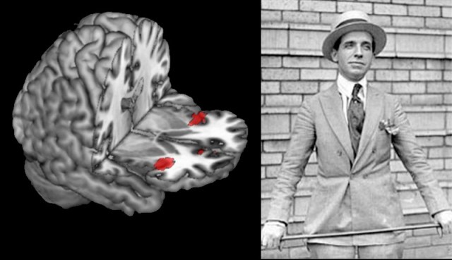
The surgery is actually performed while patients are awake in order to minimize the chances of destroying regions necessary for sensation, consciousness or movement.  Surgeons use heat to kill cells in small sections of both sides of the brain’s nucleus accumbens.  That region is saturated with neurons containing dopamine and endogenous opioids, which are involved in pleasure and desire related both to drugs and to ordinary experiences like eating, love and sex.

(MORE: A Drug to End Drug Addiction)

In the U.S. and the U.K., reports the Wall Street Journal, around two dozen stereotactic ablations are performed each year, but only in the most intractable cases of depression and obsessive-compulsive disorder and after extensive review by institutional review boards and intensive discussions with the patient, who must acknowledge the risks. Often, a different brain region is targeted, not the nucleus accumbens. Given the unpredictable and potentially harmful consequences of the procedure, experts are united in their condemnation of using the technique to treat addictions. “To lesion this region that is thought to be involved in all types of motivation and pleasure risks crippling a human being,” says Dr. Charles O’Brien, head of the Center for Studies of Addiction at the University of Pennsylvania.

David Linden, professor of neuroscience at Johns Hopkins and author of a recent book about the brain’s pleasure systems calls the surgery “horribly misguided.”  He says “This treatment will almost certainly render the subjects unable to feel pleasure from a wide range of experiences, not just drugs of abuse.”

But some neurosurgeons see it differently. Dr. John Adler, professor emeritus of neurosurgery at Stanford University, collaborated with the Chinese researchers on the publication and is listed as a co-author.  While he does not advocate the surgery and did not perform it, he believes it can provide valuable information about how the nucleus accumbens works, and how best to attempt to manipulate it. “I do think it’s worth learning from,” he says. ” As far as I’m concerned, ablation of the nucleus accumbens makes no sense for anyone.  There’s a very high complication rate. [But] reporting it doesn’t mean endorsing it. While we should have legitimate ethical concerns about anything like this, it is a bigger travesty to put our heads in the sand and not be willing to publish it,” he says.

(MORE: Anesthesia Study Opens Window Into Consciousness)

Dr. Casey Halpern, a neurosurgery resident at the University of Pennsylvania makes a similar case. He notes that German surgeons have performed experimental surgery involving placing electrodes in the same region to treat the extreme lack of pleasure and motivation associated with otherwise intractable depression.  “That had a 60% success rate, much better than [drugs like Prozac],” he says. Along with colleagues from the University of Magdeburg in Germany, Halpern has just published a paper in the Proceedings of the New York Academy of Sciences calling for careful experimental use of DBS in the nucleus accumbens to treat addictions, which have failed repeatedly to respond to other approaches. The paper cites the Chinese surgery data and notes that addiction itself carries a high mortality risk.

DBS, however, is quite different from ablation.  Although it involves the risk of any brain surgery, the stimulation itself can be turned off if there are negative side effects, while surgical destruction of brain tissue is irreversible. That permanence—along with several other major concerns — has ethicists and addiction researchers calling for a stop to the ablation surgeries, and for journals to refuse to publish related studies.

Harriet Washington, author of Medical Apartheid:  The Dark History of Medical Experimentation on Black Americans from Colonial Times to the Present, argues that by publishing the results of unethical studies, scientists are condoning the questionable conditions under which the trials are conducted. “When medical journals publish research that violates the profession’sethical guidelines, this serves not only to sanction such abuses, but to encourage them,” she says. “In doing so, this practice encourages a relaxing of moral standards that threatens all patients and subjects, but especially  the medically vulnerable.”

(MORE: Real-Time Video: First Look at a Brain Losing Consciousness Under Anesthesia)

Shi-Min Fang, a Chinese biochemist who became a freelance journalist and recently won the journal Nature‘s Maddox prize for his exposes of widespread fraud in Chinese research, has revealed some of the subpar scientific practices behind research conducted in China, facing death threats and, as the New York Times reported, a beating with a hammer. He agrees that publishing such research only perpetuates the unethical practices. Asked by TIME to comment on the addiction surgery studies, Fang writes that publishing the research, particularly in western journals, “would encourage further unethical research, particularly in China where rewards for publication in international journals are high.”

While he doesn’t have the expertise to comment specifically on the ablation data, he says “the results of clinical research in China are very often fabricated. I suspect that the approvals by Ethics Committee mentioned in these papers were made up to meet publication requirement. I also doubt if the patients were really informed in detail about the nature of the study.” Fang also notes that two of the co-authors of the paper are advertising on the internet in Chinese, offering the surgery at a cost of 35,000 renminbi, about $5,600.  That’s more than the average annual income in China, which is about $5,000.

Given the available evidence, in fact, it’s hard to find a scientific justification for even studying the technique in people at all. Carl Hart, associate professor of psychology at Columbia University and author of the leading college textbook on psychoactive drugs, says animal studies suggest the approach may ultimately fail as an effective treatment for addiction; a 1984 experiment, for example, showed that destroying the nucleus accumbens in rats does not permanently stop them from taking opioids like heroin and later research found that it similarly doesn’t work for curbing cocaine cravings. Those results alone should discourage further work in humans. “These data are clear,” he says, “If you are going to take this drastic step, you damn well better know all of the animal literature.” [Disclosure:  Hart and I have worked on a book project together].

(MORE: Top 10 Medical Breakthroughs of 2012)

Moreover, in China, where addiction is so demonized that execution has been seen as an appropriate punishment and where the most effective known treatment for heroin addiction— methadone or buprenorphine maintenance— is illegal, it’s highly unlikely that addicted people could give genuinely informed consent for any brain surgery, let alone one that risks losing the ability to feel pleasure. And even if all of the relevant research suggested that ablating the nucleus accumbens prevented animals from seeking drugs, it would be hard to tell from rats or even primates whether the change was due to an overall reduction in motivation and pleasure or to a beneficial reduction in desiring just the drug itself.

There is no question that addiction can be difficult to treat, and in the most severe cases, where patients have suffered decades of relapses and failed all available treatments multiple times, it may make sense to consider treatments that carry significant risks, just as such dangers are accepted in fighting suicidal depression or cancer.  But in the ablation surgery studies, some of the participants were reportedly as young as 19 years old and had only been addicted for three years.  Addiction research strongly suggests that such patients are likely to recover even without treatment, making the risk-benefit ratio clearly unacceptable.

The controversy highlights the tension between the push for innovation and the reality of risk. Rules on informed consent didn’t arise from fears about theoretical abuses:  they were a response to the real scientific horrors of the Holocaust. And ethical considerations become especially important when treating a condition like addiction, which is still seen by many not as an illness but as a moral problem to be solved by punishment.  Scientific innovation is the goal, but at what price?
Read more: http://healthland.time.com/2012/12/13/controversial-surgery-for-addiction-burns-away-brains-pleasure-center/#ixzz2ExzobWQq

Thanks to Dr. Lutter for bringing this to the attention of the It’s Interesting community.


Homosexuality may start in the womb

sn-homosexuality


From a strictly Darwinian viewpoint, homosexuality shouldn’t still be around. It isn’t the best way to pass along one’s genes, and to complicate the picture further, no “gay genes” have even been identified. According to a newly released hypothesis, the explanation may not lie in DNA itself. Instead, as an embryo develops, sex-related genes are turned on and off in response to fluctuating levels of hormones in the womb, produced by both mother and child. This tug of war benefits the unborn child, keeping male or female development on a steady course even amid spikes in hormones. But if these so-called epigenetic changes persist once the child is born and has children of its own, some of those offspring may be homosexual, the study proposes.

Evolutionary geneticist William Rice of the University of California, Santa Barbara, felt there had to be a reason why homosexuality didn’t just fade away down the generations. Research estimates that about 8% of the population is gay, and homosexuality is known to run in families. If one of a set of identical twins is gay, there’s a 20% probability that the other will be, too.

Furthermore, Rice notes, “homosexuality isn’t just a human thing.” Among California gulls, which he watches from his office window, about 14% of pairs are female-female. In Australian black swans, some 6% of pairs are male-male, and 8% of male sheep are attracted exclusively to male partners.

But many genetic screens have failed to turn up genes that are responsible for sexual orientation. So to find out what makes homosexuality persist, Rice and colleagues began a comprehensive survey of the literature.

According to conventional wisdom, an embryo becomes a boy when a gene on the Y chromosome triggers the development of testes, which then begin to produce male sex hormones, including testosterone, at about the 8th week of gestation. With no Y chromosome and hence no testosterone, the embryo becomes a girl.

But testosterone doesn’t explain everything, the researchers found. For one thing, female fetuses are exposed to small amounts of the hormone from their adrenal glands, the placenta, and the mother’s endocrine system. At many key points of gestation, male and female fetuses are often exposed to similar amounts of testosterone. Levels of the hormone can even be higher than normal in females and lower than normal in males without any effect on genital or brain structure.

Rice and his co-workers were more intrigued by studies showing that male and female fetuses respond differently to the hormones that surround them, even when one hormone is temporarily higher. In their study, published online today in The Quarterly Review of Biology, the authors propose that differences in sensitivity to sex hormones result from “epigenetic” changes. These are changes that affect not the structure of a gene but when, if, and how much of it is activated—by chemically altering a gene’s promoter region or “on” switch, for example. Epigenetic changes at key points in the pathway through which testosterone exerts its effects on the fetus could blunt or enhance the hormone’s activity as needed, the authors suggest.

Although epigenetic changes are usually temporary, they involve alterations in the proteins that bind together the long strands of DNA. Thus, they can sometimes be handed down to offspring. According to the hypothesis, homosexuality may be a carry-over from one’s parents’ own prenatal resistance to the hormones of the opposite sex. The “epi-marks” that adjusted parental genes to resist excess testosterone, for example, may alter gene activation in areas of the child’s brain involved in sexual attraction and preference. “These epigenetic changes protect mom and dad during their own early development,” Rice says. The initial benefit to the parents may explain why the trait of homosexuality persists throughout evolution, he says.

“The authors have done a terrific job providing a mechanism for genetic variation, especially a variation that might not be expected to persist because it’s so tightly bound to reproduction,” says evolutionary biologist Marlene Zuk of the University of Minnesota, Twin Cities. But she adds that to go from changes in gene expression to why someone is attracted to a person of the same sex is a question for which science may never fill in all the blanks.

http://news.sciencemag.org/sciencenow/2012/12/homosexuality-may-start-in-the-w.html?ref=hp

Toothy prehistoric lizard named Obamadon after smiling president

tumblr_md3o18ovd91r1z6a3o1_400

Researchers have named a newly discovered, prehistoric lizard “Obamadon gracilis” in honor of the 44th president’s toothy grin.

The small, insect-eating lizard was first discovered in eastern Montana in 1974, but a recent re-examination showed the fossil had been wrongly classified as a Leptochamops denticulatus and was in fact a new species, researchers told Reuters on Tuesday.

Obamadon gracilis was one of nine newly discovered species reported on Monday in the Proceedings of the National Academy of Sciences.

In naming the new species, scientists from Yale and Harvard universities combined the Latin “Obamadon” for “Obama’s teeth” and “gracilis,” which means slender.

“The lizard has these very tall, straight teeth and Obama has these tall, straight incisors and a great smile,” said Nick Longrich, a paleontologist at the school in New Haven, Connecticut.

It was believed to have lived during the Cretaceous period, which began 145.5 million years ago. Along with many dinosaurs from that era, the lizard died out about 65 million years ago when a giant asteroid struck earth, scientists say.

Longrich said he waited until after the recent U.S. election to name the lizard.

“It would look like we were kicking him when he’s down if he lost and we named this extinct lizard after him,” he said in an interview.

“Romneydon” was never under consideration and “Clintondon” didn’t sound good, said Longrich, who supported Hillary Clinton’s failed run against Obama in the 2008 Democratic primary.

Obama is not the first politician whose name has been used to help classify organisms. Megalonyxx jeffersonii, an extinct species of plant-eating ground sloth, was named in honor of President Thomas Jefferson, an amateur paleontologist who studied the mammal.

Earlier this year, researchers announced they had named five newly identified species of freshwater perch after Obama, Bill Clinton, Al Gore, Jimmy Carter and Theodore Roosevelt.

In 2005, entomologists named three species of North American slime-mold beetles agathidium bushi, agathidium cheneyi and agathidium rumsfeldi in honor of George W. Bush, Dick Cheney, and Donald Rumsfeld – the U.S. president, vice president and secretary of defense at the time.

Other celebrity names also have been used to name new species. A small Caribbean crustacean has been named after reggae icon Bob Marley, an Australian horsefly has been named in honor of hip-hop star Beyonce, and an endangered species of marsh rabbit has been named after Playboy magazine founder Hugh Hefner.

http://news.yahoo.com/yale-names-toothy-dinosaur-obamadon-smiling-president-200415370.html

Do we live in a computer simulation? UW researchers say idea can be tested

universesimulation

The conical (red) surface shows the relationship between energy and momentum in special relativity, a fundamental theory concerning space and time developed by Albert Einstein, and is the expected result if our universe is not a simulation. The flat (blue) surface illustrates the relationship between energy and momentum that would be expected if the universe is a simulation with an underlying cubic lattice.

A decade ago, a British philosopher put forth the notion that the universe we live in might in fact be a computer simulation run by our descendants. While that seems far-fetched, perhaps even incomprehensible, a team of physicists at the University of Washington has come up with a potential test to see if the idea holds water.

The concept that current humanity could possibly be living in a computer simulation comes from a 2003 paper published in Philosophical Quarterly by Nick Bostrom, a philosophy professor at the University of Oxford. In the paper, he argued that at least one of three possibilities is true:

  • The human species is likely to go extinct before reaching a “posthuman” stage.
  • Any posthuman civilization is very unlikely to run a significant number of simulations of its evolutionary history.
  • We are almost certainly living in a computer simulation.

He also held that “the belief that there is a significant chance that we will one day become posthumans who run ancestor simulations is false, unless we are currently living in a simulation.”

With current limitations and trends in computing, it will be decades before researchers will be able to run even primitive simulations of the universe. But the UW team has suggested tests that can be performed now, or in the near future, that are sensitive to constraints imposed on future simulations by limited resources.

Currently, supercomputers using a technique called lattice quantum chromodynamics and starting from the fundamental physical laws that govern the universe can simulate only a very small portion of the universe, on the scale of one 100-trillionth of a meter, a little larger than the nucleus of an atom, said Martin Savage, a UW physics professor.

Eventually, more powerful simulations will be able to model on the scale of a molecule, then a cell and even a human being. But it will take many generations of growth in computing power to be able to simulate a large enough chunk of the universe to understand the constraints on physical processes that would indicate we are living in a computer model.

However, Savage said, there are signatures of resource constraints in present-day simulations that are likely to exist as well in simulations in the distant future, including the imprint of an underlying lattice if one is used to model the space-time continuum.

The supercomputers performing lattice quantum chromodynamics calculations essentially divide space-time into a four-dimensional grid. That allows researchers to examine what is called the strong force, one of the four fundamental forces of nature and the one that binds subatomic particles called quarks and gluons together into neutrons and protons at the core of atoms.

“If you make the simulations big enough, something like our universe should emerge,” Savage said. Then it would be a matter of looking for a “signature” in our universe that has an analog in the current small-scale simulations.

Savage and colleagues Silas Beane of the University of New Hampshire, who collaborated while at the UW’s Institute for Nuclear Theory, and Zohreh Davoudi, a UW physics graduate student, suggest that the signature could show up as a limitation in the energy of cosmic rays.

In a paper they have posted on arXiv, an online archive for preprints of scientific papers in a number of fields, including physics, they say that the highest-energy cosmic rays would not travel along the edges of the lattice in the model but would travel diagonally, and they would not interact equally in all directions as they otherwise would be expected to do.

“This is the first testable signature of such an idea,” Savage said.

If such a concept turned out to be reality, it would raise other possibilities as well. For example, Davoudi suggests that if our universe is a simulation, then those running it could be running other simulations as well, essentially creating other universes parallel to our own.

“Then the question is, ‘Can you communicate with those other universes if they are running on the same platform?’” she said.

http://www.washington.edu/news/2012/12/10/do-we-live-in-a-computer-simulation-uw-researchers-say-idea-can-be-tested/


Chinese scientists turn human urine into brain cells

NPGs_from_urine


Chinese researchers have developed a new technique for isolating kidney cells from urine and turning them into neural progenitors — –immature brain cells that can develop into various types of glial cells and neurons. Reprogramming cells has been done before, of course, but not with cells gleaned from urine and not via a method this direct. The technique could prove extremely helpful to those pursuing treatments for neurodegenerative disorders like Parkinson’s and Alzheimer’s.

The innovation here is in the source and the method. We know that embryonic stem cells offer potential treatments for neurodegenerative disorders. And we know that we can turn adult human cells–that is, non-embryonic cells gathered from adult humans–into pluripotent cells (those that can become a different type of cell) by reprogramming them, usually with genetically engineered viruses that tamper with the cells’ genetic codes.

But embryonic stem cell treatments are fraught with ethical issues and non-embryonic methods are complicated–and complexity introduces a greater chance of something going wrong (in this case that means mutations and genetic defects). The new method, which taps skin-like cells from the linings of the kidney tubes that are present in urine, converts its source cells into neurons and glia cells via a more direct route, making the process more efficient while narrowing the margin of error.

In their study, the researchers harvested kidney cells from the urine samples of three human donors and converted the cells directly to neural progenitors. Rather than using a genetically engineered virus to reprogram the cells, they used a small piece of bacterial DNA that can replicate in the cellular cytoplasm, a technique that eliminates the need to tamper directly with the chromosome (in theory, at least, this should reduce mutations) while also speeding up the entire process. After growing their progenitors into mature neurons and glial cells, the researchers transplanted the progenitors into the brains of newborn rats. A month later, the cells were still alive in the rats’ brains, though it is not yet clear that they can survive for extended periods or mesh with the brain’s wiring to become functioning parts of the neural machine.

There’s still a lot of research to be done on this method of course, but the researchers think it may provide a way to take cells gathered non-invasively and quickly and efficiently convert them into neural cells while reducing the likelihood of genetic mutations.

http://www.guardian.co.uk/science/neurophilosophy/2012/dec/09/turning-urine-into-brain-cells