Research uncovers genetic cause underlying schizophrenia

Excessive activity in complement component 4 (C4) genes linked to the development of schizophrenia may explain the excessive pruning and reduced number of synapses in the brains of patients with schizophrenia, according to a study published in Nature.

The study, co-funded by the Office of Genomics Research Coordination at the National Institute of Mental Health and the Stanley Center for Psychiatric Research at the Broad Institute in Cambridge, Massachusetts, analyzed various structurally diverse versions of the C4 gene.

Led by Steve McCarroll, PhD, of the Broad Institute of Harvard and MIT, researchers analyzed the genomes of 65 000 study participants and 700 postmortem brains, detecting a link between specific gene versions and the biological process that causes some cases of schizophrenia.

The team—including Beth Stevens, PhD; Michael Carroll, PhD; and Aswin Sekar, BBS— determined that C4 genes generate varying levels of C4A and C4B proteins; the more C4A found in a person, the higher his or her risk of developing schizophrenia. The researchers found that during critical periods of brain maturation, C4 identifies synapses for pruning. Overexpression of C4 results in higher amounts of C4A, which could cause excessive pruning during the late teens and early adulthood, “conspicuously corresponding to the age-of-onset of schizophrenia symptoms,” the researchers noted.

“It has been virtually impossible to model [schizophrenic] disorder in cells or animals,” said Dr McCarroll. “The human genome is providing a powerful new way into this disease. Understanding these genetic effects on risk is a way of prying open that black box, peering inside, and starting to see actual biological mechanisms.”

Research suggests that future schizophrenia treatments may be developed to target and suppress excessive levels of pruning, halting a process that has the potential to develop into psychotic illness.

Reference

Sekar A, Bialas AR, de Rivera H, et al. Schizophrenia risk from complex variation of complement component 4. Nature. 2016; doi: 10.1038/nature16549.

New study shows that medical marijuana cuts average number of migraine headaches in half

Marijuana may give relief to migraine sufferers, according to research published online in Pharmacotherapy.

The research included 121 patients diagnosed with migraines and treated with medical marijuana between January 2010 and September 2014. Patients in the study used both inhaled marijuana and edible marijuana. The researchers said inhaled marijuana seemed to be preferred for treating current headaches, and edibles seemed to be favored for headache prevention.

The researchers found that 103 study participants said they had a decrease in their monthly migraines. Fifteen patients said they had the same number of migraines, and 3 reported an increase in headaches. Overall, the patients’ number of migraines fell from 10.4 to 4.6 per month, which is statistically and clinically significant.

“There was a substantial improvement for patients in their ability to function and feel better,” senior author Laura Borgelt, PharmD, a professor in the School of Pharmacy and Pharmaceutical Sciences at the University of Colorado Anschutz Medical Campus in Aurora, said in a university news release. “Like any drug, marijuana has potential benefits and potential risks. It’s important for people to be aware that using medical marijuana can also have adverse effects.”

Reference

Rhyne D, Anderson SL, Gedde M, Borgelt LM. Effects of Medical Marijuana on Migraine Headache Frequency in an Adult Population. Pharmacotherapy. 2016;

Head transplant has been successfully done on a monkey, neurosurgeon Sergio Canavero claims

by Andrew Griffin

The scientist who claims to be about to carry out the first human head transplant says that he has successfully done the procedure on a monkey.

Maverick neurosurgeon Sergio Canavero has tested the procedure in experiments on monkeys and human cadavers, he told New Scientist.

Dr Canavero says that the success shows that his plan to transplant a human’s head onto a donor body is in place. He says that the procedure will be ready before the end of 2017 and could eventually become a way of treating complete paralysis.

“I would say we have plenty of data to go on,” Canavero told New Scientist. “It’s important that people stop thinking this is impossible. This is absolutely possible and we’re working towards it.”

The team behind the work has published videos and images showing a monkey with a transplanted head, as well as mice that are able to move their legs after having their spinal cords severed and then stuck back together.

Fusing the spinal cord of a person is going to be key to successfully transplanting a human head onto a donor body. The scientists claim that they have been able to do so by cleanly cutting the cord and using polyethylene glycol (PEG), which can be used to preserve cell membranes and helps the connection recover.

The monkey head transplant was carried out at Harbin Medical University in China, according to Dr Canavero. The monkey survived the procedure “without any neurological injury of whatever kind,” the surgeon said, but that it was killed 20 hours after the procedure for ethical reasons.

It isn’t the first time that a successful transplant has been carried out on a monkey. Head transplant pioneer Robert J White successfully carried out the procedure in 1970, on a monkey that initially responded well but died after nine days when the body rejected the head.

https://en.wikipedia.org/wiki/Robert_J._White

The newly-revealed success is likely to be an attempt to help generate funds for the ultimate aim of giving a head transplant to Valery Spriridonov, the Russian patient who has been chosen to be the first to undergo the procedure. Dr Canavero has said that he will need a huge amount of money to fund the team of surgeons and scientists involved, and that he intends to ask Mark Zuckerberg to help fund it.

While the scientists behind the procedure have published the pictures and the videos, they haven’t yet made any of their work available for critique from fellow scientists. That has led some to criticise the claims, arguing that it is instead “science through PR”, and an attempt to drum up publicity and distract people from “good science”.

Peers have criticised the maverick scientist for making the claims without allowing them to be reviewed or checked out. But Dr Canavero claims that he will be publishing details from the study in journals in the coming months.

http://www.independent.co.uk/news/science/head-transplant-has-been-successfully-done-on-a-monkey-maverick-neurosurgeon-sergio-canavero-claims-a6822361.html

Thanks to Kebmodee for bringing this to the It’s Interesting community.

Wheelchair-bound multiple sclerosis patients able to walk again after new stem-cell therapy


Holly Drewry, 25, of Sheffield, was wheelchair bound after the birth of her daughter Isla, now two.

A pioneering new stem cell treatment is reversing and then halting the potentially crippling effects of multiple sclerosis.

Patients embarking on a ground-breaking trial of the new treatment have found they can walk again and that the disease even appears to be stopped in its tracks.

Holly Drewry, 25, from Sheffield, was wheelchair bound after the birth of her daughter Isla, now two. But Miss Drewry claims the new treatment has transformed her life.

She told the BBC’s Panorama programme: “I couldn’t walk steadily. I couldn’t trust myself holding her (Isla) in case I fell. Being a new mum I wanted to do it all properly but my MS was stopping me from doing it.

“It is scary because you think, when is it going to end?”

The treatment is being carried out at Royal Hallamshire Hospital in Sheffield and Kings College Hospital, London and involves use a high dose of chemotherapy to knock out the immune system before rebuilding it with stem cells taken from the patient’s own blood.

Miss Drewry had the treatment in Sheffield. She said: “I started seeing changes within days of the stem cells being put in.

“I walked out of the hospital. I walked into my house and hugged Isla. I cried and cried. It was a bit overwhelming. It was a miracle.”

Her treatment has now been reviewed and her condition found to have been dramatically halted. She will need to be monitored for years but the hope is that her transplant will be a permanent fix.

She is now planning to get married.

For other patients, the results have been equally dramatic. Steven Storey was a marathon runner and triathlete before he was struck down with the disease and left completely paralysed: “I couldn’t flicker a muscle,” he said.

But within nine days of the treatment he could move his toe and after 10 months managed a mile-long swim in the Lake District. He has also managed to ride a bike and walk again.

“It was great. I felt I was back,” he said.

Mr Storey celebrated his first transplant birthday with his daughters. His treatment has been reviewed and, like Miss Drewry, there was no evidence of active disease.

The treatment – which effectively ‘reboots’ the immune systems – is the first to reverse the symptoms of MS, which has no cure, and affects around 100,000 people in Britain.

Stem cells are so effective because they can become any cell in the body based on their environment.

Although it is unclear what causes MS, some doctors believe that it is the immune system itself which attacks the brain and spinal cord, leading to inflammation and pain, disability and in severe cases, death.

Professor Basil Sharrack, a consultant neurologist at Sheffield Teaching Hospitals NHS Foundation Trust, said: “Since we started treating patients three years ago, some of the results we have seen have been miraculous.

“This is not a word I would use lightly, but we have seen profound neurological improvements.”

During the treatment, the patient’s stem cells are harvested and stored. Then doctors use aggressive drugs which are usually given to cancer patients to completely destroy the immune system.

The harvested stem cells are then infused back into the body where they start to grow new red and white blood cells within just two weeks.

Within a month the immune system is back up and running fully and that is when patients begin to notice that they are recovering.

However specialists warn that patients need to be fit to benefit from the new treatment.

The research has been published in the Journal of the American Medical Association.

http://www.telegraph.co.uk/news/health/news/12104774/Miraculous-results-from-new-MS-treatment.html

Thanks to Steve Weihing for bringing this to the It’s Interesting community.

New research finds that evidence of autism shows up in the brain’s blood vessels

by BEC CREW

Evidence of autism can be identified in the composition of blood vessels in the brain, and certain defects or malfunctions in these vessels could serve as a new basis for detection, scientists have found.

While previous research has focussed on the neurological structure and function in a patient’s brain, a team from New York University (NYU) has found evidence of the disorder in the vascular system, suggesting that this could be a new target for medical treatments.

“Our findings show that those afflicted with autism have unstable blood vessels, disrupting proper delivery of blood to the brain,” says lead researcher, Efrain Azmitia.

“In a typical brain, blood vessels are stable, thereby ensuring a stable distribution of blood,” she adds. “Whereas in the autism brain, the cellular structure of blood vessels continually fluctuates, which results in circulation that is fluctuating and, ultimately, neurologically limiting.”

Azmita and her colleagues figured this out by examining the auditory cortex region in human postmortem brain tissue from people with diagnosed autism spectrum disorder (ADS) and an age-matched control group. To mitigate bias, they stripped the samples of all identifiers so they couldn’t tell which was which when examining them at a cellular level.

They found significant increases of two types of protein, called nestin and CD34, in the autistic brain vessels, but not in the control brains, which indicated that the vessels of the autistic patients had a higher level of plasticity. This protein surge was identified in several sections of the autistic brains, including the superior temporal cortex, the fusiform cortex (or face recognition centre), the pons/midbrain, and cerebellum.

This kind of plasticity is characteristic of a process known as angiogenesis, which controls the the production of new blood vessels. Publishing in the Journal of Autism and Developmental Disorders, the researchers suggest that evidence of angiogenesis in autistic brain tissue indicates that these vessels are being formed over and over and are in a state of constant flux. This could mean that inside the brains of people with autism, there’s a significant level of instability in the blood’s delivery mechanism.

“We found that angiogenesis is correlated with more neurogenesis in other brain diseases, therefore there is the possibility that a change in brain vasculature in autism means a change in cell proliferation or maturation, or survival, and brain plasticity in general,” said one of the team, psychiatrist Maura Boldrini. “These changes could potentially affect brain networks.”

So what now? The researchers hope to continue their investigation into how blood vessels in the brain differ in people with and without ADS, and if they can confirm angiogenesis markers as a reliable indication of the disorder, they could have a new detection method on their hands, and perhaps even a new avenue of research for future treatments.

“It’s clear that there are changes in brain vascularisation in autistic individuals from two to 20 years that are not seen in normally developing individuals past the age of two years,” says Azmitia. “Now that we know this, we have new ways of looking at this disorder and, hopefully with this new knowledge, novel and more effective ways to address it.”

http://www.sciencealert.com/evidence-of-autism-can-be-found-in-the-brain-s-blood-vessels-study-finds

Migraine headaches in the NFL


Jets linebacker Lorenzo Mauldin (55) wears special contact lenses and a helmet shade to prevent migraines.

By ZACH SCHONBRUN

FWhen he woke last Sunday morning, Jeremy Kerley sensed trouble already coming on. Fitful sleep is often his trigger, he said. The migraine eventually hit him like an anvil late in last week’s game against the Giants.

His eyes grew blurry and he felt what he described as a “sharp, shooting, throbbing pain.” He wanted to sit down. He wanted to lie down. He knew he needed to leave the field.

Kerley, the Jets’ punt returner, departed to the locker room and did not return. As the Jets came from behind to beat the Giants in overtime, he was receiving intravenous fluids and oxygen to help relieve the anguish from a struggle that has afflicted him since high school.

For Kerley, migraines are the silent menace that constantly lurks. They ambush him almost once a month, even though he rarely talks about it. He knew his grandfather got them; only recently, he discovered that his dad did, too. He just never knows when they will affect him.

Though Kerley is one of approximately 38 million Americans who suffer from them, migraines are not something that is openly discussed in N.F.L. locker rooms. They are far more common in women, and often minimized as simply a headache, a stigma that Kerley acknowledged could make it difficult to pull himself out of a game.

But those who do struggle with migraines — which the Migraine Research Foundation considers a neurological disease, like epilepsy — understand the plight. When Kerley felt a severe headache coming on last season after a game at Minnesota, his teammate Percy Harvin patted him on the back.

“I know how you feel,” Harvin said quietly. He has struggled with migraines throughout his career.

Kerley did the same thing earlier this season, after linebacker Lorenzo Mauldin revealed that he had had migraines since adolescence. Kerley gave him recommendations about nutritional supplements that he found helpful, like fish oil and magnesium. Mauldin also now takes prescription medication to both relieve and prevent severe headaches.

He said that light could often trigger his migraine episodes, so Mauldin wears special contact lenses and a protective shade on his helmet.

“It hurts because it’s pulsating and you can’t really stop it,” Mauldin said. “With a bruise or something, you can put alcohol or peroxide over it and it’ll be fine. Or if you’ve hurt a muscle, you can ice it. But you can’t put ice over a migraine.”

In September, a migraine forced Ohio State quarterback Cardale Jones to the emergency room, something that is not uncommon, said Dr. Melissa Leber, the director of emergency department sports medicine at the Icahn School of Medicine at Mount Sinai. She typically treats patients intravenously. But that often cannot relieve the crippling symptoms right away.

“Some people can’t even get out of bed,” Leber said. “Others can function just while not feeling well. It really runs the gamut for how debilitating it can be.”

Migraines are thought to be related to the brain’s trigeminal nerve, which can grow hypersensitive and cause pain signals to fire throughout the brain, typically concentrated around the eyes or temples. Though migraines are strongly hereditary, showing up in people who have had no sports history, they are often clinically similar to post-traumatic headaches, like the headaches that arise after a concussion, according to Dr. Tad Seifert, a neurologist at Norton Healthcare in Louisville, Ky.

During the summer, Seifert led a study of 74 high school football players in the Louisville area and found that 33.8 percent of them suffered from migraines, a rate twice that of the normal population. The rate rose to 37.5 percent in players who reported having sustained a concussion once in their lives, and 40.7 percent in those who reported multiple concussions.

“The elephant in the room is whether there is some influence of contact sports and the development of frequent or chronic headache later in life,” Seifert said. “And if so, how much?”

Seifert, who also chairs an N.C.A.A. task force on headaches, said that he expected to publish a similar report involving 834 Division I athletes in the spring. Though he would not go into detail about the results, he said that it looked to be “very similar to what we’ve found in this sample of high school players.” Mauldin, it should be noted, sustained a concussion earlier this season.

There is no cure for migraines, and sufferers often go the rest of their lives “controlling” the issue, Seifert said, comparing it to those dealing with high blood pressure or diabetes. What concerns him, though, are the studies that have shown that people with migraines are more susceptible to concussions, and when they do sustain one, it takes them longer to recover.

“We know that the migraine brain is just wired differently,” Seifert said. “And we know that it’s a brain that is hypersensitive to external injury. And those pain receptors that are in overdrive — it takes that much longer to calm down and return to baseline.”

In the time it takes for the receptors to settle, though, the pain can bring a linebacker to his knees.

“When they pop up out of nowhere, you start to feel a sensation like in between the middle of your forehead,” Mauldin said. “But it’s in the back of your head as well. It’s like somebody’s punching you in the side of the head.”

Kerley said he had yet to receive a migraine disease diagnosis, but he thinks it could be related to difficulties he regularly has with sleeping, being someone who has sleep apnea. When he feels a headache coming on, he has a nasal spray that he said often cured his symptoms within a half-hour. But last Sunday, it was too late.

“If you don’t catch it while it’s early, it could get pretty bad,” Kerley said. “Mine got there.”

Air evacuation following traumatic brain injury may worsen outcomes for patients.


Over the past 15 years, more than 330,000 US soldiers have suffered a traumatic brain injury. Many were evacuated by air for further treatment. A new study has found evidence that such air evacuations may pose a significant added risk, potentially causing more damage to already injured brains.

Over the past 15 years, more than 330,000 U.S. soldiers have suffered a traumatic brain injury (TBI). It is one of the leading causes of death and disability connected to the country’s recent conflicts in Afghanistan and Iraq. Many of these patients were evacuated by air from these countries to Europe and the U.S. for further treatment. In general, these patients were flown quickly to hospitals outside the battle zone, where more extensive treatment was available.

But now a new study by researchers at the University of Maryland School of Medicine has found evidence that such air evacuations may pose a significant added risk, potentially causing more damage to already injured brains. The study is the first to suggest that air evacuation may be hazardous for TBI patients. The study was published in the Journal of Neurotrauma.

“This research shows that exposure to reduced barometric pressure, as occurs on military planes used for evacuation, substantially worsens neurological function and increases brain cell loss after experimental TBI — even when oxygen levels are kept in the normal range. It suggests that we need to carefully re-evaluate the cost-benefit of air transport in the first days after injury,” said lead researcher Alan Faden, MD, the David S. Brown Professor in Trauma in the Departments of Anesthesiology, Anatomy & Neurobiology, Neurology, and Neurosurgery, and director, Shock, Trauma and Anesthesiology Research Center (STAR) as well as the National Study Center for Trauma and Emergency Medical Services.

About a quarter of all injured soldiers evacuated from Afghanistan and Iraq have suffered head injuries.

Faden and his colleagues tested rats that were subjected to TBI, using a model that simulates key aspects of human brain injury. Animals were exposed to six hours of lowered air pressure, known as hypobaria, at levels that simulated conditions during transport; control animals were exposed to normal pressure. All the animals received extra oxygen to restore normal oxygen concentrations in the blood. In another study, animals received oxygen, either as in the first study or at much higher 100 percent concentration, which is often used during military air evacuations. On its own, low air pressure worsened long-term cognitive function and increased chronic brain inflammation and brain tissue loss. Pure oxygen further worsened outcomes.

Faden and his colleagues believe the findings raise concerns about the increased use of relatively early air evacuation, and suggest that this potential risk should be weighed against the benefits of improved care after evacuation. It may be necessary, he says, to change the current policy for TBI patients and delaying air evacuation in many cases.

In an accompanying editorial, Patrick Kochanek, MD, a leading expert on TBI and trauma care at the University of Pittsburgh, called the findings “highly novel and eye-opening,” and said that they could have “impactful clinical relevance for the field of traumatic brain injury in both military and civilian applications.”

Faden and colleagues believe that one of the mechanisms by which hypobaria worsens TBI is by increasing persistent brain inflammation after injury. They are currently examining how this process occurs and have tested treatments that can reduce the risks of air evacuation. Early results are promising. Scientists suspect that breathing pure oxygen could worsen TBI by increasing production of dangerous free radicals in the brain. After brain injury, these free radicals flood the site of injury, and pure oxygen may further boost these levels. Several recent studies from trauma centers, including from the R Adams Cowley Shock Trauma Center at the University of Maryland Medical Center, have found evidence that using 100 percent oxygen in trauma patients may be counterproductive.

Journal Reference:

Jacob W Skovira, Shruti V Kabadi, Junfang Wu, Zaorui Zhao, Joseph DuBose, Robert E Rosenthal, Gary Fiskum, Alan I Faden. Simulated Aeromedical Evacuation Exacerbates Experimental Brain Injury. Journal of Neurotrauma, 2015; DOI: 10.1089/neu.2015.4189

http://www.sciencedaily.com/releases/2015/11/151130110013.htm

The Power of Music in Alleviating Dementia Symptoms

by Tori Rodriguez, MA, LPC

As the search continues for effective drug treatments for dementia, patients and caregivers may find some measure of relief from a common, non-pharmaceutical source. Researchers have found that music-related memory appears to be exempt from the extent of memory impairment generally associated with dementia, and several studies report promising results for several different types of musical experiences across a variety of settings and formats.

“We can say that perception of music can be intact, even when explicit judgments and overt recognition have been lost,” Manuela Kerer, PhD, told Psychiatry Advisor. “We are convinced that there is a specialized memory system for music, which is distinct from other domains, like verbal or visual memory, and may be very resilient against Alzheimer’s disease.”

Kerer is a full-time musical composer with a doctoral degree in psychology who co-authored a study on the topic while working at the University of Innsbruck in Austria. She and her colleagues investigated explicit memory for music among ten patients with early-state Alzheimer’s disease (AD) and ten patients with mild cognitive impairment (MCI), and compared their performance to that of 23 healthy participants. Not surprisingly, the patient group demonstrated worse performance on tasks involving verbal memory, but they did significantly better than controls on the music-perceptional tasks of detecting distorted tunes and judging timbre.

“The temporal brain structures necessary for verbal musical memory were mildly affected in our clinical patients, therefore attention might have shifted to the discrimination tasks which led to better results in this area,” she said. “Our results enhance the notion of an explicit memory for music that can be distinguished from other types of explicit memory — that means that memory for music could be spared in this patient group.”

Other findings suggest that music might even improve certain aspects of memory among people with dementia. In a randomized controlled trial published in last month in the Journal of Alzheimer’s Disease, music coaching interventions improved multiple outcomes for both patients with dementia and their caregivers. The researchers divided 89 pairs of patients with dementia and their caregivers into three groups: two groups were assigned to caregiver-led interventions that involved either singing or listening to music, while a third group received standard care. Before and after the 10-week intervention, and six months after the intervention, participants were assessed on measures of mood, quality of life and neuropsychological functioning.

Results showed that the singing intervention improved working memory among patients with mild dementia and helped to preserve executive function and orientation among younger patients, and it also improved the well-being of caregivers. The listening intervention was found to have a positive impact on general cognition, working memory and quality of life, particularly among patients in institutional care with moderate dementia not caused by AD. Both interventions led to reductions in depression.

The findings suggest that “music has the power to improve mood and stimulate cognitive functions in dementia, most likely by engaging limbic and medial prefrontal brain regions, which are often preserved in the early stages of the illness,” study co-author Teppo Särkämö, PhD, a researcher at the University of Helsinki, Finland, told Psychiatry Advisor. “The results indicate that when used regularly, caregiver-implemented musical activities can be an important and easily applicable way to maintain the emotional and cognitive well-being of persons with dementia and also to reduce the psychological burden of family caregivers.”

Singing has also been shown to increase learning and retention of new verbal material in patients with AD, according to research published this year in the Journal of Clinical & Experimental Neuropsychology, and findings published in 2013 show that listening to familiar music improves the verbal narration of autobiographical memories in such patients. Another study found that a music intervention delivered in a group format reduced depression and delayed the deterioration of cognitive functions, especially short-term recall, in patients with mild and moderate dementia. Group-based music therapy appears to also decrease agitation among patients in all stages of dementia, as described in a systematic review published in 2014 in Nursing Times.

n addition to the effects of singing and listening to music on patients who already have dementia, playing a musical instrument may also offer some protection against the condition, according to a population-based twin study reported in 2014 in the International Journal of Alzheimer’s Disease. Researchers at the University of Southern California found that older adults who played an instrument were 64% less likely than their non-musician twin to develop dementia or cognitive impairment.

“Playing an instrument is a unique activity in that it requires a wide array of brain regions and cognitive functions to work together simultaneously, throughout both the right and left hemispheres,” co-author Alison Balbag, PhD, told Psychiatry Advisor. While the study did not examine causal mechanisms, “playing an instrument may be a very effective and efficient way to engage the brain, possibly granting older musicians better maintained cognitive reserve and possibly providing compensatory abilities to mitigate age-related cognitive declines.”

She notes that clinicians might consider suggesting that patients incorporate music-making into their lives as a preventive activity, or encouraging them to keep it up if they already play an instrument.
Further research, particularly neuroimaging studies, is needed to elucidate the mechanisms behind the effects of music on dementia, but in the meantime it could be a helpful supplement to patients’ treatment plans. “Music has considerable potential and it should be introduced much more in rehabilitation and neuropsychological assessment,” Kerer said.

http://www.psychiatryadvisor.com/alzheimers-disease-and-dementia/neurocognitive-neurodegenerative-memory-musical-alzheimers/article/452120/3/

References

Kerer M, Marksteiner J, Hinterhuber H, et al. Explicit (semantic) memory for music in patients with mild cognitive impairment and early-stage Alzheimer’s disease. Experimental Aging Research; 2013; 39(5):536-64.

Särkämö T, Laitinen S, Numminen A, et al. Clinical and Demographic Factors Associated with the Cognitive and Emotional Efficacy of Regular Musical Activities in Dementia. Journal of Alzheimer’s Disease; 2015; published online ahead of print.

Palisson J, Roussel-Baclet C, Maillet D, et al. Music enhances verbal episodic memory in Alzheimer’s disease. Journal of Clinical & Experimental Neuropsychology; 2015; 37(5):503-17.

El Haj M, Sylvain Clément, Luciano Fasotti, Philippe Allain. Effects of music on autobiographical verbal narration in Alzheimer’s disease. Journal of Neurolinguistics; 2013; 26(6): 691–700.

Chu H, Yang CY, Lin Y, et al. The impact of group music therapy on depression and cognition in elderly persons with dementia: a randomized controlled study. Biological Research for Nursing; 2014; 16(2):209-17.

Craig J. Music therapy to reduce agitation in dementia. Nursing Times; 2014; 110(32-33):12-5.
Balbag MA, Pedersen NL, Gatz M. Playing a Musical Instrument as a Protective Factor against Dementia and Cognitive Impairment: A Population-Based Twin Study. International Journal of Alzheimer’s Disease; 2014; 2014: 836748.

Exploring the Biology of Eating Disorders

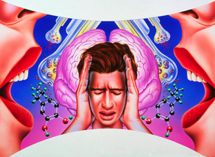
With the pressure for a certain body type prevalent in the media, eating disorders are on the rise. But these diseases are not completely socially driven; researchers have uncovered important genetic and biological components as well and are now beginning to tease out the genes and pathways responsible for eating disorder predisposition and pathology.

As we enter the holiday season, shoppers will once again rush into crowded department stores searching for the perfect gift. They will be jostled and bumped, yet for the most part, remain cheerful because of the crisp air, lights, decorations, and the sound of Karen Carpenter’s contralto voice ringing out familiar carols.

While Carpenter is mainly remembered for her musical talents, unfortunately, she is also known for introducing the world to anorexia nervosa (AN), a severe life-threatening mental illness characterized by altered body image and stringent eating patterns that claimed her life just before her 33rd birthday in 1983.

Even though eating disorders (ED) carry one of the highest mortality rates of any mental illness, many researchers and clinicians still view them as socially reinforced behaviors and diagnose them based on criteria such as “inability to maintain body weight,” “undue influence of body weight or shape on self-evaluation,” and “denial of the seriousness of low body weight” (1). This way of thinking was prevalent when Michael Lutter, then an MD/PhD student at the University of Texas Southwestern Medical Center, began his psychiatry residency in an eating disorders unit. “I just remember the intense fear of eating that many patients exhibited and thought that it had to be biologically driven,” he said.

Lutter carried this impression with him when he established his own research laboratory at the University of Iowa. Although clear evidence supports the idea that EDs are biologically driven—they predominantly affect women and significantly alter energy homeostasis—a lack of well-defined animal models combined with the view that they are mainly behavioral abnormalities have hindered studies of the neurobiology of EDs. Still, Lutter is determined to find the biological roots of the disease and tease out the relationship between the psychiatric illness and metabolic disturbance using biochemistry, neuroscience, and human genetics approaches.

We’ve Only Just Begun

Like many diseases, EDs result from complex interactions between genes and environmental risk factors. They tend to run in families, but of course, for many family members, genetics and environment are similar enough that teasing apart the influences of nature and nurture is not easy. Researchers estimate that 50-80% of the predisposition for developing an ED is genetic, but preliminary genome-wide analyses and candidate gene studies failed to identify specific genes that contribute to the risk.

According to Lutter, finding ED study participants can be difficult. “People are either reluctant to participate, or they don’t see that they have a problem,” he reported. Set on finding the genetic underpinnings of EDs, his team began recruiting volunteers and found 2 families, 1 with 20 members, 10 of whom had an ED and another with 5 out of 8 members affected. Rather than doing large-scale linkage and association studies, the team decided to characterize rare single-gene mutations in these families, which led them to identify mutations in the first two genes, estrogen-related receptor α (ESRRA) and histone deacetylase 4 (HDAC4), that clearly associated with ED predisposition in 2013 (1).

“We have larger genetic studies on-going, including the collection of more families. We just happened to publish these two families first because we were able to collect enough individuals and because there is a biological connection between the two genes that we identified,” Lutter explained.

ESRRA appears to be a transcription factor upregulated by exercise and calorie restriction that plays a role in energy balance and metabolism. HDAC4, on the other hand, is a well-described histone deacteylase that has previously been implicated in locomotor activity, body weight homeostasis, and neuronal plasticity.

Using immunoprecipitation, the researchers found that ESRRA interacts with HDAC4, in both the wild type and mutant forms, and transcription assays showed that HDAC4 represses ESRRA activity. When Lutter’s team repeated the transcription assays using mutant forms of the proteins, they found that the ESRRA mutation seen in one family significantly reduced the induction of target gene transcription compared to wild type, and that the mutation in HDAC4 found in the other family increased transcriptional repression for ESRRA target genes.

“ESRRA is a well known regulator of mitochondrial function, and there is an emerging view that mitochondria in the synapse are critical for neurotransmission,” Lutter said. “We are working on identifying target pathways now.”

Bless the Beasts and the Children

Finding genes associated with EDs provides the groundwork for molecular studies, but EDs cannot be completely explained by the actions of altered transcription factors. Individuals suffering these disorders often experience intense anxiety, intrusive thoughts, hyperactivity, and poor coping strategies that lead to rigid and ritualized behaviors and severe crippling perfectionism. They are less aware of their emotions and often try to avoid emotion altogether. To study these complex behaviors, researchers need animal models.

Until recently, scientists relied on mice with access to a running wheel and restricted access to food. Under these conditions, the animals quickly increase their locomotor activity and reduce eating, frequently resulting in death. While some characteristics of EDs—excessive exercise and avoiding food—can be studied in these mice, the model doesn’t allow researchers to explore how the disease actually develops. However, Lutter’s team has now introduced a promising new model (3).

Based on their previous success with identifying the involvement of ESRRA and HDAC4 in EDs, the researchers wondered if mice lacking ESRRA might make suitable models for studies on ED development. To find out, they first performed immunohistochemistry to understand more about the potential cognitive role of ESRRA.

“ESRRA is not expressed very abundantly in areas of the brain typically implicated in the regulation of food intake, which surprised us,” Lutter said. “It is expressed in many cortical regions that have been implicated in the etiology of EDs by brain imaging like the prefrontal cortex, orbitofrontal cortex, and insula. We think that it probably affects the activity of neurons that modulate food intake instead of directly affecting a core feeding circuit.”

With these data, the team next tried providing only 60% of the normal daily calories to their mice for 10 days and looked again at ESRRA expression. Interestingly, ESRRA levels increased significantly when the mice were insufficiently fed, indicating that the protein might be involved in the response to energy balance.

Lutter now believes that upregulation of ESRRA helps organisms adapt to calorie restriction, an effect possibly not happening in those with ESRRA or HDAC4 mutations. “This makes sense for the clinical situation where most individuals will be doing fine until they are challenged by something like a diet or heavy exercise for a sporting event. Once they start losing weight, they don’t adapt their behaviors to increase calorie intake and rapidly spiral into a cycle of greater and greater weight loss.”

When Lutter’s team obtained mice lacking ESRRA, they found that these animals were 15% smaller than their wild type littermates and put forth less effort to obtain food both when fed restricted calorie diets and when they had free access to food. These phenotypes were more pronounced in female mice than male mice, likely due to the role of estrogen signaling. Loss of ESRRA increased grooming behavior, obsessive marble burying, and made mice slower to abandon an escape hole after its relocation, indicating behavioral rigidity. And the mice demonstrated impaired social functioning and reduced locomotion.

Some people with AN exercise extensively, but this isn’t seen in all cases. “I would say it is controversial whether or not hyperactivity is due to a genetic predisposition (trait), secondary to starvations (state), or simply a ritual that develops to counter the anxiety of weight related obsessions. Our data would suggest that it is not due to genetic predisposition,” Lutter explained. “But I would caution against over-interpretation of mouse behavior. The locomotor activity of mice is very different from people and it’s not clear that you can directly translate the results.”

For All We Know

Going forward, Lutter’s group plans to drill down into the behavioral phenotypes seen in their ESRRA null mice. They are currently deleting ESRRA from different neuronal cell types to pair individual neurons with the behaviors they mediate in the hope of working out the neural circuits involved in ED development and pathology.

In addition, the team has created a mouse line carrying one of the HDAC4 mutations previously identified in their genetic study. So far, this mouse “has interesting parallels to the ESRRA-null mouse line,” Lutter reported.

The team continues to recruit volunteers for larger-scale genetic studies. Eventually, they plan to perform RNA-seq to identify the targets of ESRRA and HDAC4 and look into their roles in mitochondrial biogenesis in neurons. Lutter suspects that this process is a key target of ESRRA and could shed light on the cognitive differences, such as altered body image, seen in EDs. In the end, a better understanding of the cells and pathways involved with EDs could create new treatment options, reduce suffering, and maybe even avoid the premature loss of talented individuals to the effects of these disorders.

References

1. Lutter M, Croghan AE, Cui H. Escaping the Golden Cage: Animal Models of Eating Disorders in the Post-Diagnostic and Statistical Manual Era. Biol Psychiatry. 2015 Feb 12.

2. Cui H, Moore J, Ashimi SS, Mason BL, Drawbridge JN, Han S, Hing B, Matthews A, McAdams CJ, Darbro BW, Pieper AA, Waller DA, Xing C, Lutter M. Eating disorder predisposition is associated with ESRRA and HDAC4 mutations. J Clin Invest. 2013 Nov;123(11):4706-13.

3. Cui H, Lu Y, Khan MZ, Anderson RM, McDaniel L, Wilson HE, Yin TC, Radley JJ, Pieper AA, Lutter M. Behavioral disturbances in estrogen-related receptor alpha-null mice. Cell Rep. 2015 Apr 21;11(3):344-50.

http://www.biotechniques.com/news/Exploring-the-Biology-of-Eating-Disorders/biotechniques-361522.html

Cancer drug nilotinib may reverse Parkinson’s disease

by Jon Hamilton

A drug that’s already approved for treating leukemia appears to dramatically reduce symptoms in people who have Parkinson’s disease with dementia, or a related condition called Lewy body dementia.

A pilot study of 12 patients given small doses of nilotinib found that movement and mental function improved in all of the 11 people who completed the six-month trial, researchers reported Saturday at the Society for Neuroscience meeting in Chicago.

And for several patients the improvements were dramatic, says Fernando Pagan, an author of the study and director of the Movement Disorders Program at Georgetown University Medical Center. One woman regained the ability to feed herself, one man was able to stop using a walker, and three previously nonverbal patients began speaking again, Pagan says.

“After 25 years in Parkinson’s disease research, this is the most excited I’ve ever been,” Pagan says.

If the drug’s effectiveness is confirmed in larger, placebo-controlled studies, nilotinib could become the first treatment to interrupt a process that kills brain cells in Parkinson’s and other neurodegenerative diseases, including Alzheimer’s.

One of the patients in the pilot study was Alan Hoffman, 74, who lives with his wife, Nancy, in Northern Virginia.

Hoffman was diagnosed with Parkinson’s in 1997. At first, he had trouble moving his arms. Over time, walking became more difficult and his speech became slurred. And by 2007, the disease had begun to affect his thinking.

“I knew I’d dropped off in my ability to read,” Hoffman says. “People would keep giving me books and I’d have read the first chapter of about 10 of them. I had no ability to focus on it.”

“He had more and more difficulty making sense,” Nancy Hoffman says. He also became less active, less able to have conversations, and eventually stopped doing even household chores, she says.

But after a few weeks on nilotinib, Hoffman “improved in every way,” his wife says. “He began loading the dishwasher, loading the clothes in the dryer, things he had not done in a long time.”

Even more surprising, Hoffman’s scores on cognitive tests began to improve. At home, Nancy Hoffman says her husband was making sense again and regained his ability to focus. “He actually read the David McCullough book on the Wright Brothers and started reading the paper from beginning to end,” she says.

The idea of using nilotinib to treat people like Alan Hoffman came from Charbel Moussa, an assistant professor of neurology at Georgetown University and an author of the study.

Moussa knew that in people who have Parkinson’s disease with dementia or a related condition called Lewy body dementia, toxic proteins build up in certain brain cells, eventually killing them. Moussa thought nilotinib might be able to reverse this process.

His reasoning was that nilotinib activates a system in cells that works like a garbage disposal — it clears out unwanted proteins. Also, Moussa had shown that while cancer cells tend to die when exposed to nilotinib, brain cells actually become healthier.

So Moussa had his lab try the drug on brain cells in a Petri dish. “And we found that, surprisingly, with a very little amount of the drug we can clear all these proteins that are supposed to be neurotoxic,” he says.

Next, Moussa had his team give the drug to transgenic mice that were almost completely paralyzed from Parkinson’s disease. The treatment “rescued” the animals, he says, allowing them to move almost as well as healthy mice.

Moussa’s mice got the attention of Pagan from Georgetown’s Movement Disorders Program. “When Dr. Moussa showed them to me,” Pagan says, “it looked like, hey, this is type of drug that we’ve been looking for because it goes to the root of the problem.”

The pilot study was designed to determine whether nilotinib was safe for Parkinson’s patients and to determine how much drug from the capsules they were taking was reaching their brains. “But we also saw efficacy, which is really unheard of in a safety study,” Pagan says.

The study found that levels of toxic proteins in blood and spinal fluid decreased once patients began taking nilotinib. Also, tests showed that the symptoms of Parkinson’s including tremor and “freezing” decreased. And during the study patients were able to use lower doses of Parkinson’s drugs, suggesting that the brain cells that produce dopamine were working better.

But there are some caveats, Pagan says. For one thing, the study was small, not designed to measure effectiveness, and included no patients taking a placebo.

Also, nilotinib is very expensive. The cost of providing it to leukemia patients is thousands of dollars a month.

And finally, Parkinson’s and dementia patients would have to keep taking nilotinib indefinitely or their symptoms would continue to get worse.

Alan Hoffman was okay for about three weeks after the study ended and he stopped taking the drug. Since then, “There’s (been) a pretty big change,” his wife says. “He does have more problems with his speech, and he has more problems with cognition and more problems with mobility.”

The Hoffmans hope to get more nilotinib from the drug’s maker, Novartis, through a special program for people who improve during experiments like this one.

Meanwhile, the Georgetown team plans to try nilotinib in patients with another brain disease that involves toxic proteins: Alzheimer’s.

http://www.npr.org/sections/health-shots/2015/10/17/448323916/can-a-cancer-drug-reverse-parkinsons-disease-and-dementia