New research shows that infants need to be able to freely move their tongues in order to distinguish sounds.

A team of researchers led by Dr Alison Bruderer, a postdoctoral fellow at the University of British Columbia, has discovered a direct link between tongue movements of infants and their ability to distinguish speech sounds.

“Until now, research in speech perception development and language acquisition has primarily used the auditory experience as the driving factor. Researchers should actually be looking at babies’ oral-motor movements as well,” said Dr Bruderer, who is the lead author on a study published in the Proceedings of the National Academy of Sciences on October 12, 2015.

In the study, teething toys were placed in the mouths of six-month-old English-learning infants while they listened to speech sounds – two different Hindi ‘d’ sounds that infants at this age can readily distinguish.

When the teethers restricted movements of the tip of the tongue, the infants were unable to distinguish between the two sounds.

But when their tongues were free to move, the babies were able to make the distinction.

“Before infants are able to speak, their articulatory configurations affect the way they perceive speech, suggesting that the speech production system shapes speech perception from early in life,” the scientists said.

“These findings implicate oral-motor movements as more significant to speech perception development and language acquisition than current theories would assume and point to the need for more research.”

http://www.sci-news.com/othersciences/psychology/science-infants-tongue-movement-speech-sounds-03336.html?utm_source=feedburner&utm_medium=feed&utm_campaign=Feed%3A+BreakingScienceNews+%28Breaking+Science+News%29

Discovery of fat breakdown trigger opens door for new obesity treatments

While it’s known that the brain is responsible for instructing our fat stores to break down and release energy as we need it, scientists haven’t yet been able to pin down exactly how this process plays out. Leptin, a hormone produced by our fat cells, travels to the brain to regulate appetite, metabolism and energy, but it hasn’t been clear what communication was coming back the other way. New research has now uncovered this missing link for the first time, revealing a set of nerves that connect with fat tissue to stimulate the process in a development that could lead to new types of anti-obesity treatments.

The leptin hormone was identified around 20 years ago as a regulator of the body’s metabolism. Low levels of the hormone serve to boost one’s appetite and slow metabolism, while conversely, high leptin levels dull the appetite and facilitate better fat breakdown. Using a combination of techniques, a research team led by Ana Domingos from Portugal’s Instituto Gulbenkian de Ciência were able to shed light on how leptin behaves when sending signals back to the fat by finding the nerves that meet with white fat tissue to prompt its breakdown.

“We dissected these nerve fibers from mouse fat, and using molecular markers identified these as sympathetic neurons,” explains Domingos. “When we used an ultra sensitive imaging technique, on the intact white fat tissue of a living mouse, we observed that fat cells can be encapsulated by these sympathetic neural terminals.”

But to determine the extent of these neurons’ role in obesity, the team carried out further research on mice. The rodents were genetically engineered so that these neurons could be switched on and off through optogenetics, where brain cells are made to behave differently by exposing them to light. Optogenetics is an emerging technique we have seen explored as a means of treating blindness and altering our pain threshold, among other things.

Domingos’ team found that flicking the switch on the neurons locally triggered the release of a neurotransmitter called norepinephrine, which in turn flooded the fat cells with signals that brought about fat breakdown. The team report that without these sympathetic neurons, leptin was not able to stimulate fat breakdown on its own. Therefore the findings suggest that these sympathetic neurons offer a potential target for obesity treatments other than leptin, which the brains of many obese people have a resistance to.

“This result provides new hopes for treating central leptin resistance, a condition in which the brains of obese people are insensitive to leptin,” says Domingos.

The team’s research was published in the journal Cell.

http://www.gizmag.com/neural-mechanism-fat-breakdown-anti-obesity-therapies/39601/

The human brain is particularly vulnerable to trauma at two distinct ages

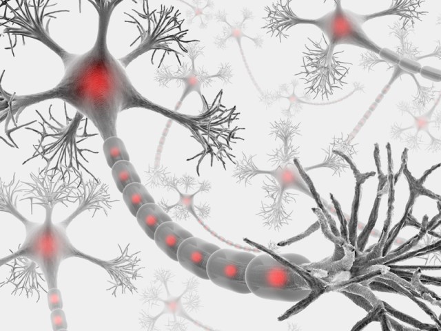
Our brain’s ability to process information and adapt effectively is dependent on a number of factors, including genes, nutrition, and life experiences. These life experiences wield particular influence over the brain during a few sensitive periods when our most important muscle is most likely to undergo physical, chemical, and functional remodeling.

According to Tara Swart, a neuroscientist and senior lecturer at MIT, your “terrible twos” and those turbulent teen years are when the brain’s wiring is most malleable. As a result, traumatic experiences that occur during these time periods can alter brain activity and ultimately change gene expressions—sometimes for good.

Throughout the first two years of life, the brain develops at a rapid pace. However, around the second year, something important happens—babies begin to speak.

“We start to understand speech first, then we start to articulate speech ourselves and that’s a really complex thing that goes on in the brain,” Swart, who conducts ongoing research on the brain and how it affects how we become leaders, told Quartz. “Additionally, children start to walk—so from a physical point of view, that’s also a huge achievement for the brain.

Learning and understanding a new language forces your brain to work in new ways, connecting neurons and forming new pathways. This is a mentally taxing process, which is why learning a new language or musical instrument often feels exhausting.

With so many important changes happening to the brain in such a short period of time, physical or emotional trauma can cause potentially momentous interruptions to neurological development. Even though you won’t have any memories of the interruptions (most people can’t remember much before age five), any kind of traumatic event—whether it’s abuse, neglect, ill health, or separation from your loved ones—can lead to lasting behavioral and cognitive deficits later in life, warns Swart.

To make her point, Swart points to numerous studies on orphans in Romania during the 1980s and 1990s. After the nation’s communist regime collapsed, an economic decline swept throughout the region and 100,000 children found themselves in harsh, overcrowded government institutions.

“[The children] were perfectly well fed, clothed, washed, but for several reasons—one being that people didn’t want to spread germs—they were never cuddled or played with,” explains Swart. “There was a lot of evidence that these children grew up with some mental health problems and difficulty holding down jobs and staying in relationships.”

Swart continues: “When brain scanning became possible, they scanned the brains of these children who had grown up into adults and showed that they had issues in the limbic system, the part of the brain [that controls basic emotions].”

In short, your ability to maintain proper social skills and develop a sense of empathy is largely dependent on the physical affection, eye contact, and playtime of those early years. Even something as simple as observing facial expressions and understanding what those expressions mean is tied to your wellbeing as a toddler.

The research also found that the brains of the Romanian orphans had lower observable brain activity and were physically smaller than average. As a result, researchers concluded that children adopted into loving homes by age two have a much better chance of recovering from severe emotional trauma or disturbances.

The teenage years

By the time you hit your teenage years, the brain has typically reached its adult weight of about three pounds. Around this same time, the brain is starting to eliminate, or “prune” fragile connections and unused neural pathways. The process is similar to how one would prune a garden—cutting back the deadwood allows other plants to thrive.

During this period, the brain’s frontal lobes, especially the prefrontal cortex, experience increased activity and, for the first time, the brain is capable of comparing and analyzing several complex concepts at once. Similar to a baby learning how to speak, this period in an adolescent’s life is marked by a need for increasingly advanced communication skills and emotional maturity.

“At that age, they’re starting to become more understanding of social relationships and politics. It’s really sophisticated,” Swart noted. All of this brain activity is also a major reason why teenagers need so much sleep.

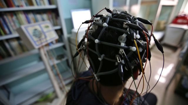
Swart’s research dovetails with the efforts of many other scientists who have spent decades attempting to understand how the brain develops, and when. The advent of MRIs and other brain-scanning technology has helped speed along this research, but scientists are still working to figure out what exactly the different parts of the brain do.

What is becoming more certain, however, is the importance of stability and safety in human development, and that such stability is tied to cognitive function. At any point in time, a single major interruption has the ability to throw off the intricate workings of our brain. We may not really understand how these events affect our lives until much later.

http://qz.com/470751/your-brain-is-particularly-vulnerable-to-trauma-at-two-distinct-ages/

New evidence that sleeping on your side may be better for the brain

Removal of waste, including soluble amyloid β (Aβ), from the brain may be most efficient in the lateral vs. the prone position, according to an experimental study published in the August 5 issue of the Journal of Neuroscience.

Hedok Lee, PhD, from Stony Brook University in New York, and colleagues examined whether body posture impacts cerebrospinal fluid (CSF)-interstitial fluid (ISF) exchange efficiency. They quantified CSF-ISF exchange rates using dynamic-contrast-enhanced magnetic resonance imaging (MRI) and kinetic modeling in the brains of rodents in supine, prone, or lateral positions. Fluorescence microscopy and radioactive tracers were used to validate the MRI data and assess the influence of body posture on clearance of Aβ.

The researchers found that glymphatic transport was most efficient in the lateral vs. the supine or prone positions. Transport was characterized by “retention” of the tracer, slower clearance, and more CSF efflux along larger caliber cervical vessels in the prone position, in which the rat’s head was in the most upright position (mimicking posture during the awake state). Glymphatic transport and Aβ clearance were superior in the lateral and supine positions in optical imaging and radiotracer studies.

“We propose that the most popular sleep posture (lateral) has evolved to optimize waste removal during sleep and that posture must be considered in diagnostic imaging procedures developed in the future to assess CSF-ISF transport in humans,” the authors write.

FDA grants fast-track status to promising new drug for treating the cognitive symptoms of schizophrenia

Forum Pharmaceuticals announced that the FDA has granted Fast Track designation to encenicline for the treatment of cognitive impairment in schizophrenia.

Forum recently completed patient enrollment for the COGNITIV SZ phase 3 clinical trial program which includes two randomized, double-blind, placebo-controlled studies. The program is evaluating the safety and efficacy of two oral doses of once-daily treatment with encenicline as a pro-cognitive treatment compared to placebo when added to chronic, stable, atypical antipsychotic therapy in people with schizophrenia.

Primary endpoints of the trials include effect on cognitive function and effect on clinical function. The two global 26-week trials enrolled a total of more than 1,500 patients at approximately 200 clinical sites.

Encenicline is an orally administered, selective, and potent agonist of the alpha 7 receptor found in hippocampal and cortical neurons involved in cognition.

In a phase 2 trial, which was sponsored by Forum and results of which were released in March, 319 schizophrenia patients were randomized to receive either encenicline in one of two doses daily, or a placebo, for 12 weeks.

Patients in both encenicline dose groups showed significant cognitive improvement based on various measures, according to a presentation made at the 15th International Congress on Schizophrenia Research. In a subset of 154 patients, the improvement was greater in the higher-dose group (0.9 mg) than the lower-dose cohort (0.27 mg).

New research shows that people with ‘O’ blood type have decreased risk of cognitive decline

A pioneering study conducted by leading researchers at the University of Sheffield has revealed blood types play a role in the development of the nervous system and may impact the risk of developing cognitive decline.

The research, carried out in collaboration with the IRCCS San Camillo Hospital Foundation in Venice, shows that people with an ‘O’ blood type have more grey matter in their brain, which helps to protect against diseases such as Alzheimer’s, than those with ‘A’, ‘B’ or ‘AB’ blood types.

Research fellow Matteo De Marco and Professor Annalena Venneri, from the University’s Department of Neuroscience, made the discovery after analysing the results of 189 Magnetic Resonance Imaging (MRI) scans from healthy volunteers.

The researchers calculated the volumes of grey matter within the brain and explored the differences between different blood types.

The results, published in the Brain Research Bulletin, show that individuals with an ‘O’ blood type have more grey matter in the posterior proportion of the cerebellum.

In comparison, those with ‘A’, ‘B’ or ‘AB’ blood types had smaller grey matter volumes in temporal and limbic regions of the brain, including the left hippocampus, which is one of the earliest part of the brain damaged by Alzheimer’s disease.

These findings indicate that smaller volumes of grey matter are associated with non-‘O’ blood types.

As we age a reduction of grey matter volumes is normally seen in the brain, but later in life this grey matter difference between blood types will intensify as a consequence of ageing.

“The findings seem to indicate that people who have an ‘O’ blood type are more protected against the diseases in which volumetric reduction is seen in temporal and mediotemporal regions of the brain like with Alzheimer’s disease for instance,” said Matteo DeMarco.

“However additional tests and further research are required as other biological mechanisms might be involved.”

Professor Annalena Venneri added: “What we know today is that a significant difference in volumes exists, and our findings confirm established clinical observations. In all likelihood the biology of blood types influences the development of the nervous system. We now have to understand how and why this occurs.”

More information: “‘O’ blood type is associated with larger grey-matter volumes in the cerebellum,” Brain Research Bulletin, Volume 116, July 2015, Pages 1-6, ISSN 0361-9230, dx.doi.org/10.1016/j.brainresbull.2015.05.005

An axon self-destruct mechanism that kills neurons

Just as losing a limb can spare a life, parting with a damaged axon by way of Wallerian degeneration can spare a neuron. A protein called SARM1 acts as the self-destruct button, and now researchers led by Jeffrey Milbrandt of Washington University Medical School in St. Louis believe they have figured out how. They report in the April 24 Science that SARM1 forms dimers that trigger the destruction of NAD+. Basic biochemistry dictates that this enzyme cofactor is essential for cell survival.

ARM1 and NAD+ have emerged as key players in the complex, orderly process underlying Wallerian degeneration. Scientists are still filling in other parts of the pathway. SARM1, short for sterile alpha and TIR motif-containing 1, seems to act as a damage sensor, but researchers are not sure how. Recently, researchers led by Marc Tessier-Lavigne at Rockefeller University, New York, found that SARM1 turns on a mitogen-activated protein (MAP) kinase cascade that is involved. Loss of NAD+ may also contribute to axon degeneration, because its concentration drops in dying axons, and Wlds mutant mice that overproduce an NAD+ synthase have slower Wallerian degeneration.

Now, first author Josiah Gerdts confirms that SARM1 is the self-destruct switch. He engineered a version of the protein with a target sequence for tobacco etch virus (TEV) protease embedded in it. Using a rapamycin-activated form of TEV, he eliminated SARM1 from axons he had sliced off of mouse dorsal root ganglion (DRG) neurons. Without SARM1, the severed axons survived.

SARM1 contains SAM and TIR domains, which promote protein-protein interactions. Previously, Gerdts discovered that the TIR domain was sufficient to induce degeneration, even in healthy axons, but it relied on the SAM region to bring multiple SARM1 molecules together. He hypothesized that axonal SARM1 multimerizes upon axon damage. To test this idea, he used a standard biochemical technique to force the SARM1 TIR domains together. He fused domains to one or another of the rapamycin-binding peptides Frb and Fkbp and expressed them in DRG neurons. When he added rapamycin to the cultures, the Frb and Fkbp snapped the TIR domains together within minutes. As Gerdts had predicted, this destroyed axons, confirming that SARM1 activates via dimerization.

Next, the authors investigated what happens to NAD+ during that process. Using high-performance liquid chromatography, Gerdts measured the concentration of NAD+ in the disembodied axons. Normally, its level dropped by about two-thirds within 15 minutes of severing. In axons from SARM1 knockout mice, however, the NAD+ concentration stayed unchanged. In neurons carrying the forced-dimerization constructs, adding rapamycin was sufficient to knock down NAD+ levels—Gerdts did not even have to cut the axons. Ramping up NAD+ production by overexpressing its synthases, NMNAT and NAMPT, overcame the effects of TIR dimerization, and the axons survived. Gerdts concluded that loss of NAD+ was a crucial, SARM1-controlled step on the way to degeneration.

He still wondered what caused the loss of NAD+. It might be that the axon simply stopped making it, or maybe the Wallerian pathway actively destroyed it. To distinguish between these possibilities, Gerdts added radiolabeled exogenous NAD+ to human embryonic kidney HEK293 cultures expressing the forced-dimerization TIR domains. Rapamycin caused them to rapidly degrade the radioactive NAD+, confirming that the cell actively disposes of it.

Gerdts suspects that with this essential cofactor gone, the axon runs out of energy and can no longer survive. He speculated that the MAP kinase cascade reportedly turned on by SARM1 might lead to NAD+ destruction. Alternatively, SARM1 might induce distinct MAP kinase and NAD+ destruction pathways in parallel, he suggested.

“Demonstrating how NAD+ is actively and locally degraded in the axon is a big advance,” commented Andrew Pieper of the Iowa Carver College of Medicine in Iowa City, who was not involved in the study. Jonathan Gilley and Michael Coleman of the Babraham Institute in Cambridge, U.K., predict that there will be more to the story. They note that a drug called FK866, which prevents NAD+ production, protects axons in some instances. Gerdts suggested that FK866 acts on processes upstream of SARM1, delaying the start of axon degeneration. In contrast, his paper only addressed what happens after SARM1 activates. “It will be fascinating to see how the apparent contradictions raised by this new study will be resolved,” wrote Gilley and Coleman.

Could these findings help researchers looking for ways to prevent neurodegeneration? “The study supports the notion that augmenting NAD+ levels is potentially a valuable approach,” said Pieper. He and his colleagues developed a small molecule that enhances NAD+ synthesis, now under commercial development. It improved symptoms in ALS model mice, and protected neurons in mice mimicking Parkinson’s. NAD+ also activates sirtuin, an enzyme important for longevity and stress resistance as well as learning and memory.

However, both Pieper and Gerdts cautioned that they cannot clearly predict which conditions might benefit from an anti-SARM1 or NAD+-boosting therapy. At this point, Gerdts said, researchers do not fully understand how much axon degeneration contributes to symptoms of diseases like Alzheimer’s and Parkinson’s. He suggested that crossing SARM1 knockout mice with models for various neurodegenerative conditions would indicate how well an anti-Wallerian therapy might work.

—Amber Dance

http://www.alzforum.org/news/research-news/axon-self-destruct-button-triggers-energy-woes

Scientists manage to give mice ‘eating disorders’ by knocking out one gene

By Rachel Feltman

If you give a mouse an eating disorder, you might just figure out how to treat the disease in humans. In a new study published Thursday in Cell Press, researchers created mice who lacked a gene associated with disordered eating in humans. Without it, the mice showed behaviors not unlike those seen in humans with eating disorders: They tended to be obsessive compulsive and have trouble socializing, and they were less interested in eating high-fat food than the control mice. The findings could lead to novel drug treatments for some of the 24 million Americans estimated to suffer from eating disorders.

In a 2013 study, the same researchers went looking for genes that might contribute to the risk of an eating disorder. Anorexia nervosa and bulimia nervosa aren’t straightforwardly inherited — there’s definitely more to an eating disorder than your genes — but it does seem like some families might have higher risks than others. Sure enough, the study of two large families, each with several members who had eating disorders, yielded mutations in two interacting genes. In one family, the estrogen-related receptor α (ESRRA) gene was mutated. The other family had a mutation on another gene that seemed to affect how well ESRRA could do its job.

So in the latest study, they created mice that didn’t have ESRRA in the parts of the brain associated with eating disorders.

“You can’t go testing this kind of gene expression in a human,” lead author and University of Iowa neuroscientist Michael Lutter said. “But in mice, you can manipulate the expression of the gene and then look at how it changes their behavior.”

It’s not a perfect analogy to what the gene mutation might do in a human, but the similarities can allow researchers to figure out the mechanism that causes the connection between your DNA and your eating habits.

The mice without ESRRA were tested for several eating-disorder-like behaviors: The researchers tested how hard they were willing to work for high fat food when they were hungry (less, it seemed, so much so that they weighed 15 percent less than their unaltered littermates), how compulsive they were, and how they behaved socially.

In general, the ESRRA-lacking mice were twitchier: They tended to overgroom, a common sign of anxiety in mice, and they were more wary of novelty, growing anxious when researchers put marbles into their cages. They also showed an inability to adapt: When researchers taught the mice how to exit a maze and then changed where the exit was, the mice without ESRRA spent way more time checking out the area where the exit should have been before looking for where it had gone.

The social changes were even more striking: Mice will usually show more interest in a new mouse than one they’ve met before, but in tests the modified mice showed the opposite preference, socializing with a familiar mouse when a new one was also presented.

They were also universally submissive to other mice, something the researchers detected with a sort of scientific game of chicken. Two mice are placed at either end of a tube, and one always plows past the other to get to the opposite side. It’s just the way mice size each other up — someone has to be on top. But every single one of the modified mice let themselves get pushed around.

“100% of the mice lacking this gene were subordinate,” Lutter said. “I’ve never seen an experiment before that produced a 0% verses 100% result.”

The avoidance of fats has an obvious connection to human disorders. But the social anxiety and rigidity are also close analogies to disordered eating in humans.

Now that Lutter and his colleagues know that the gene does something similar in mice, they can start looking for the actual mechanism that’s tripping these switches in the brain. They know that the gene’s pathway is very important for energy metabolism, especially in the breakdown of glucose. It’s possible that mutations in the gene cause some kind of impairment in neurons’ ability to get and process energy, but they can’t be sure yet.

They’ll see if they can pinpoint affected neurons and fix them. They’re also going to test some drugs that are known to affect this gene and its pathways. It’s possible that they’ll land on a treatment that helps calm these negative behaviors in affected mice, leading to treatments for humans with the mutation.

http://www.washingtonpost.com/news/speaking-of-science/wp/2015/04/09/scientists-manage-to-give-mice-eating-disorders-by-knocking-out-one-gene/

Open Access Article here: http://www.cell.com/cell-reports/abstract/S2211-1247(15)00301-0

Mind-controlled drones promise a future of hands-free flying

There have been tentative steps into thought-controlled drones in the past, but Tekever and a team of European researchers just kicked things up a notch. They’ve successfully tested Brainflight, a project that uses your mental activity (detected through a cap) to pilot an unmanned aircraft. You have to learn how to fly on your own, but it doesn’t take long before you’re merely thinking about where you want to go. And don’t worry about crashing because of distractions or mental trauma, like seizures — there are “algorithms” to prevent the worst from happening.

You probably won’t be using Brainflight to fly anything larger than a small drone, at least not in the near future. There’s no regulatory framework that would cover mind-controlled aircraft, after all. Tekever is hopeful that its technology will change how we approach transportation, though. It sees brain power reducing complex activities like flying or driving to something you can do instinctively, like walking — you’d have freedom to focus on higher-level tasks like navigation. The underlying technology would also let people with injuries and physical handicaps steer vehicles and their own prosthetic limbs. Don’t be surprised if you eventually need little more than some headgear to take to the skies.

http://www.engadget.com/2015/02/25/tekever-mind-controlled-drone/?ncid=rss_truncated

Brain decoder can eavesdrop on your inner voice

brainy_2758840b

Talking to yourself used to be a strictly private pastime. That’s no longer the case – researchers have eavesdropped on our internal monologue for the first time. The achievement is a step towards helping people who cannot physically speak communicate with the outside world.

“If you’re reading text in a newspaper or a book, you hear a voice in your own head,” says Brian Pasley at the University of California, Berkeley. “We’re trying to decode the brain activity related to that voice to create a medical prosthesis that can allow someone who is paralysed or locked in to speak.”

When you hear someone speak, sound waves activate sensory neurons in your inner ear. These neurons pass information to areas of the brain where different aspects of the sound are extracted and interpreted as words.

In a previous study, Pasley and his colleagues recorded brain activity in people who already had electrodes implanted in their brain to treat epilepsy, while they listened to speech. The team found that certain neurons in the brain’s temporal lobe were only active in response to certain aspects of sound, such as a specific frequency. One set of neurons might only react to sound waves that had a frequency of 1000 hertz, for example, while another set only cares about those at 2000 hertz. Armed with this knowledge, the team built an algorithm that could decode the words heard based on neural activity alone (PLoS Biology, doi.org/fzv269).

The team hypothesised that hearing speech and thinking to oneself might spark some of the same neural signatures in the brain. They supposed that an algorithm trained to identify speech heard out loud might also be able to identify words that are thought.

Mind-reading

To test the idea, they recorded brain activity in another seven people undergoing epilepsy surgery, while they looked at a screen that displayed text from either the Gettysburg Address, John F. Kennedy’s inaugural address or the nursery rhyme Humpty Dumpty.

Each participant was asked to read the text aloud, read it silently in their head and then do nothing. While they read the text out loud, the team worked out which neurons were reacting to what aspects of speech and generated a personalised decoder to interpret this information. The decoder was used to create a spectrogram – a visual representation of the different frequencies of sound waves heard over time. As each frequency correlates to specific sounds in each word spoken, the spectrogram can be used to recreate what had been said. They then applied the decoder to the brain activity that occurred while the participants read the passages silently to themselves.

Despite the neural activity from imagined or actual speech differing slightly, the decoder was able to reconstruct which words several of the volunteers were thinking, using neural activity alone (Frontiers in Neuroengineering, doi.org/whb).

The algorithm isn’t perfect, says Stephanie Martin, who worked on the study with Pasley. “We got significant results but it’s not good enough yet to build a device.”

In practice, if the decoder is to be used by people who are unable to speak it would have to be trained on what they hear rather than their own speech. “We don’t think it would be an issue to train the decoder on heard speech because they share overlapping brain areas,” says Martin.

The team is now fine-tuning their algorithms, by looking at the neural activity associated with speaking rate and different pronunciations of the same word, for example. “The bar is very high,” says Pasley. “Its preliminary data, and we’re still working on making it better.”

The team have also turned their hand to predicting what songs a person is listening to by playing lots of Pink Floyd to volunteers, and then working out which neurons respond to what aspects of the music. “Sound is sound,” says Pasley. “It all helps us understand different aspects of how the brain processes it.”

“Ultimately, if we understand covert speech well enough, we’ll be able to create a medical prosthesis that could help someone who is paralysed, or locked in and can’t speak,” he says.

Several other researchers are also investigating ways to read the human mind. Some can tell what pictures a person is looking at, others have worked out what neural activity represents certain concepts in the brain, and one team has even produced crude reproductions of movie clips that someone is watching just by analysing their brain activity. So is it possible to put it all together to create one multisensory mind-reading device?

In theory, yes, says Martin, but it would be extraordinarily complicated. She says you would need a huge amount of data for each thing you are trying to predict. “It would be really interesting to look into. It would allow us to predict what people are doing or thinking,” she says. “But we need individual decoders that work really well before combining different senses.”

http://www.newscientist.com/article/mg22429934.000-brain-decoder-can-eavesdrop-on-your-inner-voice.html