Scientists Have Figured Out How to Recover Forgotten Memories Still Lurking in the Brain

memory

All might not be lost. Researchers recently announced a discovery that could have significant implications later down the road for helping people with severe amnesia or Alzheimer’s disease.

The research tackles a highly debated topic of whether memory loss due to damaged brain cells means that memories cannot be stored anymore or if just accessing that memory is inhibited in some way.

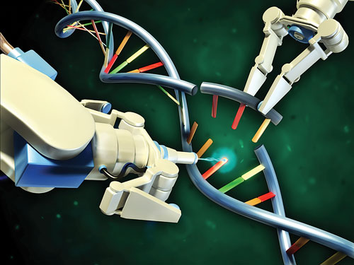
Scientists from MIT found in new research that the latter is most likely the case, demonstrating how lost memories could be recovered using technology known as optogenetics, which a news release about the study described as when “proteins are added to neurons to allow them to be activated with light.”

“The majority of researchers have favored the storage theory, but we have shown in this paper that this majority theory is probably wrong,” Susumu Tonegawa, a professor in MIT’s biology department and director of the RIKEN-MIT Center at the Picower Institute for Learning and Memory, said in a statement. “Amnesia is a problem of retrieval impairment.”

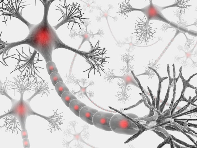
First, the scientists demonstrated how “memory engram cells” — brain cells that trigger a memory upon experiencing a related sight or smell, for example — could be strengthened in mice.

The researchers then gave the mice anisomycin, which blocked protein synthesis in neurons, after they had formed a new memory. In doing so, the researchers prevented the engram cells from strengthening.

A day later, the scientists tried to trigger the memory in mice, but couldn’t see any activation that would indicate the mice were remembering it.

“So even though the engram cells are there, without protein synthesis those cell synapses are not strengthened, and the memory is lost,” Tonegawa explained of this part of the research.

The team first developed a clever technique to selectively label the neurons representing what is known as a memory engram – in other words, the brain cells involved in forming a specific memory. They did this by genetically engineering mice so they had extra genes in all their neurons. As a result, when neurons fire as a memory is formed, they produce red proteins visible under a microscope, allowing the researchers to tell which cells were part of the engram. They also inserted a gene that made the neurons fire when illuminated by blue light.

After the researchers induced amnesia, they used optogenetic tools on the mice and witnessed the animals experiencing full recollection.

“If you test memory recall with natural recall triggers in an anisomycin-treated animal, it will be amnesiac, you cannot induce memory recall. But if you go directly to the putative engram-bearing cells and activate them with light, you can restore the memory,” Tonegawa said.

With this discovery, the researchers wrote in the study published this week in the journal Science that they believe a “specific pattern of connectivity of engram cells may be crucial for memory information storage and that strengthened synapses in these cells critically contribute to the memory retrieval process.”

James Bisby, a neuroscientist at University College London, told New Scientist that it’s “not surprising that they could trigger the memories, but it is a cool way to do it.”

http://www.newscientist.com/article/dn27618-lost-memories-recovered-in-mice-with-a-flash-of-light.html

Thanks to Steven Weihing for bringing this to the It’s Interesting community.

Ex-prof set up company to re-sell lab equipment to Colorado University at 300% markup


Donald Cooper with scientific colleagues.


Donald Cooper mug shot.

A former University of Colorado professor has been arrested on suspicion of creating a company to sell marked-up lab equipment to the Boulder campus in what prosecutors call a theft “scheme.”

Donald Cooper, 44, was arrested at his home in Boulder on Tuesday afternoon, according to Boulder County District Attorney’s Office officials. It was unclear late Tuesday if Cooper had posted bond, which was set at $5,000.

He is facing a felony charge of theft between $20,000 and $100,000. Prosecutors allege that he created Boulder Science Resource to buy lasers and other lab equipment that he marked up 300 percent and then resold to his university laboratory, according to an arrest affidavit.

The arrangement also benefitted the professor’s father, who received a salary and a car from Boulder Science Resource, according to the arrest affidavit.

In total, CU paid Boulder Science Resource $97,554.03 between Jan. 1, 2009, and April 30, 2013, according to the affidavit.

According to CU’s calculations, Cooper’s markups cost the university $65,036.

Cooper resigned in July 2014 as part of a settlement deal with the university, which had begun the process of firing him on suspicion of fiscal misconduct. He had been director of the molecular neurogenetics and optophysiology laboratory in CU’s Institute for Behavioral Genetics, where he was a tenured associate professor.

After he learned about the university’s internal investigation, Cooper filed a notice of claim in September 2013 seeking $20 million in damages. Any person who wishes to sue a state entity must first file a notice of claim.

Cooper’s attorney Seth Benezra wrote in the notice of claim that Gary Cooper, the professor’s father, was the sole owner of Boulder Science Resource. He also wrote that the company sold CU equipment “at prices that were greatly discounted.”

Donald Cooper also complained that CU investigators had obtained an email about his father’s “alleged mental impairment,” according to the notice of claim.

“(The investigator’s) theory is that Gary Cooper lacks the mental capacity to run (Boulder Science Resource) and so Dr. Cooper must really be in charge,” Benezra wrote. “This assertion was pure speculation based on entirely private information and was rebutted by Dr. Cooper in multiple meetings with investigators.”

Benezra did not return messages from the Daily Camera on Tuesday. It’s unclear who is representing Cooper in the criminal case.

Though Cooper claims that his father was in charge of the company, prosecutors assert that the professor “employed a scheme” to deceive the university for his own gain, according to the affidavit.

“It is alleged that (Boulder Science Resource) was created to defraud the University of Colorado Boulder by acting as a middleman to generate income to employ Gary and to provide personal benefit for Cooper,” wrote Alisha Baurer, an investigator in the District Attorney’s Office.

‘Fake business’

CU was tipped off about Boulder Science Resource by another employee in Cooper’s department, who told investigators that he heard about the arrangement from Cooper’s ex-wife, according to the arrest affidavit.

The ex-wife told the CU employee that Cooper had created a “fake business” using “dirty money” from grants and start-up funds, according to the affidavit.

The financial manager for Cooper’s department told investigators that he never mentioned that his dad owned Boulder Science Resource, and said Cooper only referred to “Gary” by his first name, according to the affidavit.

The DA’s Office determined that Gary Cooper received $23,785.80 from Boulder Science Resource in the form of a salary and a car. They also found that $31,974.89 was paid from the company’s accounts to Donald Cooper’s personal credit card and that $14,733.54 was paid to his personal PayPal account from the business, according to the affidavit.

CU’s internal audit found that Boulder Science Resource had no customers other than the university and Mobile Assay, a company founded by Donald Cooper based on a technology he developed at the university.

Some of the money CU paid to Boulder Science Resource came from federal grants, including $7,220 from the National Institutes of Health and $15,288 from the National Institute on Drug Abuse, according to the internal audit report.

CU’s investigation found that although Cooper claimed his father purchased the lab equipment for Boulder Science Resource, the professor used his university email account to negotiate with the manufacturers.

“It is internal audit’s conclusion that the forgoing acts/failures to act were done with intent to gain an unauthorized benefit,” according to the audit report.

Boulder Science Resource was dissolved in December 2013, according to the Secretary of State’s Office.

Settlement terms

Reached by phone Tuesday afternoon, Patrick O’Rourke, CU’s chief legal officer, said the university was aware of Cooper’s arrest and will cooperate with prosecutors.

CU settled with Cooper last summer after initiating termination proceedings. In exchange for his resignation, the university agreed to provide the professor with a letter of reference “acknowledging his significant achievement in creating a neuroscience undergraduate program,” according to the settlement document.

CU also paid $20,000 to partially reimburse Cooper’s attorney and forgave an $80,000 home loan. CU provides down payment-assistance loans to some faculty members.

Had the university continued the termination process, which is lengthy, Cooper would have continued to receive his full salary of $89,743 and all benefits during the proceedings.

O’Rourke said the university instead opted to accept Cooper’s resignation and saved money with the settlement.

http://www.dailycamera.com/cu-news/ci_28056525/former-cu-boulder-professor-arrested-theft-case

An axon self-destruct mechanism that kills neurons

Just as losing a limb can spare a life, parting with a damaged axon by way of Wallerian degeneration can spare a neuron. A protein called SARM1 acts as the self-destruct button, and now researchers led by Jeffrey Milbrandt of Washington University Medical School in St. Louis believe they have figured out how. They report in the April 24 Science that SARM1 forms dimers that trigger the destruction of NAD+. Basic biochemistry dictates that this enzyme cofactor is essential for cell survival.

ARM1 and NAD+ have emerged as key players in the complex, orderly process underlying Wallerian degeneration. Scientists are still filling in other parts of the pathway. SARM1, short for sterile alpha and TIR motif-containing 1, seems to act as a damage sensor, but researchers are not sure how. Recently, researchers led by Marc Tessier-Lavigne at Rockefeller University, New York, found that SARM1 turns on a mitogen-activated protein (MAP) kinase cascade that is involved. Loss of NAD+ may also contribute to axon degeneration, because its concentration drops in dying axons, and Wlds mutant mice that overproduce an NAD+ synthase have slower Wallerian degeneration.

Now, first author Josiah Gerdts confirms that SARM1 is the self-destruct switch. He engineered a version of the protein with a target sequence for tobacco etch virus (TEV) protease embedded in it. Using a rapamycin-activated form of TEV, he eliminated SARM1 from axons he had sliced off of mouse dorsal root ganglion (DRG) neurons. Without SARM1, the severed axons survived.

SARM1 contains SAM and TIR domains, which promote protein-protein interactions. Previously, Gerdts discovered that the TIR domain was sufficient to induce degeneration, even in healthy axons, but it relied on the SAM region to bring multiple SARM1 molecules together. He hypothesized that axonal SARM1 multimerizes upon axon damage. To test this idea, he used a standard biochemical technique to force the SARM1 TIR domains together. He fused domains to one or another of the rapamycin-binding peptides Frb and Fkbp and expressed them in DRG neurons. When he added rapamycin to the cultures, the Frb and Fkbp snapped the TIR domains together within minutes. As Gerdts had predicted, this destroyed axons, confirming that SARM1 activates via dimerization.

Next, the authors investigated what happens to NAD+ during that process. Using high-performance liquid chromatography, Gerdts measured the concentration of NAD+ in the disembodied axons. Normally, its level dropped by about two-thirds within 15 minutes of severing. In axons from SARM1 knockout mice, however, the NAD+ concentration stayed unchanged. In neurons carrying the forced-dimerization constructs, adding rapamycin was sufficient to knock down NAD+ levels—Gerdts did not even have to cut the axons. Ramping up NAD+ production by overexpressing its synthases, NMNAT and NAMPT, overcame the effects of TIR dimerization, and the axons survived. Gerdts concluded that loss of NAD+ was a crucial, SARM1-controlled step on the way to degeneration.

He still wondered what caused the loss of NAD+. It might be that the axon simply stopped making it, or maybe the Wallerian pathway actively destroyed it. To distinguish between these possibilities, Gerdts added radiolabeled exogenous NAD+ to human embryonic kidney HEK293 cultures expressing the forced-dimerization TIR domains. Rapamycin caused them to rapidly degrade the radioactive NAD+, confirming that the cell actively disposes of it.

Gerdts suspects that with this essential cofactor gone, the axon runs out of energy and can no longer survive. He speculated that the MAP kinase cascade reportedly turned on by SARM1 might lead to NAD+ destruction. Alternatively, SARM1 might induce distinct MAP kinase and NAD+ destruction pathways in parallel, he suggested.

“Demonstrating how NAD+ is actively and locally degraded in the axon is a big advance,” commented Andrew Pieper of the Iowa Carver College of Medicine in Iowa City, who was not involved in the study. Jonathan Gilley and Michael Coleman of the Babraham Institute in Cambridge, U.K., predict that there will be more to the story. They note that a drug called FK866, which prevents NAD+ production, protects axons in some instances. Gerdts suggested that FK866 acts on processes upstream of SARM1, delaying the start of axon degeneration. In contrast, his paper only addressed what happens after SARM1 activates. “It will be fascinating to see how the apparent contradictions raised by this new study will be resolved,” wrote Gilley and Coleman.

Could these findings help researchers looking for ways to prevent neurodegeneration? “The study supports the notion that augmenting NAD+ levels is potentially a valuable approach,” said Pieper. He and his colleagues developed a small molecule that enhances NAD+ synthesis, now under commercial development. It improved symptoms in ALS model mice, and protected neurons in mice mimicking Parkinson’s. NAD+ also activates sirtuin, an enzyme important for longevity and stress resistance as well as learning and memory.

However, both Pieper and Gerdts cautioned that they cannot clearly predict which conditions might benefit from an anti-SARM1 or NAD+-boosting therapy. At this point, Gerdts said, researchers do not fully understand how much axon degeneration contributes to symptoms of diseases like Alzheimer’s and Parkinson’s. He suggested that crossing SARM1 knockout mice with models for various neurodegenerative conditions would indicate how well an anti-Wallerian therapy might work.

—Amber Dance

http://www.alzforum.org/news/research-news/axon-self-destruct-button-triggers-energy-woes

Chinese researchers report first-ever gene editing of human embryos

In an ethically charged first, Chinese researchers have used gene editing to modify human embryos obtained from an in-vitro fertilization clinic.

The 16-person scientific team, based at the Sun Yat-Sen University in Guangzhou, China, set out to see whether it could correct the gene defect that causes beta-thalassemia, a blood disease, by editing the DNA of fertilized eggs.

The team’s report showed the method is not yet very accurate, confirming scientific doubts around whether gene editing could be practical in human embryos, and whether genetically engineered people are going to be born anytime soon.

The authors’ report appeared on April 18 in a low-profile scientific journal called Protein & Cell. The authors, led by Junjiu Huang, say there is a “pressing need” to improve the accuracy of gene editing before it can be applied clinically, for instance to produce children with repaired genes.

The team did not try to establish a pregnancy and say for ethical reasons they did their tests only in embryos that were abnormal.

“These authors did a very good job pointing out the challenges,” says Dieter Egli, a researcher at the New York Stem Cell Foundation in Manhattan. “They say themselves this type of technology is not ready for any kind of application.”

The paper had previously circulated among researchers and had provoked concern by highlighting how close medical science may be to tinkering with the human gene pool.

n March, an industry group called for a complete moratorium on experiments of the kind being reported from China, citing risks and the chance they would open the door to eugenics, or changing nonmedical traits in embryos, such as stature or intelligence.

Other scientists recommended high-level meetings of experts, regulators, and ethicists to debate if there are acceptable uses for such engineering.

The Chinese team reported editing the genes of more than 80 embryos using a technology called CRISPR-Cas9. While in some cases they were successful, in others the CRISPR technology didn’t work or introduced unexpected mutations. Some of the embryos ended up being mosaics, with a repaired gene in some cells, but not in others.

Parents who are carriers of beta-thalassemia could choose to test their IVF embryos, selecting those that have not inherited the disease-causing mutation. However, gene editing opens the possibility of germline modification, or permanently repairing the gene in an embryo, egg, or sperm in a way that is passed onto the offspring and to future generations.

That idea is the subject of intense debate, since some think the human gene pool is sacrosanct and should never be the subject of technological alteration, even for medical reasons. Others allow that germline engineering might one day be useful, but needs much more testing. “You can’t discount it,” says Egli. “It’s very interesting.”

The Chinese team performed the gene editing in eggs that had been fertilized in an IVF clinic but were abnormal because they had been fertilized by two sperm, not one. “Ethical reasons precluded studies of gene editing in normal embryos,” they said.

Abnormal embryos are widely available for research, both in China and the U.S. At least one U.S. genetics center is also using CRISPR in abnormal embryos rejected by IVF clinics. That group described aspects of its work on the condition that it would not be identified, since the procedure remains controversial.

Making repairs using CRISPR harnesses a cell’s own DNA repair machinery to correct genes. The technology guides a cutting protein to a particular site on the DNA molecule, chopping it open. If a DNA “repair template” is provided—in this case a correct version of the beta-globin gene—the DNA will mend itself using the healthy sequence.

The Chinese group says that among the problems they encountered, the embryo sometimes ignored the template, and instead repaired itself using similar genes from its own genome, “leading to untoward mutations.”

Huang said he stopped the research after the poor results. “If you want to do it in normal embryos, you need to be close to 100 percent,” Huang told Nature News. “That’s why we stopped. We still think it’s too immature.”

http://www.technologyreview.com/news/536971/chinese-team-reports-gene-editing-human-embryo/

Thanks to Michael Moore for bringing this to the It’s Interesting community.

New typing test may help diagnose Parkinson’s disease

Whether it’s on a keyboard, a smartphone, or even a credit card reader, you spend a lot of your day typing. Well, researchers at MIT noticed the value of this daily habit, and are putting it to a secondary use; they’ve developed software that can gauge the speed at which a typist is tapping the keyboard to help diagnose Parkinson’s disease.

In order to type a word, your brain has to send signals down through your spinal cord to the nerves that operate your fingers. If your central nervous system is functioning perfectly, then you should be able to tap most of the keys at a fairly constant rate. But a number of conditions might slow the signal from the brain to the fingers, such as sleep deprivation (which slows all motor skills) and diseases that affect the central nervous system, including Parkinson’s.

For the first version of this study, the researchers were looking at typing patterns that indicated whether a person was sleep-deprived or well rested. They created a browser plug-in that detected the timing at which the volunteers hit they keys and found that the people who were sleepy had a much wider variation in their typing speed. They found similar results in their preliminary test with Parkinson’s patients; the 21 typists with Parkinson’s tapped the keys at much more variable rates than the 15 healthy volunteers. The researchers called it a “window into the brain.”

Right now, the algorithm they’ve developed is not refined enough to distinguish Parkinson’s patients from people who are sleep deprived, though the results might be clearer after a number of trials. The researchers plan to conduct a study with a larger group of subjects, but they hope that this type test could eventually lead to earlier diagnoses of Parkinson’s–today most people are diagnosed after they have had symptoms for 5-10 years–and to distinguish Parkinson’s from other conditions that might affect a person’s motor skills, like rheumatoid arthritis. They are currently developing a smartphone app that can test participants even more easily.

http://www.popsci.com/type-test-diagnose-parkinsons

Scientists manage to give mice ‘eating disorders’ by knocking out one gene

By Rachel Feltman

If you give a mouse an eating disorder, you might just figure out how to treat the disease in humans. In a new study published Thursday in Cell Press, researchers created mice who lacked a gene associated with disordered eating in humans. Without it, the mice showed behaviors not unlike those seen in humans with eating disorders: They tended to be obsessive compulsive and have trouble socializing, and they were less interested in eating high-fat food than the control mice. The findings could lead to novel drug treatments for some of the 24 million Americans estimated to suffer from eating disorders.

In a 2013 study, the same researchers went looking for genes that might contribute to the risk of an eating disorder. Anorexia nervosa and bulimia nervosa aren’t straightforwardly inherited — there’s definitely more to an eating disorder than your genes — but it does seem like some families might have higher risks than others. Sure enough, the study of two large families, each with several members who had eating disorders, yielded mutations in two interacting genes. In one family, the estrogen-related receptor α (ESRRA) gene was mutated. The other family had a mutation on another gene that seemed to affect how well ESRRA could do its job.

So in the latest study, they created mice that didn’t have ESRRA in the parts of the brain associated with eating disorders.

“You can’t go testing this kind of gene expression in a human,” lead author and University of Iowa neuroscientist Michael Lutter said. “But in mice, you can manipulate the expression of the gene and then look at how it changes their behavior.”

It’s not a perfect analogy to what the gene mutation might do in a human, but the similarities can allow researchers to figure out the mechanism that causes the connection between your DNA and your eating habits.

The mice without ESRRA were tested for several eating-disorder-like behaviors: The researchers tested how hard they were willing to work for high fat food when they were hungry (less, it seemed, so much so that they weighed 15 percent less than their unaltered littermates), how compulsive they were, and how they behaved socially.

In general, the ESRRA-lacking mice were twitchier: They tended to overgroom, a common sign of anxiety in mice, and they were more wary of novelty, growing anxious when researchers put marbles into their cages. They also showed an inability to adapt: When researchers taught the mice how to exit a maze and then changed where the exit was, the mice without ESRRA spent way more time checking out the area where the exit should have been before looking for where it had gone.

The social changes were even more striking: Mice will usually show more interest in a new mouse than one they’ve met before, but in tests the modified mice showed the opposite preference, socializing with a familiar mouse when a new one was also presented.

They were also universally submissive to other mice, something the researchers detected with a sort of scientific game of chicken. Two mice are placed at either end of a tube, and one always plows past the other to get to the opposite side. It’s just the way mice size each other up — someone has to be on top. But every single one of the modified mice let themselves get pushed around.

“100% of the mice lacking this gene were subordinate,” Lutter said. “I’ve never seen an experiment before that produced a 0% verses 100% result.”

The avoidance of fats has an obvious connection to human disorders. But the social anxiety and rigidity are also close analogies to disordered eating in humans.

Now that Lutter and his colleagues know that the gene does something similar in mice, they can start looking for the actual mechanism that’s tripping these switches in the brain. They know that the gene’s pathway is very important for energy metabolism, especially in the breakdown of glucose. It’s possible that mutations in the gene cause some kind of impairment in neurons’ ability to get and process energy, but they can’t be sure yet.

They’ll see if they can pinpoint affected neurons and fix them. They’re also going to test some drugs that are known to affect this gene and its pathways. It’s possible that they’ll land on a treatment that helps calm these negative behaviors in affected mice, leading to treatments for humans with the mutation.

http://www.washingtonpost.com/news/speaking-of-science/wp/2015/04/09/scientists-manage-to-give-mice-eating-disorders-by-knocking-out-one-gene/

Open Access Article here: http://www.cell.com/cell-reports/abstract/S2211-1247(15)00301-0

Guatemalans deliberately infected with STDs sue Johns Hopkins University and Rockefeller Foundation for $1billion dollars


Marta Orellana was experimented on when she was nine. Photograph: Rory Carroll/Guardian

Lawsuit with 800 plaintiffs seeks damages for individuals, spouses and children of people deliberately infected with STDs through US government program.

Nearly 800 plaintiffs have launched a billion-dollar lawsuit against Johns Hopkins University over its alleged role in the deliberate infection of hundreds of vulnerable Guatemalans with sexually transmitted diseases, including syphilis and gonorrhea, during a medical experiment program in the 1940s and 1950s.

The lawsuit, which also names the philanthropic Rockefeller Foundation, alleges that both institutions helped “design, support, encourage and finance” the experiments by employing scientists and physicians involved in the tests, which were designed to ascertain if penicillin could prevent the diseases.

Researchers at Johns Hopkins School of Medicine held “substantial influence” over the commissioning of the research program by dominating panels that approved federal funding for the research, the suit claims.

The lawsuit asserts that a researcher paid by the Rockefeller Foundation was assigned to the experiments, which he travelled to inspect on at least six occasions.

The suit also claims that predecessor companies of the pharmaceutical giant Bristol-Myers Squibb supplied penicillin for use in the experiments, which they knew to be both secretive and non-consensual.

The experiments, which occurred between 1945 and 1956, were kept secret until they were discovered in 2010 by a college professor, Susan Reverby. The program published no findings and did not inform Guatemalans who were infected of the consequences of their participation, nor did it provide them with follow up medical care or inform them of ways to prevent the infections spreading, the lawsuit states.

Orphans, prisoners and mental health patients were deliberately infected in the experiments.

The plaintiffs’ case quotes the correspondence from one of the program’s lead researchers who tells another doctor that if it were discovered by “some goody organization” that the program was testing people who were mentally ill it would “raise a lot of smoke”. The manager continues: “I see no reason to say where the work was done and the type of volunteer.”

Baltimore-based attorney for the plaintiffs Paul Bekman told the Guardian that of the 774 claimants, about 60 were direct survivors of the program. Many have died as a result of deliberate infection and others had passed on disease to family members and partners.

“The people who are responsible [for carrying out the research] now are long dead,” said Bekman “But the records are there, and we have detailed documentation that supports the allegations in our complaint.”

Marta Orellana was a nine-year-old orphan when she was included in the experiments. In an interview with the Guardian in 2011 she recalled being forcibly examined by light-complexioned foreigners and a Guatemalan doctor in the orphanage infirmary.

“They never told me what they were doing, never gave me a chance to say no,” Orellana said. “I’ve lived almost my whole life without knowing the truth. May God forgive them.”

Included within the legal claim are graphic descriptions of some of the methods used by the researchers to infect their subjects:

During the experiments, the following occurred:
1.Prostitutes were infected with venereal disease and then provided for sex to subjects for intentional transmission of the disease;
2.Subjects were inoculated by injection of syphilis spirochaetes into the spinal fluid that bathes the brain and spinal cord, under the skin, and on mucous membranes;
3.An emulsion containing syphilis or gonorrhoea was spread under the foreskin of the penis in male subjects;
4.The penis of male subjects was scraped and scarified and then coated with the emulsion containing syphilis or gonorrhea;
5.A woman from the psychiatric hospital was injected with syphilis, developed skin lesions and wasting, and then had gonorrhoeal pus from a male subject injected into both of her eyes and;
6.Children were subjected to blood studies to check for the presence of venereal disease.

The then secretary of state Hillary Clinton apologised for the programme in 2010 after a presidential bioethics commission investigation found the experiments “involved unconscionable basic violations of ethics”.

A federal lawsuit for damages under the Federal Tort Claims Act failed in 2012 after a judge determined the US government cannot be held liable for actions outside the US. Bekman told the Guardian he believed the new lawsuit stood a greater chance of success as it was lodged in the state court of Maryland and against private entities.

Both Johns Hopkins University and the Rockefeller Foundation have vigorously denied any involvement in the experiments.

A spokeswoman for Johns Hopkins School of Medicine said the institute expressed “profound sympathy” for the victims of the experiments and their families, but added: “Johns Hopkins did not initiate, pay for, direct of conduct the study in Guatemala. No nonprofit university or hospital has ever been held liable for a study conducted by the US government.”

The university stated it would “vigorously defend” the lawsuit.

The Rockefeller Foundation issued a detailed response to the claim online, which it described as seeking to “improperly to assign ‘guilt by association’ in the absence of compensation from the United States federal government”.

The statement continued: “In the absence of a connection to the Rockefeller Foundation, the lawsuit attempts to connect the Foundation to the experiments through misleading characterizations of relationships between the Foundation and individuals who were in some way associated with the experiments.”

A spokeswoman for Bristol-Myers Squibb declined to comment.

http://www.theguardian.com/us-news/2015/apr/02/johns-hopkins-lawsuit-deliberate-std-infections-guatemala

Thank to Kebmodee.

Art informing science: possible medieval remedy for modern day superbugs?

A one thousand year old Anglo-Saxon remedy for eye infections which originates from a manuscript in the British Library has been found to kill the modern-day superbug MRSA in an unusual research collaboration at The University of Nottingham.

Dr Christina Lee, an Anglo-Saxon expert from the School of English has enlisted the help of microbiologists from University’s Centre for Biomolecular Sciences to recreate a 10th century potion for eye infections from Bald’s Leechbook an Old English leatherbound volume in the British Library, to see if it really works as an antibacterial remedy. The Leechbook is widely thought of as one of the earliest known medical textbooks and contains Anglo-Saxon medical advice and recipes for medicines, salves and treatments.

Early results on the ‘potion’, tested in vitro at Nottingham and backed up by mouse model tests at a university in the United States, are, in the words of the US collaborator, “astonishing”. The solution has had remarkable effects on Methicillin-resistant Staphylococcus aureus (MRSA) which is one of the most antibiotic-resistant bugs costing modern health services billions.

The team now has good, replicated data showing that Bald’s eye salve kills up to 90% of MRSA bacteria in ‘in vivo’ wound biopsies from mouse models. They believe the bactericidal effect of the recipe is not due to a single ingredient but the combination used and brewing methods/container material used. Further research is planned to investigate how and why this works.

The testing of the ancient remedy was the idea of Dr Christina Lee, Associate Professor in Viking Studies and member of the University’s Institute for Medieval Research. Dr Lee translated the recipe from a transcript of the original Old English manuscript in the British Library.

The recipe calls for two species of Allium (garlic and onion or leek), wine and oxgall (bile from a cow’s stomach). It describes a very specific method of making the topical solution including the use of a brass vessel to brew it in, a straining to purify it and an instruction to leave the mixture for nine days before use.

The scientists at Nottingham made four separate batches of the remedy using fresh ingredients each time, as well as a control treatment using the same quantity of distilled water and brass sheet to mimic the brewing container but without the vegetable compounds.

The remedy was tested on cultures of the commonly found and hard to treat bacteria, Staphylococcus aureus, in both synthetic wounds and in infected wounds in mice.

The team made artificial wound infections by growing bacteria in plugs of collagen and then exposed them to each of the individual ingredients, or the full recipe. None of the individual ingredients alone had any measurable effect, but when combined according to the recipe the Staphylococcus populations were almost totally obliterated: about one bacterial cell in a thousand survived.

The team then went on to see what happened if they diluted the eye salve – as it is hard to know just how much of the medicine bacteria would be exposed to when applied to a real infection. They found that when the medicine is too dilute to kill Staphylococcus aureus, it interfered with bacterial cell-cell communication (quorum sensing). This is a key finding, because bacteria have to talk to each other to switch on the genes that allow them to damage infected tissues. Many microbiologists think that blocking this behaviour could be an alternative way of treating infection.

Dr Lee said: “We were genuinely astonished at the results of our experiments in the lab. We believe modern research into disease can benefit from past responses and knowledge, which is largely contained in non-scientific writings. But the potential of these texts to contribute to addressing the challenges cannot be understood without the combined expertise of both the arts and science.

“Medieval leech books and herbaria contain many remedies designed to treat what are clearly bacterial infections (weeping wounds/sores, eye and throat infections, skin conditions such as erysipelas, leprosy and chest infections). Given that these remedies were developed well before the modern understanding of germ theory, this poses two questions: How systematic was the development of these remedies? And how effective were these remedies against the likely causative species of bacteria? Answering these questions will greatly improve our understanding of medieval scholarship and medical empiricism, and may reveal new ways of treating serious bacterial infections that continue to cause illness and death.”

University microbiologist, Dr Freya Harrison has led the work in the laboratory at Nottingham with Dr Steve Diggle and Research Associate Dr Aled Roberts. She will present the findings at the Annual Conference of the Society for General Microbiology which starts on Monday 30th March 2015 in Birmingham.

Dr Harrison commented: “We thought that Bald’s eyesalve might show a small amount of antibiotic activity, because each of the ingredients has been shown by other researchers to have some effect on bacteria in the lab – copper and bile salts can kill bacteria, and the garlic family of plants make chemicals that interfere with the bacteria’s ability to damage infected tissues. But we were absolutely blown away by just how effective the combination of ingredients was. We tested it in difficult conditions too; we let our artificial ‘infections’ grow into dense, mature populations called ‘biofilms’, where the individual cells bunch together and make a sticky coating that makes it hard for antibiotics to reach them. But unlike many modern antibiotics, Bald’s eye salve has the power to breach these defences.”

Dr Steve Diggle added: “When we built this recipe in the lab I didn’t really expect it to actually do anything. When we found that it could actually disrupt and kill cells in S. aureus biofilms, I was genuinely amazed. Biofilms are naturally antibiotic resistant and difficult to treat so this was a great result. The fact that it works on an organism that it was apparently designed to treat (an infection of a stye in the eye) suggests that people were doing carefully planned experiments long before the scientific method was developed.”

Dr Kendra Rumbaugh carried out in vivo testing of the Bald’s remedy on MRSA infected skin wounds in mice at Texas Tech University in the United States. Dr Rumbaugh said: “We know that MRSA infected wounds are exceptionally difficult to treat in people and in mouse models. We have not tested a single antibiotic or experimental therapeutic that is completely effective; however, this ‘ancient remedy’ performed as good if not better than the conventional antibiotics we used.”

Dr Harrison concludes: “The rise of antibiotic resistance in pathogenic bacteria and the lack of new antimicrobials in the developmental pipeline are key challenges for human health. There is a pressing need to develop new strategies against pathogens because the cost of developing new antibiotics is high and eventual resistance is likely. This truly cross-disciplinary project explores a new approach to modern health care problems by testing whether medieval remedies contain ingredients which kill bacteria or interfere with their ability to cause infection”.

http://www.nottingham.ac.uk/news/pressreleases/2015/march/ancientbiotics—a-medieval-remedy-for-modern-day-superbugs.aspx

Blueberries may be effective in the treatment for post-traumatic stress disorder (PTSD)

Researchers from Louisiana State University have found that blueberries may be effective in the treatment for post-traumatic stress disorder (PTSD). Findings from the study have been presented at the Experimental Biology Meeting in Boston, MA.

Presently, the only therapy approved by the Food and Drug Administration (FDA) for PTSD is selective serotonin reuptake inhibitors (SSRIs) such as sertraline and paroxetine. Study authors have previously shown that SSRIs increase the level of serotonin (5-HT) and norepinephrine, and that the increased norepinephrine be a possible reason for the reduced efficacy of SSRI therapy.

For this study, the team studied the ability of blueberries to modulate neurotransmitter levels in a rat model of PTSD. Some of the rats received a 2% blueberry-enriched supplement diet and others received a control diet. A third control group consisted of rats without PTSD and received a standard diet without blueberries. Scientists used high-performance liquid chromatography to to measure monoamines and related metabolite levels.

Rats with PTSD who did not receive blueberries showed a predictable increase in 5-HT and norepinephrine level compared with the control group. But rats with PTSD that received blueberries showed a beneficial increase in 5-HT levels with no impact on norepinephrine levels, which suggest that blueberries can alter neurotransmitter levels in PTSD. More studies are needed to understand the protective effects of blueberries and its potential target as a treatment for PTSD.

http://www.empr.com/benefits-of-blueberries-for-post-traumatic-stress-disorder-explored-in-study/article/405810/

Scientists achieve implantation of memory into the brains of mice while they sleep

Sleeping minds: prepare to be hacked. For the first time, conscious memories have been implanted into the minds of mice while they sleep. The same technique could one day be used to alter memories in people who have undergone traumatic events.

When we sleep, our brain replays the day’s activities. The pattern of brain activity exhibited by mice when they explore a new area during the day, for example, will reappear, speeded up, while the animal sleeps. This is thought to be the brain practising an activity – an essential part of learning. People who miss out on sleep do not learn as well as those who get a good night’s rest, and when the replay process is disrupted in mice, so too is their ability to remember what they learned the previous day.

Karim Benchenane and his colleagues at the Industrial Physics and Chemistry Higher Educational Institution in Paris, France, hijacked this process to create new memories in sleeping mice. The team targeted the rodents’ place cells – neurons that fire in response to being in or thinking about a specific place. These cells are thought to help us form internal maps, and their discoverers won a Nobel prize last year.

Benchenane’s team used electrodes to monitor the activity of mice’s place cells as the animals explored an enclosed arena, and in each mouse they identified a cell that fired only in a certain arena location. Later, when the mice were sleeping, the researchers monitored the animals’ brain activity as they replayed the day’s experiences. A computer recognised when the specific place cell fired; each time it did, a separate electrode would stimulate brain areas associated with reward.

When the mice awoke, they made a beeline for the location represented by the place cell that had been linked to a rewarding feeling in their sleep. A brand new memory – linking a place with reward – had been formed.

It is the first time a conscious memory has been created in animals during sleep. In recent years, researchers have been able to form subconscious associations in sleeping minds – smokers keen to quit can learn to associate cigarettes with the smells of rotten eggs and fish in their sleep, for example.

Previous work suggested that if this kind of subconscious learning had occurred in Benchenane’s mice, they would have explored the arena in a random manner, perhaps stopping at the reward-associated location. But these mice headed straight for the location, suggesting a conscious memory. “The mouse develops a goal-directed behaviour to go towards the place,” says Benchenane. “It proves that it’s not an automatic behaviour. What we create is an association between a particular place and a reward that can be consciously accessed by the mouse.”

“The mouse is remembering enough abstract information to think ‘I want to go to a certain place’, and go there when it wakes up,” says neuroscientist Neil Burgess at University College London. “It’s a bigger breakthrough [than previous studies] because it really does show what the man in the street would call a memory – the ability to bring to mind abstract knowledge which can guide behaviour in a directed way.”

Benchenane doesn’t think the technique can be used to implant many other types of memories, such as skills – at least for the time being. Spatial memories are easier to modify because they are among the best understood.

His team’s findings also provide some of the strongest evidence for the way in which place cells work. It is almost impossible to test whether place cells function as an internal map while animals are awake, says Benchenane, because these animals also use external cues, such as landmarks, to navigate. By specifically targeting place cells while the mouse is asleep, the team were able to directly test theories that specific cells represent specific places.

“Even when those place cells fire in sleep, they still convey spatial information,” says Benchenane. “That provides evidence that when you’ve got activation of place cells during the consolidation of memories in sleep, you’ve got consolidation of the spatial information.”

Benchenane hopes that his technique could be developed to help alter people’s memories, perhaps of traumatic events (see “Now it’s our turn”, below).

Loren Frank at the University of California, San Francisco, agrees. “I think this is a really important step towards helping people with memory impairments or depression,” he says. “It is surprising to me how many neurological and psychiatric illnesses have something to do with memory, including schizophrenia and obsessive compulsive disorder.”

“In principle, you could selectively change brain processing during sleep to soften memories or change their emotional content,” he adds.

Journal reference: Nature Neuroscience, doi:10.1038/nn.3970

http://www.newscientist.com/article/dn27115-new-memories-implanted-in-mice-while-they-sleep.html#.VP_L9uOVquD

Thanks to Kebmodee for bringing this to the attention of the It’s Interesting community.