Huntington’s breakthrough may stop disease


Prof Sarah Tabrizi , from the UCL Institute of Neurology, led the trials

By James Gallagher

The defect that causes the neurodegenerative disease Huntington’s has been corrected in patients for the first time, the BBC has learned. An experimental drug, injected into spinal fluid, safely lowered levels of toxic proteins in the brain. The research team, at University College London, say there is now hope the deadly disease can be stopped.

Experts say it could be the biggest breakthrough in neurodegenerative diseases for 50 years.

Huntington’s is one of the most devastating diseases. Some patients described it as Parkinson’s, Alzheimer’s and motor neurone disease rolled into one.

Peter Allen, 51, is in the early stages of Huntington’s and took part in the trial: “You end up in almost a vegetative state, it’s a horrible end.”

Huntington’s blights families. Peter has seen his mum Stephanie, uncle Keith and grandmother Olive die from it. Tests show his sister Sandy and brother Frank will develop the disease. The three siblings have eight children – all young adults, each of whom has a 50-50 chance of developing the disease.

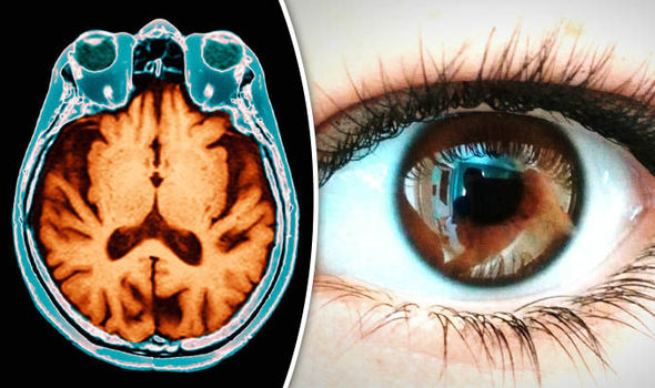
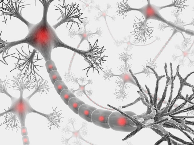
The unstoppable death of brain cells in Huntington’s leaves patients in permanent decline, affecting their movement, behaviour, memory and ability to think clearly.

Peter, from Essex, told me: “It’s so difficult to have that degenerative thing in you.

“You know the last day was better than the next one’s going to be.”
Huntington’s generally affects people in their prime – in their 30s and 40s
Patients die around 10 to 20 years after symptoms start
About 8,500 people in the UK have Huntington’s and a further 25,000 will develop it when they are older

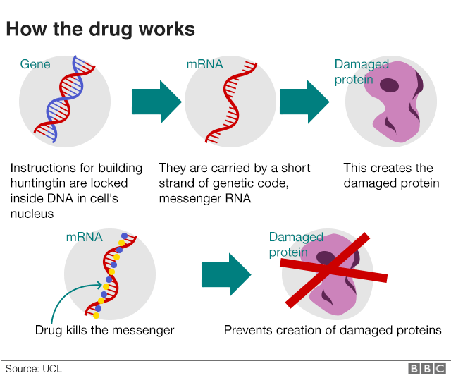
Huntington’s is caused by an error in a section of DNA called the huntingtin gene. Normally this contains the instructions for making a protein, called huntingtin, which is vital for brain development. But a genetic error corrupts the protein and turns it into a killer of brain cells.

The treatment is designed to silence the gene.

On the trial, 46 patients had the drug injected into the fluid that bathes the brain and spinal cord. The procedure was carried out at the Leonard Wolfson Experimental Neurology Centre at the National Hospital for Neurology and Neurosurgery in London. Doctors did not know what would happen. One fear was the injections could have caused fatal meningitis. But the first in-human trial showed the drug was safe, well tolerated by patients and crucially reduced the levels of huntingtin in the brain.

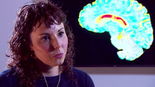
Prof Sarah Tabrizi, the lead researcher and director of the Huntington’s Disease Centre at UCL, told the BBC: “I’ve been seeing patients in clinic for nearly 20 years, I’ve seen many of my patients over that time die. For the first time we have the potential, we have the hope, of a therapy that one day may slow or prevent Huntington’s disease . This is of groundbreaking importance for patients and families.”

Doctors are not calling this a cure. They still need vital long-term data to show whether lowering levels of huntingtin will change the course of the disease. The animal research suggests it would. Some motor function even recovered in those experiments.

Peter, Sandy and Frank – as well as their partners Annie, Dermot and Hayley – have always promised their children they will not need to worry about Huntington’s as there will be a treatment in time for them. Peter told the BBC: “I’m the luckiest person in the world to be sitting here on the verge of having that. “Hopefully that will be made available to everybody, to my brothers and sisters and fundamentally my children.”

He, along with the other trial participants, can continue taking the drug as part of the next wave of trials. They will set out to show whether the disease can be slowed, and ultimately prevented, by treating Huntington’s disease carriers before they develop any symptoms.

Prof John Hardy, who was awarded the Breakthrough Prize for his work on Alzheimer’s, told the BBC: “I really think this is, potentially, the biggest breakthrough in neurodegenerative disease in the past 50 years. That sounds like hyperbole – in a year I might be embarrassed by saying that – but that’s how I feel at the moment.”

The UCL scientist, who was not involved in the research, says the same approach might be possible in other neurodegenerative diseases that feature the build-up of toxic proteins in the brain. The protein synuclein is implicated in Parkinson’s while amyloid and tau seem to have a role in dementias.

Off the back of this research, trials are planned using gene-silencing to lower the levels of tau.

Prof Giovanna Mallucci, who discovered the first chemical to prevent the death of brain tissue in any neurodegenerative disease, said the trial was a “tremendous step forward” for patients and there was now “real room for optimism”.

But Prof Mallucci, who is the associate director of UK Dementia Research Institute at the University of Cambridge, cautioned it was still a big leap to expect gene-silencing to work in other neurodegenerative diseases.

She told the BBC: “The case for these is not as clear-cut as for Huntington’s disease, they are more complex and less well understood. But the principle that a gene, any gene affecting disease progression and susceptibility, can be safely modified in this way in humans is very exciting and builds momentum and confidence in pursuing these avenues for potential treatments.”

The full details of the trial will be presented to scientists and published next year.

The therapy was developed by Ionis Pharmaceuticals, which said the drug had “substantially exceeded” expectations, and the licence has now been sold to Roche.

http://www.bbc.com/news/health-42308341

Examining the eye to spot early signs of Alzheimer’s disease

Last year, doctors of optometry detected more than 320,000 cases of diabetes. Imagine if they could make the same impact when it comes to exposing early signs of Alzheimer’s disease.

November is National Alzheimer’s Disease Awareness Month. An estimated 5.4 million Americans are affected by Alzheimer’s disease, according to the Centers for Disease Control and Prevention (CDC). Projections put the number at 13.8 million by 2050.

Maryke Nijhuis Neiberg, O.D., associate professor in the School of Optometry at Massachusetts College of Pharmacy and Heath Sciences, in Worcester, Massachusetts, considers this an unrealized patient education opportunity for doctors of optometry.

“The earlier diagnoses give doctors and patients a better chance at managing the progressive brain disease and preserving the patient’s quality of life,” Dr. Neiberg says. “There has been some increase in Alzheimer’s awareness over the years, particularly in the eye community, but not enough yet.

“Alzheimer’s is a significant future public health issue,” she adds. “It is still a terminal disease.”

Early intervention

Much of the research on Alzheimer’s disease seeks to slow the disease’s progression. For instance, a study in Biological Psychiatry on Nov. 6 by researchers at the University of Iowa and the University of Texas Southwestern Medical Center in Dallas reports that there may be a new treatment that can slow the depression and cognitive decline associated with Alzheimer’s disease, without affecting amyloid plaque deposits or reactive glia in rats.

Among the early signs of Alzheimer’s, the researchers say, are anxiety, depression and irritability-long before the devastating effects of memory loss.

“Thus, P7C3 compounds may form the basis for a new class of neuroprotective drugs for mitigating the symptoms in patients with Alzheimer’s disease by preserving neuronal cell survival, irrespective of other pathological events,” researchers say. “P7C3 compounds represent a novel route to treating depression, and new-onset depression in elderly patients may herald the development of Alzheimer’s disease with later cognitive impairments to follow.”

Another study in JAMA Ophthalmology in September by researchers at Stanford University and Veterans Affairs Palo Alto Health Care System linked visual impairment and cognition in older adults and also stressed the “potential importance” of vision screening in identifying these patients’ eye disease and cognitive deficits. The AOA strongly recommends comprehensive eye examinations and stresses the limitations of screenings.

Optometry’s role

According to the CDC:

The rate of Alzheimer’s jumped 50 percent between 1999 and 2014.

Americans fear losing their mental capacity more than losing their physical abilities.

More than $230 billion is estimated to be spent in 2017 on providing health care, long-term care, hospice plus unpaid care for relatives with Alzheimer’s and other dementias.

More large-scale research on Alzheimer’s needs to be done, but progress is being made. Dr. Neiberg pointed to research linking optical coherence tomography (OCT) of the macula to Alzheimer’s and Parkinson’s diseases.

“With the advent of OCT, we now know that the retinal ganglion cell layer thins and that the optic nerve cup-to-disc ratio increases in size, not unlike glaucoma,” Dr. Neiberg says. “Alzheimer’s produces visual field defects that are easily confused with glaucoma. What we need is large-scale research to determine how much of the normal tension glaucoma we diagnose and treat is ultimately related to Alzheimer’s disease.”

She adds, “The early perceptual changes that occur in early Alzheimer’s are startling and measurable. One of the earliest signs is a decline in the Benton Visual Retention Test, a test of visual memory. This test requires the duplication of shapes on paper with a pencil, and is scored.

“Research has shown that this test is able to predict high risk for Alzheimer’s 15 years before diagnosis,” she says. “It’s a simple test many developmental and pediatric optometrists already have on their shelves. If we combine that test and the ocular findings we see, we have a very strong indication that something is indeed amiss. Armed with this information, the patient can then consult with their primary care physician, initiate lifestyle modification and request a referral if necessary.”

There is no cure for Alzheimer’s disease. But doctors of optometry can engage patients in conversation about Alzheimer’s disease and how they can manage their own risk factors, including:

Smoking
Mid-life obesity
Sedentary lifestyle
High-cholesterol diet|
Vascular disease (i.e., diabetes and hypertension)

“Lifestyle modification and early access to medication, which can delay the progression of dementia, might be enough to keep the disease at bay for longer,” Dr. Neiberg says. “We should include the Alzheimer’s disease connection when we educate our patients about lifestyle diseases.”

https://finchannel.com/society/health-beauty/69483-doctors-of-optometry-can-spot-early-signs-of-alzheimer-s-disease

Adding lithium to tap water may decrease rates of dementia

by CHRIS SMYTH

People living in areas with high levels of lithium in tap water are 17 per cent less likely to get dementia, according to a large study that suggests the naturally occurring metal could help to prevent mental decline.

The findings raise the possibility that lithium could one day be added to drinking water to protect the brain in the same way as fluoride is added to protect teeth.

Lithium is already widely available as a psychiatric drug and experts said the findings suggested that it could be used as a treatment to prevent dementia if further trials proved successful. Lithium is known to affect neurological signalling and has long been used as a treatment for conditions such as bipolar disorder. It occurs naturally in water and previous studies have found lower suicide rates in areas with higher levels.

Scientists studied 74,000 older people with dementia and 734,000 without across Denmark, comparing illness rates with lithium levels, which were 15 times higher in some areas.

Scientists at the University of Copenhagen found that dementia rates increased slightly with low levels of lithium before falling sharply above 10 micrograms per litre. At 15 to 27 micrograms/l, dementia rates were 17 per cent lower than for 2-5 micrograms/l, according to results published in JAMA Psychiatry.

The authors acknowledged that other factors could explain the results, including worse healthcare in the remoter areas that had less lithium in water, but they said it was plausible that tiny amounts in tap water could have a significant effect on dementia.

In a linked editorial John McGrath, of the University of Queensland, and Michael Berk, of the University of Melbourne, wrote: “In the spirit of alchemy, could we convert lithium, a simple metal used as a mood stabiliser, into a golden public health intervention that could prevent dementia?

They added: “That a relatively safe, simple, and cheap intervention (ie optimising lithium concentrations in the drinking water) could lead to the primary prevention of dementia is a tantalising prospect.”

David Smith, emeritus professor of pharmacology at the University of Oxford, said the findings tallied with MRI studies showing that lithium salts increased the volume of areas of the brain involved in Alzheimer’s. However, he added: “We should not be adding lithium salts to our tap water because we would not know what amount to use.”

David Reynolds, chief scientific officer at Alzheimer’s Research UK, said: “It is potentially exciting that low doses of a drug already available in the clinic could help limit the number of people who develop dementia.”

Rob Howard, professor of old-age psychiatry at University College London, said: “These results represent another important piece of evidence for lithium’s potential as a treatment for Alzheimer’s disease. We now need clinical trials of lithium in patients with Alzheimer’s disease to determine once and for all whether this cheap and well-tolerated element can slow dementia progression.”

http://www.theaustralian.com.au/news/world/the-times/lithium-in-tap-water-could-lower-dementia-risk/news-story/c40599203eca195402c03c0a168961a6

A metabolic shift in neurons may provide insight into neurodegenerative diseases


A key metabolic pathway must be switched off during neuron development or fewer neurons (green, on the right) survive.

by Jennifer Hicks

Researchers at the Salk Institute of Biological Studies released a study in the July 12 issue of eLife, which identifies the point at which there’s a dramatic metabolic shift in developing neurons. This discovery of the path a neuron takes during development could help provide insight into neurodegenerative diseases such as Alzheimer’s and Parkinson’s disease.

In a press release, Tony Hunter, American Cancer Society Professor, Salk Molecular and Cell Biology Laboratory said there’s relatively little understanding about how neuron metabolism is first established.

Oxidative stress leads to disruptions in neural cells which are key players in neurodegenerative diseases like Parkinson’s or ALS. The brain needs oxygen to survive but by knowing when and how neuron metabolism goes off track and mitochondria fail to function properly in these diseases, researchers can begin to devise ways to re-route metabolic processes to prevent degeneration.

“Aside from enabling us to understand this process during neuronal development, the work also allows us to better understand neurodegenerative disease,” added Hunter.

What the researchers found in the study was that while neurons shut off the aerobic glycolysis to survive during the metabolic process at the same time neurons also had to kick-start oxidative phosphorylation in order to survive. When the researchers stopped that metabolic process from happening, the neurons died. A neuron dysfunction of any kind can potentially lead to neurodegenerative disease for a number of reasons.

http://www.forbes.com/sites/jenniferhicks/2016/07/31/a-look-at-the-metabolic-shift-in-neurons-for-insight-into-neurodegenerative-disease/#14296174e07b

Having a socially interactive job helps protect from Alzheimer’s disease.

By Patrick Foster

Lawyers, teachers and doctors have a better chance of fighting off the effects of Alzheimer’s disease, because of the complex nature of their jobs, scientists reported this week.

Researchers found that people whose jobs combined complex thinking with social engagement with others – such as social workers and engineers – were better protected against the onset of Alzheimer’s, compared to those in manual work.

The study came as another report suggested that people with a poor diet could protect themselves against cognitive decline by adopting a mentally stimulating lifestyle.

Both pieces of research, published at the international conference of the Alzheimer’s Association, in Toronto, examined the impact of complex thinking on the onset of the disease.

In the first study, carried out by scientists at the Alzheimer’s Disease Research Centre, in Wisconsin, researchers examined white matter hyperintensities (WMHs) – white spots that appear on brain scans and are associated with Alzheimer’s – in 284 late-middle-aged patients considered at risk of contracting the disease.

They found that people who worked primarily with other people, as opposed to with “things or data”, were less likely to be affected by brain damage indicated by WMHs.

While lawyers, social workers, teachers and doctors were best protected, those who enjoyed the least protection included shelf-stackers, machine operators and labourers.

Elizabeth Boots, a researcher on the project, said: “These findings indicate that participants with higher occupational complexity are able to withstand pathology associated with Alzheimer’s and cerebrovascular disease and perform at a similar cognitive level as their peers.

“This association is primarily driven by work with people, rather than data or things. These analyses underscore the importance of social engagement in the work setting for building resilience to Alzheimer’s disease.”

The second study, carried out by Baycrest Health Sciences, in Toronto, examined the diet of 351 older adults.

Researchers found that those who had a traditional Western diet of red and processed meat, white bread, potatoes and sweets were more likely to experience cognitive decline.

However, those who adhered to such a diet but who had a mentally stimulating lifestyle enjoyed some protection from such decline.

Dr Matthew Parrott, one member of the team, said: “Our results show the role higher educational attainment, mentally stimulating work and social engagement can play in protecting your brain from cognitive decline, counteracting some negative effects of an unhealthy diet.

“This adds to the growing body of evidence showing how various lifestyle factors may combine to increase or protect against vulnerability to Alzheimer’s disease.”

Other research put forward at the convention included a study showing that digital brain training exercises can help stave of Alzheimer’s, and another paper that suggested that some newly-identified genes may also increase resilience to the disease.

Maria C. Carrillo, the chief science officer at the Alzheimer’s Association, said: “These new data add to a growing body of research that suggests more stimulating lifestyles, including more complex work environments with other people, are associated with better cognitive outcomes in later life.

“As each new study emerges, we further understand just how powerful cognitive reserve can be in protecting the brain from disease. Formal education and complex occupation could potentially do more than just slow cognitive decline – they may actually help compensate for the cognitive damage done by bad diet and small vessel disease in the brain.

“It is becoming increasingly clear that in addition to searching for pharmacological treatments, we need to address lifestyle factors to better treat and ultimately prevent Alzheimer’s and other dementias.”

http://www.telegraph.co.uk/news/2016/07/24/stressful-job-it-might-help-you-fight-off-alzheimers/

Urinary biomarker of Parkinson’s disease identified

New findings indicate that phosphorylated LRRK2 (leucine-rich repeat kinase 2) protein levels in urine are elevated in patients diagnosed with idiopathic Parkinson Disease (PD), and that urinary phosphorylated LRRK2 levels correlate with the presence and severity of symptoms such as cognitive impairment in individuals with PD. Researchers affiliated with the University of Alabama at Birmingham published their findings in Neurology and in Movement Disorders (1,2).

The etiology of PD is currently unknown and mechanisms of action are still not completely clarified. It is well established, however, that aging is the single most important risk factor. PD is the second most frequent age-related neurodegenerative disorder, and one of the key pathogenic features is slow and progressive neuronal death that is concomitant with cognitive dysfunction. Current therapeutic modalities are inadequate and clinical need is significant. More than 6 million individuals worldwide are diagnosed with PD.

To date, several common genetic variants, or single nucleotide polymorphisms (SNPs), have been identified that influence the risk for disease. For example, polymorphic variants in LRRK2 gene have previously been validated as genetic factors that confer susceptibility to PD.

Although the gene remains poorly characterized, five different mutations in the gene encoding LRRK2 are considered a common cause of inherited PD (3). One of the five mutations that are causal is the G2019S mutation in the LRRK2 kinase domain, a mutation that significantly increases phosphorylation activity (1,3).

“There are currently no known ways to predict which G2019S mutation carriers will develop PD,” the authors wrote in the Neurology publication. Investigators purified LRRK2 protein from urinary exosomes collected from a total of 76 men. (Exosomes are membrane vesicles of endosomal origin that are secreted by most cells in culture, and are present in most biological fluids such as urine, blood, and saliva.) Then, they compared the ratio of phosphorylated LRRK2 to total LRRK2 in urine exosomes. Results show that “elevated … phosphorylated LRRK2 predicted the risk” for onset of PD in LRRK2 G2019S mutation carriers (1).

In their follow-up study, which was published in Movement Disorders, investigators compared phosphorylated LRRK2 levels in urine samples of 79 individuals diagnosed with PD to those of 79 healthy control participants. Results show that phosphorylated LRRK2 levels were significantly elevated in patients with PD when compared to those of controls. Also, phosphorylated LRRK2 levels correlated with the severity of cognitive impairment in patients with PD (2).

“Because few viable biomarkers for PD exist … phosphorylated LRRK2 levels may be a promising candidate for further exploration,” the authors concluded in their publication.

References
1. Fraser KB, Moehle MS, Alcalay RN, et al. Urinary LRRK2 phosphorylation predicts parkinsonian phenotypes in G2019S LRRK2 carriers. Neurology. 2016;86:994-999.
2. Fraser KB, Rawlins AB, Clar RG, et al. Ser(P)-1292 LRRK2 in urinary exosomes is elevated in idiopathic Parkinson’s disease. Mov Disord. 2016. doi: 10.1002/mds.26686.
3. Greggio E, Cookson MR. Leucine-rich repeat kinase 2 mutations and Parkinson’s disease: three questions. ASN Neuro. 2009;1:e00002.

http://www.psychiatryadvisor.com/neurocognitive-disorders/urinary-biomarker-of-parkinson-disease-identified/article/508195/?DCMP=EMC-PA_Update_RD&cpn=psych_md,psych_all&hmSubId=&hmEmail=5JIkN8Id_eWz7RlW__D9F5p_RUD7HzdI0&NID=1710903786&dl=0&spMailingID=14919209&spUserID=MTQ4MTYyNjcyNzk2S0&spJobID=820575619&spReportId=ODIwNTc1NjE5S0

Toxoplasma infection might trigger neurodegenerative disease


Infection with the common parasite Toxoplasma gondii promotes accumulation of a neurotransmitter in the brain called glutamate, triggering neurodegenerative diseases in individuals predisposed to such conditions.

Written by Honor Whiteman

This is the finding of a new study conducted by researchers from the University of California-Riverside (UC-Riverside), recently published in PLOS Pathogens.

T. gondii is a single-celled parasite that can cause a disease known as toxoplasmosis.

Infection with the parasite most commonly occurs through eating undercooked, contaminated meat or drinking contaminated water.

It may also occur through accidentally swallowing the parasite after coming into contact with cat feces – by cleaning a litter tray, for example.

Though more than 60 million people in the United States are believed to be infected with T. gondii, few people become ill from it; a healthy immune system can normally stave it off.

As such, most people who become infected with the parasite are unaware of it.

Those who do become ill from T. gondii infection may experience flu-like symptoms – such as swollen lymph glands or muscle aches – that last for at least a month.

In severe cases, toxoplasmosis can cause damage to the eyes, brain, and other organs, though such complications usually only arise in people with weakened immune systems.

The new study, however, suggests there may be another dark side to T. gondii infection: it may lead to development of neurodegenerative disease in people who are predisposed to it.

To reach their findings, lead author Emma Wilson – an associate professor in the Division of Biomedical Sciences at the UC-Riverside School of Medicine – and colleagues focused on how T. gondii infection in mice affects glutamate production

How a build-up of glutamate can damage the brain

Glutamate is an amino acid released by nerve cells, or neurons. It is one of the brain’s most abundant excitatory neurotransmitters, aiding communication between neurons.

However, previous studies have shown that too much glutamate may cause harm; a build-up of glutamate is often found in individuals with traumatic brain injury (TBI) and people with certain neurodegenerative diseases, such as multiple sclerosis (MS) and amyotrophic lateral sclerosis (ALS).

The researchers explain that excess glutamate accumulates outside of neurons, and this build-up is regulated by astrocytes – cells in the central nervous system (CNS).

Astrocytes use a glutamate transporter called GLT-1 in an attempt to remove excess glutamate from outside of neurons and convert it into a less harmful substance called glutamine, which cells use for energy.

“When a neuron fires, it releases glutamate into the space between itself and a nearby neuron,” explains Wilson. “The nearby neuron detects this glutamate, which triggers a firing of the neuron. If the glutamate isn’t cleared by GLT-1 then the neurons can’t fire properly the next time and they start to die.”


T. gondii increases glutamate by inhibiting GLT-1

n mice infected with T. gondii, the researchers identified an increase in glutamate levels.

They found that the parasite causes astrocytes to swell, which impairs their ability to regulate glutamate accumulation outside of neurons.

Furthermore, the parasite prevents GLT-1 from being properly expressed, which causes an accumulation of glutamate and misfiring of neurons. This may lead to neuronal death, and ultimately, neurodegenerative disease.

“These results suggest that in contrast to assuming chronic Toxoplasma infection as quiescent and benign, we should be aware of the potential risk to normal neurological pathways and changes in brain chemistry.” – Emma Wilson

Next, the researchers gave the infected mice an antibiotic called ceftriaxone, which has shown benefits in mouse models of ALS and a variety of CNS injuries.

They found the antibiotic increased expression of GLT-1, which led to a reduction in glutamate build-up and restored neuronal function.

Wilson says their study represents the first time that T. gondii has been shown to directly disrupt a key neurotransmitter in the brain.

“More direct and mechanistic research needs to be performed to understand the realities of this very common pathogen,” she adds.

While their findings indicate a link between T. gondii infection and neurodegenerative disease, Wilson says they should not be cause for panic.

“We have been living with this parasite for a long time,” she says. “It does not want to kill its host and lose its home. The best way to prevent infection is to cook your meat and wash your hands and vegetables. And if you are pregnant, don’t change the cat litter.”

The team now plans to further investigate what causes the reduced expression of GLT-1 in T. gondii infection.

http://www.medicalnewstoday.com/articles/310865.php

7 habits to avoid in order to have a healthy brain

Why are some people sharp as a tack at 95 years old, while others begin struggling with mental clarity in their 50s?

A lot of it has to do with genetics, but certain lifestyle factors also play an important role in how our brain ages. So while you can’t control your genes, you can take advantage of the latest science and avoid these seven big brain mistakes:

Mistake No. 1: Eating a standard American diet

Foods high in sugar, unhealthy fats and processed foods — i.e., the typical American diet — can wreak havoc on your brain over time. Studies have shown that excess sugar consumption can impair learning and memory, and increase your vulnerability to neurodegenerative diseases like Alzheimer’s. Some scientists have even referred to Alzheimer’s as “Type 3 Diabetes,” suggesting that diet may have some role in an individual’s risk for developing the disease.

A Mediterranean-based diet, on the other hand, can help protect the brain from signs of aging and ward off cognitive decline. A recent study showed that following this type of diet — which is a good source of brain-healthy nutrients and includes a lot of fish, healthy fats, whole grains and vegetables — could slash Alzheimer’s risk by up to 50 percent.

Mistake No. 2: Living next to a highway

Living in a smoggy city might be bad news for your brain. According to research published this month in the journal Stroke, exposure to air pollution is linked with premature aging of the brain.

The researchers found that people who lived closer to a major highway had greater markers of pollution in their lungs and blood, which increased their risk for a form of brain damage known as “silent strokes,” or symptomless strokes. Increased pollution volume was also linked to decreased brain volume — a major sign of aging.

Mistake No. 3: Drinking a few evening cocktails

Don Draper’s daily cigarettes and two-martini lunches might seem glamorous on “Mad Men,” but research suggests that they’re a fast track to neurodegeneration.

It should come as no surprise that excessive drinking and cigarette smoking at any stage of life can have a negative effect on the brain, damaging brain tissue and leading to cognitive impairment. Alcoholism can cause or accelerate aging of the brain.

But just a couple of glasses of wine a night could pose a risk to brain health, even though there are some cardiovascular benefits. A 2012 Rutgers University study found that moderate to binge drinking — drinking relatively lightly during the week and then more on the weekends — can decrease adult brain cell production by 40 percent.

“In the short term there may not be any noticeable motor skills or overall functioning problems, but in the long term this type of behavior could have an adverse effect on learning and memory,” one of the study’s authors, Rutgers neuroscience graduate student Megan Anderson, said in a statement.

Mistake No. 4: Giving in to stress

Living a stressful lifestyle may be the worst thing you can do for your health as you age. Chronic stress is known to shorten the length of telomeres, the sequences at the end of DNA strands that help determine how fast (or slow) the cells in our body age. By shortening telomeres, stress can accelerate the onset of age-related health problems.

What about the brain? Well, some research has suggested that high levels of stress hormones can increase an individual’s risk for age-related brain damage.

“Over the course of a lifetime, the effects of chronic stress can accumulate and become a risk factor for cognitive decline and Alzheimer’s disease,” Howard Fillit, a clinical professor of geriatric medicine at The Mount Sinai School of Medicine, wrote in Psychology Today. “Several studies have shown that stress, and particularly one’s individual way of reacting to stress (the propensity to become ‘dis-stressed’ often found in neurotic people for example), increases the risk for Alzheimer’s disease.”

If you’re feeling stressed out, try picking up a meditation practice. Research has shown that meditation is effective in lowering levels of the stress hormone cortisol and protecting the brain from aging.

Mistake No. 5: Getting by on less sleep than you need

There are a number of scary health effects associated with sleep deprivation, from a higher risk of stroke and diabetes to impaired cognitive functioning. Over the years, losing shut-eye can also accelerate brain aging. In a study conducted last year, researchers from Singapore found that the less that older adults slept, the faster their brains aged.

The study’s lead author explained in a statement that among older adults, “sleeping less will increase the rate their brain ages and speed up the decline in their cognitive functions.”

Mistake No. 6: Sitting all day

It’s a well-established fact that sitting for long periods is terrible for your health. A growing body of research has linked a sedentary lifestyle with health risks including heart disease, diabetes, cancer and early death, even among people who get the recommended daily amount of exercise.

And it turns out that sitting is also pretty bad for your brain. Research has linked physical inactivity with cognitive decline. Moreover, weight gain in older adults — which may result from too much sitting — has been linked with shrinkage in brain areas associated with memory.

So when in doubt, move around. Physical activity has been linked with a number of brain health benefits, including improved learning and memory.

Mistake No. 7: Zoning out

Use it or lose it! If you want to keep your brain sharp, keep it engaged. It doesn’t have to be a challenging intellectual task or a brain-training game, either — simply engaging in everyday activities like reading, cooking or having a conversation (as opposed to vegging out in front of the TV or computer) can make a difference.

But mental exercises like crossword puzzles and sudoku can help, too. A 2013 study published in the Canadian Medical Association Journal found that brain exercises are more effective than drugs in preventing cognitive decline.

The bottom line? Doing new and novel things promotes neurogenesis, the creation of new neurons in the brain. So get outside, learn, discover and try something new to keep your brain sharp through the decades.

http://www.huffingtonpost.com/2015/04/30/brain-aging-risk-factors_n_7169912.html

An axon self-destruct mechanism that kills neurons

Just as losing a limb can spare a life, parting with a damaged axon by way of Wallerian degeneration can spare a neuron. A protein called SARM1 acts as the self-destruct button, and now researchers led by Jeffrey Milbrandt of Washington University Medical School in St. Louis believe they have figured out how. They report in the April 24 Science that SARM1 forms dimers that trigger the destruction of NAD+. Basic biochemistry dictates that this enzyme cofactor is essential for cell survival.

ARM1 and NAD+ have emerged as key players in the complex, orderly process underlying Wallerian degeneration. Scientists are still filling in other parts of the pathway. SARM1, short for sterile alpha and TIR motif-containing 1, seems to act as a damage sensor, but researchers are not sure how. Recently, researchers led by Marc Tessier-Lavigne at Rockefeller University, New York, found that SARM1 turns on a mitogen-activated protein (MAP) kinase cascade that is involved. Loss of NAD+ may also contribute to axon degeneration, because its concentration drops in dying axons, and Wlds mutant mice that overproduce an NAD+ synthase have slower Wallerian degeneration.

Now, first author Josiah Gerdts confirms that SARM1 is the self-destruct switch. He engineered a version of the protein with a target sequence for tobacco etch virus (TEV) protease embedded in it. Using a rapamycin-activated form of TEV, he eliminated SARM1 from axons he had sliced off of mouse dorsal root ganglion (DRG) neurons. Without SARM1, the severed axons survived.

SARM1 contains SAM and TIR domains, which promote protein-protein interactions. Previously, Gerdts discovered that the TIR domain was sufficient to induce degeneration, even in healthy axons, but it relied on the SAM region to bring multiple SARM1 molecules together. He hypothesized that axonal SARM1 multimerizes upon axon damage. To test this idea, he used a standard biochemical technique to force the SARM1 TIR domains together. He fused domains to one or another of the rapamycin-binding peptides Frb and Fkbp and expressed them in DRG neurons. When he added rapamycin to the cultures, the Frb and Fkbp snapped the TIR domains together within minutes. As Gerdts had predicted, this destroyed axons, confirming that SARM1 activates via dimerization.

Next, the authors investigated what happens to NAD+ during that process. Using high-performance liquid chromatography, Gerdts measured the concentration of NAD+ in the disembodied axons. Normally, its level dropped by about two-thirds within 15 minutes of severing. In axons from SARM1 knockout mice, however, the NAD+ concentration stayed unchanged. In neurons carrying the forced-dimerization constructs, adding rapamycin was sufficient to knock down NAD+ levels—Gerdts did not even have to cut the axons. Ramping up NAD+ production by overexpressing its synthases, NMNAT and NAMPT, overcame the effects of TIR dimerization, and the axons survived. Gerdts concluded that loss of NAD+ was a crucial, SARM1-controlled step on the way to degeneration.

He still wondered what caused the loss of NAD+. It might be that the axon simply stopped making it, or maybe the Wallerian pathway actively destroyed it. To distinguish between these possibilities, Gerdts added radiolabeled exogenous NAD+ to human embryonic kidney HEK293 cultures expressing the forced-dimerization TIR domains. Rapamycin caused them to rapidly degrade the radioactive NAD+, confirming that the cell actively disposes of it.

Gerdts suspects that with this essential cofactor gone, the axon runs out of energy and can no longer survive. He speculated that the MAP kinase cascade reportedly turned on by SARM1 might lead to NAD+ destruction. Alternatively, SARM1 might induce distinct MAP kinase and NAD+ destruction pathways in parallel, he suggested.

“Demonstrating how NAD+ is actively and locally degraded in the axon is a big advance,” commented Andrew Pieper of the Iowa Carver College of Medicine in Iowa City, who was not involved in the study. Jonathan Gilley and Michael Coleman of the Babraham Institute in Cambridge, U.K., predict that there will be more to the story. They note that a drug called FK866, which prevents NAD+ production, protects axons in some instances. Gerdts suggested that FK866 acts on processes upstream of SARM1, delaying the start of axon degeneration. In contrast, his paper only addressed what happens after SARM1 activates. “It will be fascinating to see how the apparent contradictions raised by this new study will be resolved,” wrote Gilley and Coleman.

Could these findings help researchers looking for ways to prevent neurodegeneration? “The study supports the notion that augmenting NAD+ levels is potentially a valuable approach,” said Pieper. He and his colleagues developed a small molecule that enhances NAD+ synthesis, now under commercial development. It improved symptoms in ALS model mice, and protected neurons in mice mimicking Parkinson’s. NAD+ also activates sirtuin, an enzyme important for longevity and stress resistance as well as learning and memory.

However, both Pieper and Gerdts cautioned that they cannot clearly predict which conditions might benefit from an anti-SARM1 or NAD+-boosting therapy. At this point, Gerdts said, researchers do not fully understand how much axon degeneration contributes to symptoms of diseases like Alzheimer’s and Parkinson’s. He suggested that crossing SARM1 knockout mice with models for various neurodegenerative conditions would indicate how well an anti-Wallerian therapy might work.

—Amber Dance

http://www.alzforum.org/news/research-news/axon-self-destruct-button-triggers-energy-woes

New typing test may help diagnose Parkinson’s disease

Whether it’s on a keyboard, a smartphone, or even a credit card reader, you spend a lot of your day typing. Well, researchers at MIT noticed the value of this daily habit, and are putting it to a secondary use; they’ve developed software that can gauge the speed at which a typist is tapping the keyboard to help diagnose Parkinson’s disease.

In order to type a word, your brain has to send signals down through your spinal cord to the nerves that operate your fingers. If your central nervous system is functioning perfectly, then you should be able to tap most of the keys at a fairly constant rate. But a number of conditions might slow the signal from the brain to the fingers, such as sleep deprivation (which slows all motor skills) and diseases that affect the central nervous system, including Parkinson’s.

For the first version of this study, the researchers were looking at typing patterns that indicated whether a person was sleep-deprived or well rested. They created a browser plug-in that detected the timing at which the volunteers hit they keys and found that the people who were sleepy had a much wider variation in their typing speed. They found similar results in their preliminary test with Parkinson’s patients; the 21 typists with Parkinson’s tapped the keys at much more variable rates than the 15 healthy volunteers. The researchers called it a “window into the brain.”

Right now, the algorithm they’ve developed is not refined enough to distinguish Parkinson’s patients from people who are sleep deprived, though the results might be clearer after a number of trials. The researchers plan to conduct a study with a larger group of subjects, but they hope that this type test could eventually lead to earlier diagnoses of Parkinson’s–today most people are diagnosed after they have had symptoms for 5-10 years–and to distinguish Parkinson’s from other conditions that might affect a person’s motor skills, like rheumatoid arthritis. They are currently developing a smartphone app that can test participants even more easily.

http://www.popsci.com/type-test-diagnose-parkinsons