MRI scans from more than 800 incarcerated men pinpoint distinct structural features of people who have committed homicide, compared with those who carried out other crimes.

by NICOLETTA LANESE

Kent Kiehl and his research team regularly park their long, white trailer just outside the doors of maximum-security prisons across the US. Inside the vehicle sits the bulky body of a mobile MRI machine. During each visit, people from the prison make their way to and from the vehicle in hourly shifts to have their brains scanned and help to answer an age-old question: What makes a murderer?

“It’s not an uncommon thing for [incarcerated people], while they’re getting a scan, to be like, ‘I’ve always been different. Can you tell me why I’ve always been so different?’” says Kiehl, a neuroscientist at the University of New Mexico and the Albuquerque-based nonprofit Mind Research Network (MRN) who helped design the mobile MRI system back in the early 2000s.


SCAN-MOBILE: Kiehl and his colleagues made more than 75 modifications to a trailer and the MRI system inside to outfit both for the team’s unique research.

The author of The Psychopath Whisperer: The Science of Those Without a Conscience, Kiehl has been fascinated by the criminal mind since he was an undergraduate at the University of California, Davis. Now, as director of mobile imaging at MRN, he oversees efforts to gather brain scans from thousands of people held in US prisons to learn what features, if any, might differ from scans of the general population.

This massive dataset recently allowed Kiehl to examine the brain structures of more than 800 men held in state prisons in New Mexico and Wisconsin in an attempt to distinguish incarcerated people who have committed homicide from those who have committed other crimes.

First, Kiehl and his colleagues laboriously sorted the pool of people who had volunteered for the study into three categories based on their crimes: homicide, violent offenses that were not homicide, or non-violent or minimally violent transgressions. The team relied on official convictions, self-reported homicides, and confidential interviews with participants to determine who attempted or committed murder—both offenses that got a “homicide” label in their dataset.

People charged with felony murder—meaning that they had committed a serious felony that was in some way connected to a person’s death, even though they hadn’t intended to kill the victim—and people whose cases indicated considerable doubt about a judgment of homicide were not counted among murderers. And occasionally, people were moved from another category into the homicide group, Kiehl says. The researchers excluded people with abnormal radiology reports, traumatic brain injury, or diagnosed psychotic disorders from the study.

Controlling for substance use severity, time in prison, age, and IQ, the team analyzed the MRI data to look for differences among the study participants. Compared with the other two groups, the 200 men who had committed homicide showed significantly reduced gray matter in several brain regions that play important roles in behavioral control and social cognition.

“I think that the intriguing thing was, first, that they found a difference,” says Hannes Vogel, a neuropathologist at Stanford University Medical Center who was not involved in the work. “And second of all, that it correlates with some of the brain centers that deal with behavior and social interaction.”

Lora Cope, a neuroscientist who studies substance disorders at the University of Michigan, notes in an email to The Scientist that the team’s mobile MRI system has now been used in correctional facilities all over New Mexico and Wisconsin, and “has really revolutionized this area of research.” Indeed, the MRN has now used the equipment to collect roughly 6,500 scans from more than 3,000 research participants since its first outing in 2007.

Although Cope wasn’t involved in the current project, she worked with Kiehl a few years ago while earning her doctorate at the University of New Mexico. After speaking with members of the Avielle Foundation, named for a six-year-old victim of the 2012 Sandy Hook Elementary School shooting, the two researchers spearheaded a study of more than 150 incarcerated young males, 20 of whom had been convicted of homicide, held at a maximum-security detention facility within the state. “Jeremy, [Avielle’s] father, really wanted to know if there was anything neuroscience could tell us about boys who commit homicide,” says Kiehl.

As in the current study, Cope and Kiehl deployed the mobile scanner to collect MRI scans of the incarcerated teens in New Mexico and discovered differences between those who had committed homicide and their imprisoned peers. The homicide offenders “had significantly less gray matter volume in parts of their temporal lobes,” Cope says. When Kiel compared the data from that study with the results of his latest project, he found a high degree of overlap. “Lo and behold . . . we found and replicated every region that was different in the boys and was different in the adult males, and in the same way,” he says.

The latest study’s finding that MRI data can distinguish homicide offenders not only from people who committed non-violent crimes, but also from those who performed other violent crimes, is particularly interesting, says Harold Koenigsberg, a psychiatrist at Icahn School of Medicine at Mount Sinai. “I would have thought there would be more of an overlap between [homicide and violent non-homicide offenders],” he says. “I’m surprised that it was so specific to homicide.”


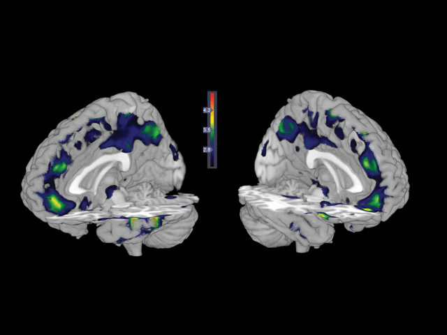
ANATOMY OF A MURDERER: Homicide offenders exhibited reduced gray matter density compared with other violent offenders in the regions of the brain highlighted blue and green above.

Koenigsberg notes that homicidal violence can itself be split into two categories: impulsive and instrumental. Impulsive violence is born of unbridled emotions and overblown reactions, a brand of behavior linked to poor frontal lobe functioning and abnormal serotonin levels. Instrumental violence, on the other hand, is premeditated and is associated with other brain changes, such as reduced amygdala activation during emotion processing. “These two groups, we think that they have different biologies,” says Koenigsberg. Kiehl’s dataset could be enriched by adding measures of neurotransmitter release and electrical activity, along with related behavioral assessments, he suggests, and with both functional and structural data, psychologists might learn more about what gives rise to these distinct behavioral phenotypes.

Koenigsberg, Vogel, and Kiehl all note that the structural data collected in the current study cannot on its own be used to predict who has committed homicide, let alone who might in the future. Nonetheless, the paper may find its way into the courtroom, says Vogel. If lawyers felt so inclined, they could try to “find an expert on one side who will quote this [paper]” in defense of someone who has committed a homicide, by arguing a client’s actions were due to brain abnormalities and thus out of his or her control. Or, a prosecutor could potentially use the paper to argue that MRI findings should be admissible as evidence that a defendant has committed a homicide, says Vogel, who has served as a consultant for court cases in California and Nevada, and helped investigate the brain of the Route 91 Harvest music festival shooter in 2017. “But then you’re [also] going to find an expert that will tear that [testimony] to pieces.”

Kiehl notes that his MRI study could also someday contribute to new evidence-based measures of homicidal risk. These measures could supplement current measures of violent behavior, such as psychological questionnaires, if future studies demonstrated they carried predictive weight, he says. Beyond courts of law, he also suggests that understanding how violent behavior arises could pave the way to better psychological treatment aimed at both rehabilitation and prevention.

https://www.the-scientist.com/notebook/secrets-in-the-brains-of-people-who-have-committed-murder-66589

Scientists are now able to take an MRI Scan of an atom

By Knvul Sheikh

As our devices get smaller and more sophisticated, so do the materials we use to make them. That means we have to get up close to engineer new materials. Really close.

Different microscopy techniques allow scientists to see the nucleotide-by-nucleotide genetic sequences in cells down to the resolution of a couple atoms as seen in an atomic force microscopy image. But scientists at the IBM Almaden Research Center in San Jose, Calif., and the Institute for Basic Sciences in Seoul, have taken imaging a step further, developing a new magnetic resonance imaging technique that provides unprecedented detail, right down to the individual atoms of a sample.

The technique relies on the same basic physics behind the M.R.I. scans that are done in hospitals.

When doctors want to detect tumors, measure brain function or visualize the structure of joints, they employ huge M.R.I. machines, which apply a magnetic field across the human body. This temporarily disrupts the protons spinning in the nucleus of every atom in every cell. A subsequent, brief pulse of radio-frequency energy causes the protons to spin perpendicular to the pulse. Afterward, the protons return to their normal state, releasing energy that can be measured by sensors and made into an image.

But to gather enough diagnostic data, traditional hospital M.R.I.s must scan billions and billions of protons in a person’s body, said Christopher Lutz, a physicist at IBM. So he and his colleagues decided to pack the power of an M.R.I. machine into the tip of another specialized instrument known as a scanning tunneling microscope to see if they could image individual atoms.


Four M.R.I. scans, combined, of a single titanium atom, showing the magnetic field of the atom at different strengths.CreditWillke et al.

The tip of a scanning tunneling microscope is just a few atoms wide. And it moves along the surface of a sample, it picks up details about the size and conformation of molecules.

The researchers attached magnetized iron atoms to the tip, effectively combining scanning-tunneling microscope and M.R.I. technologies.

When the magnetized tip swept over a metal wafer of iron and titanium, it applied a magnetic field to the sample, disrupting the electrons (rather than the protons, as a typical M.R.I. would) within each atom. Then the researchers quickly turned a radio-frequency pulse on and off, so that the electrons would emit energy that could be visualized. The results were described Monday in the journal Nature Physics.

“It’s a really magnificent combination of imaging technologies,” said A. Duke Shereen, director of the M.R.I. Core Facility at the Advanced Science Research Center in New York. “Medical M.R.I.s can do great characterization of samples, but not at this small scale.”

The atomic M.R.I. provides subångström-level resolution, meaning it can distinguish neighboring atoms from one another, as well as reveal which types of atoms are visible based on their magnetic interactions.

“It is the ultimate way to miniaturization,” Dr. Lutz said. He hopes the new technology could one day be used to design atomic-scale methods of storing information, for quantum computers.

Current transistors are thousands of atoms wide and need to switch on and off to store a single bit of information in a computer. The ability to corral individual atoms could drastically increase computing power and enable researchers to tackle complex calculations such as predicting weather patterns or diagnosing illnesses with artificial intelligence.

Moving an atom from one location to another in a composite could also change and lead to the development of new ones.

The technique might also help scientists study how proteins fold and develop new drugs that bind to specific curves in a biological structure.

“We can now see something that we couldn’t see before,” Dr. Lutz said. “So our imagination can go to a whole bunch of new ideas that we can test out with this technology.”

AI and MRIs at birth can predict cognitive development at age 2


Researchers at the University of North Carolina School of Medicine used MRI brain scans and machine learning techniques at birth to predict cognitive development at age 2 years with 95 percent accuracy.

“This prediction could help identify children at risk for poor cognitive development shortly after birth with high accuracy,” said senior author John H. Gilmore, MD, Thad and Alice Eure Distinguished Professor of psychiatry and director of the UNC Center for Excellence in Community Mental Health. “For these children, an early intervention in the first year or so of life – when cognitive development is happening – could help improve outcomes. For example, in premature infants who are at risk, one could use imaging to see who could have problems.”

The study, which was published online by the journal NeuroImage, used an application of artificial intelligence called machine learning to look at white matter connections in the brain at birth and the ability of these connections to predict cognitive outcomes.

Gilmore said researchers at UNC and elsewhere are working to find imaging biomarkers of risk for poor cognitive outcomes and for risk of neuropsychiatric conditions such as autism and schizophrenia. In this study, the researchers replicated the initial finding in a second sample of children who were born prematurely.

“Our study finds that the white matter network at birth is highly predictive and may be a useful imaging biomarker. The fact that we could replicate the findings in a second set of children provides strong evidence that this may be a real and generalizable finding,” he said.

Jessica B. Girault, PhD, a postdoctoral researcher at the Carolina Institute for Developmental Disabilities, is the study’s lead author. UNC co-authors are Barbara D. Goldman, PhD, of UNC’s Frank Porter Graham Child Development Institute, Juan C. Prieto, PhD, assistant professor, and Martin Styner, PhD, director of the Neuro Image Research and Analysis Laboratory in the department of psychiatry.

AI and MRIs at birth can predict cognitive development at age 2

Playing video games may increase your brain’s gray matter and improve how it communicates

According to the results of a study published in Nature, gaming could possibly increase the volume of gray matter in the brain.
Researchers recently studied the insular cortex regions of frequent gamers and those who didn’t play video games as regularly.
The study found a correlation between playing action video games and increased gray matter volume in the brain.

Do you ever feel you could do with polishing up on your cognitive skills?

Well, according to the results of a study published in Nature, gaming could possibly be the way forward.

Researchers from the Chinese University of Electronic Science and Technology and the Australian Macquarie University in Sydney joined forces, and recently found a correlation between playing action video games and increased gray matter volume in the brain.

How video games stimulate the gray matter in your brain

The focus of the team’s research was on the insular cortex, a part of the cerebral cortex folded deep in the brain that has been the subject of very few studies to date.

It’s thought that a large part of linguistic processing takes place in this region of the brain, and that other processes relating to taste and smell, compassion and empathy, and interpersonal experiences are also managed here.

The study looked at 27 regular video game players described in the study as “Action Video Game experts” as well as 30 amateurs who played less frequently and didn’t perform as well in games.

The participants in the “expert” group were all recognised participants of regional or national championships of League of Legends and Dota 2. Using an MRI scanner, the scientists took detailed pictures of the participants’ insular cortices.

“By comparing AVG experts and amateurs, we found that AVG experts had enhanced functional connectivity and gray matter volume in insular subregions,” wrote the research team.

Gaming actually promotes networking within the brain

The gray matter in your brain is part of your central nervous system and essentially controls all your brain’s functions.

It follows that better connectivity in this region will lead to faster thought processes and correspondingly higher intelligence.

If you want to improve your cognitive performance, you don’t necessarily have to resort to hours of video games; sports and art-based recreation are just two among many activities that promote connectivity in the brain.

However it does mean that those who still like to sit in front of their console from time to time no longer need to feel guilty about being sat in front of a screen — after all, it is exercise — just for the brain.

https://www.businessinsider.com/video-games-may-increase-your-brains-gray-matter-2018-12

‘Mindful people’ feel less pain; MRI imaging pinpoints supporting brain activity

mindfulpeopl
Greater deactivation of the posterior cingulate cortex, a brain region associated with processing self-related thoughts, was associated with lower pain and higher trait mindfulness. Credit: Wake Forest Baptist Medical Center

Ever wonder why some people seem to feel less pain than others? A study conducted at Wake Forest School of Medicine may have found one of the answers—mindfulness. “Mindfulness is related to being aware of the present moment without too much emotional reaction or judgment,” said the study’s lead author, Fadel Zeidan, Ph.D., assistant professor of neurobiology and anatomy at the medical school, part of Wake Forest Baptist Medical Center. “We now know that some people are more mindful than others, and those people seemingly feel less pain.”

The study is an article in press, published ahead-of-print in the journal Pain.

The researchers analyzed data obtained from a study published in 2015 that compared mindfulness meditation to placebo analgesia. In this follow-up study, Zeidan sought to determine if dispositional mindfulness, an individual’s innate or natural level of mindfulness, was associated with lower pain sensitivity, and to identify what brain mechanisms were involved.

In the study, 76 healthy volunteers who had never meditated first completed the Freiburg Mindfulness Inventory, a reliable clinical measurement of mindfulness, to determine their baseline levels. Then, while undergoing functional magnetic resonance imaging, they were administered painful heat stimulation (120°F).

Whole brain analyses revealed that higher dispositional mindfulness during painful heat was associated with greater deactivation of a brain region called the posterior cingulate cortex, a central neural node of the default mode network. Further, in those that reported higher pain, there was greater activation of this critically important brain region.

The default mode network extends from the posterior cingulate cortex to the medial prefrontal cortex of the brain. These two brain regions continuously feed information back and forth. This network is associated with processing feelings of self and mind wandering, Zeidan said.

“As soon as you start performing a task, the connection between these two brain regions in the default mode network disengages and the brain allocates information and processes to other neural areas,” he said.

“Default mode deactivates whenever you are performing any kind of task, such as reading or writing. Default mode network is reactivated whenever the individual stops performing a task and reverts to self-related thoughts, feelings and emotions. The results from our study showed that mindful individuals are seemingly less caught up in the experience of pain, which was associated with lower pain reports.”

The study provided novel neurobiological information that showed people with higher mindfulness ratings had less activation in the central nodes (posterior cingulate cortex) of the default network and experienced less pain. Those with lower mindfulness ratings had greater activation of this part of the brain and also felt more pain, Zeidan said.

“Now we have some new ammunition to target this brain region in the development of effective pain therapies. Importantly this work shows that we should consider one’s level of mindfulness when calculating why and how one feels less or more pain,” Zeidan said. “Based on our earlier research, we know we can increase mindfulness through relatively short periods of mindfulness meditation training, so this may prove to be an effective way to provide pain relief for the millions of people suffering from chronic pain.”

https://medicalxpress.com/news/2018-09-mindful-people-pain-mri-imaging.html

Superslow Brain Waves May Play a Critical Role in Consciousness


Signals long thought to be “noise” appear to represent a distinct form of brain activity.

By Tanya Lewis

Every few seconds a wave of electrical activity travels through the brain, like a large swell moving through the ocean. Scientists first detected these ultraslow undulations decades ago in functional magnetic resonance imaging (fMRI) scans of people and other animals at rest—but the phenomenon was thought to be either electrical “noise” or the sum of much faster brain signals and was largely ignored.

Now a study that measured these “infraslow” (less than 0.1 hertz) brain waves in mice suggests they are a distinct type of brain activity that depends on an animal’s conscious state. But big questions remain about these waves’ origin and function.

An fMRI scan detects changes in blood flow that are assumed to be linked to neural activity. “When you put someone in a scanner, if you just look at the signal when you don’t ask the subject to do anything, it looks pretty noisy,” says Marcus Raichle, a professor of radiology and neurology at Washington University School of Medicine in St. Louis and senior author of the new study, published in April in Neuron. “All this resting-state activity brought to the forefront: What is this fMRI signal all about?”

To find out what was going on in the brain, Raichle’s team employed a combination of calcium/hemoglobin imaging, which uses fluorescent molecules to detect the activity of neurons at the cellular level, and electrophysiology, which can record signals from cells in different brain layers. They performed both measurements in awake and anesthetized mice; the awake mice were resting in tiny hammocks in a dark room.

The team found that infraslow waves traveled through the cortical layers of the awake rodents’ brains—and changed direction when the animals were anesthetized. The researchers say these waves are distinct from so-called delta waves (between 1 and 4 Hz) and other higher-frequency brain activity.

These superslow waves may be critical to how the brain functions, Raichle says. “Think of, say, waves on the water of Puget Sound. You can have very rough days where you have these big groundswells and then have whitecaps sitting on top of them,” he says. These “swells” make it easier for brain areas to become active—for “whitecaps” to form, in other words.

Other researchers praised the study’s general approach but were skeptical that it shows the infraslow waves are totally distinct from other brain activity. “I would caution against jumping to a conclusion that resting-state fMRI is measuring some other property of the brain that’s got nothing to do with the higher-frequency fluctuations between areas of the cortex,” says Elizabeth Hillman, a professor of biomedical engineering at Columbia University’s Zuckerman Institute, who was not involved in the work. Hillman published a study in 2016 finding that resting-state fMRI signals represent neural activity across a range of frequencies, not just low ones.

More studies are needed to tease apart how these different types of brain signals are related. “These kinds of patterns are very new,” Hillman notes. “We haven’t got much of a clue what they are, and figuring out what they are is really, really difficult.”

https://www.scientificamerican.com/article/superslow-brain-waves-may-play-a-critical-role-in-consciousness1/

New research links subgroups of schizophrenia to specific visualized brain anomalies

An international team of researchers has linked specific symptoms of schizophrenia with various anatomical characteristics in the brain, according to research published in NeuroImage.

By analyzing the brain’s anatomy with magnetic resonance imaging (MRI), researchers from the University of Granada, Washington University in St. Louis, and the University of South Florida have demonstrated the existence of distinctive subgroups among patients with schizophrenia who suffer from different symptoms.

These findings could herald a significant step forward in diagnosing and treating schizophrenia.

To perform the study, the researchers conducted the MRI technique “diffusion tensor imaging” on 36 healthy participants and 47 schizophrenic participants.

The researchers found that tests on schizophrenic participants revealed various abnormalities in parts of the corpus callosum, a bundle of neural fibers that connects the left and right cerebral hemispheres and is essential for effective interhemispheric communication.

Different anomalies in the corpus callosum were associated with different symptoms in the schizophrenic participants. An anomaly in one part of the brain structure was associated with strange and disorganized behavior; another anomaly was associated with disorganized thought and speech, as well as negative symptoms such as a lack of emotion; and other anomalies were associated with hallucinations.

In 2014, this same research group proved that schizophrenia is not a single illness. The team demonstrated the existence of 8 genetically distinct disorders, each with its own symptoms. Igor Zwir, PhD, and Javier Arnedo from the University of Granada’s Department of Computer Technology and Artificial Intelligence found that different sets of genes were strongly linked with different clinical symptoms.

“The current study provides further evidence that schizophrenia is a heterogeneous group of disorders, as opposed to a single illness, as was previously thought to be case,” Dr Zwir said in a statement.

While current treatments for schizophrenia tend to be generic regardless of the symptoms exhibited by each patient, the researchers believe that in the future, analyzing how specific gene networks are linked to various brain features and specific symptoms will help develop treatments that are adapted to each patient’s individual disorder.

To conduct the analysis of the gene groups and brain scans, the researchers developed a new, complex analysis of the relationships between different types of data and recommendations regarding new data. The system is similar to that used by companies such as Netflix to determine what movies they want to broadcast.

“To conduct the research, we did not begin by studying individuals who had certain schizophrenic symptoms in order to determine whether they had the corresponding brain anomalies,” said Dr Zwir in a statement. “Instead, we first analyzed the data, and that’s how we discovered these patterns. This type of information, combined with data on the genetics of schizophrenia, will someday be of vital importance in helping doctors treat the disorders in a more precise and effective way.”

Reference
Arnedo J, Mamah D, Baranger DA, et al. Decomposition of brain diffusion imaging data uncovers latent schizophrenias with distinct patterns of white matter anisotropy. NeuroImage. 2015; doi:10.1016/j.neuroimage.2015.06.083.

http://www.psychiatryadvisor.com/schizophrenia-and-psychoses/types-subgroups-schizophrenia-linked-various-different-brain-anomalies-corpus-callosum/article/470226/?DCMP=EMC-PA_Update_rd&cpn=psych_md&hmSubId=&hmEmail=5JIkN8Id_eWz7RlW__D9F5p_RUD7HzdI0&NID=&dl=0&spMailingID=13630678&spUserID=MTQ4MTYyNjcyNzk2S0&spJobID=720090900&spReportId=NzIwMDkwOTAwS0

Mother-child MRI

While most new moms get their children’s first portrait done at, say, the local mall’s JC Penney Portrait Studio, neuroscientist Rebecca Saxe opted for a slightly different location: the tube of an MRI scanner.

“No one, to my knowledge, had ever made an MR image of a mother and child,” she wrote in a article for Smithsonian magazine.

“We made this one because we wanted to see it.”

A Professor of Cognitive Neuroscience at the Massachusetts Institute of Technology, Saxe told Mic that the inspiration behind the photo had little to do with the typical medical or research-based uses of MRI technology.

“We see brain scan images on TV and in subways advertisements as a proxy for technology and progress… [and] the Madonna is one of the oldest tropes in human art making,” she said of trying to capture the union between science and art in the image.

“These brain scanners are extremely modern technology, only available here and now, to the wealthiest place and time in human history,” she added. “[Yet] the image you see would look the same if it had been made on any continent or in any century, because the biology of human mothers and children you see in the picture has been the same for thousands, probably tens of thousands of years.”

In an interview with Today, Saxe suggested that the image may be indicative of how a child’s brain development is strengthened by a mother’s love. “Some people look at it and see mostly the differences: how thin his skull is, how little space there is between the outside world and his brain. It’s just this very fragile, very thin little shell,” she said. “On the other hand, you can look at it and see how similar it is to his mother’s brain. How close in size — so much closer in size than his hand is.”

Past MRI scans have also suggested that the bond between a child and mother can indeed have a major impact on brain size. Back in 2012, a side-by-side image of two three-year-olds’ brain scans indicated that the size of a neglected child’s brain is significantly smaller than one who was nurtured by his or her mother. Of that particular image, neurology professor Allan Schore told the Telegraph that the development of brain cells is a “consequence of an infant’s interaction with the main caregiver [usually the mother].”

Meanwhile, Saxe believes that the image can also help generate an interest in science. “I hope the main takeaway is that people who don’t normally feel a human connection to science and scientists, have a moment to pause and feel touched, and recognize that the scientific pursuit of self-knowledge is being done for, and by, people like us,” she told Mic.

http://mic.com/articles/130456/this-brain-scan-image-illustrates-the-powerful-bond-between-mother-and-child#.tkqP2UYTE

Mummified monk revealed inside 1,000-year-old Buddha statue

Scientific tests have revealed that an ancient Buddhist statue contains the perfectly preserved remains of a 1,000-year-old mummified monk, in what is believed to be the only such example in the world.

The monk, who is sitting in the lotus position, is thought to have starved himself to death in an act of extreme spiritual devotion in China or Tibet in the 10th century. His preserved remains were displayed in his monastery.

Some 200 years later, perhaps after his remains started to deteriorate, his mummified body was placed inside the elaborate, lacquered statue of Buddha.

The unusual contents of the statue were discovered in the 1990s when the statue underwent restoration. Experts were unable to remove the mummy due to the risk of disintegration, so they could do little more than peer into the darkened cavity of the Buddha.

Now, an international team of German, Dutch and Italian scientists has conducted a CAT scan which revealed the monk’s skeleton in perfect detail.

“It was not uncommon for monks to practise self-mummification but to find a mummified monk inside a statue is really extraordinary,” said Wilfrid Rosendahl, a German palaeontologist who led the research. “It’s the only known example in the world.

“Using a CAT scan, we saw that there was a perfectly preserved body with skin and muscles inside the statue. It’s a complete mummy, not just a skeleton. He was aged between 30 and 50.”

The mummy has been studied by an interdisciplinary team of experts, including radio carbon dating specialists and textile analysts, at the Meander Medical Centre in Amersfoort, the Netherlands.

Using an endoscope, experts took samples from inside the mummy’s thoracic and abdominal cavities and discovered that the monk’s organs had been removed and replaced with ancient wads of paper printed with Chinese characters.

Samples of bone were also taken for DNA testing.

The Buddha statue was bought several decades ago on the art market by a Dutch private collector, who had no idea that the mummy was hidden inside.

It will go on display in museums around Europe, and is currently in the Natural History Museum in Budapest.

“The monk died in a process of self-mummification,” said Dr Rosendahl.

“During the last weeks he would have started eating less food and drinking only water. Eventually he would have gone into a trance, stopped breathing and died. He basically starved himself to death.

“The other monks would have put him close to a fire to dry him out and put him on display in the monastery, we think somewhere in China or Tibet.

“He was probably sitting for 200 years in the monastery and the monks then realised that he needed a bit of support and preservation so they put him inside the statue.”

Mummified monks were not only the focus of religious devotion, but important for the economy of the monastery because they attracted pilgrims who would offer donations.

Mummified-monk-revealed-inside-1000-year-old-Buddha-statue

Thanks to Steven Weihing for bringing this to the attention of the It’s Interesting community.

New research may help explain why curiosity promotes better memory

Everyone knows it’s easier to learn about a topic you’re curious about. Now, a new study reveals what’s going on in the brain during that process, revealing that such curiosity may give a person a memory boost.

When participants in the study were feeling curious, they were better at remembering information even about unrelated topics, and brain scans showed activity in areas linked to reward and memory.

The results, detailed October 2 in the journal Neuron, hint at ways to improve learning and memory in both healthy people and those with neurological disorders, the researchers said.

“Curiosity may put the brain in a state that allows it to learn and retain any kind of information, like a vortex that sucks in what you are motivated to learn, and also everything around it,” Matthias Gruber, a memory researcher at the University of California, Davis, said in a statement. “These findings suggest ways to enhance learning in the classroom and other settings.”

Gruber and his colleagues put people in a magnetic resonance imaging (MRI) scanner and showed them a series of trivia questions, asking them to rate their curiosity about the answers to those questions. Later, the participants were shown selected trivia questions, then a picture of a neutral face during a 14-second delay, followed by the answer. Afterward, the participants were given a surprise memory test of the faces, and then a memory test of the trivia answers.

Not surprisingly, the study researchers found that people remembered more information about the trivia when they were curious about the trivia answers. But unexpectedly, when the participants were curious, they were also better at remembering the faces, an entirely unrelated task. Participants who were curious were also more likley than others to remember both the trivia information and unrelated faces a day later, the researchers found.

The brain scans showed that, compared with when their curiosity wasn’t piqued, when people were curious, they showed more activation of brain circuits in the nucleus accumbens, an area involved in reward. These same circuits, mediated by the neurochemical messenger dopamine, are involved in forms of external motivation, such as food, sex or drug addiction.

Finally, being curious while learning seemed to produce a spike of activity in the hippocampus, an area involved in forming new memories, and strengthened the link between memory and reward brain circuits.

The study’s findings not only highlight the importance of curiosity for learning in healthy people, but could also give insight into neurological conditions. For example, as people age, their dopamine circuits tend to deteriorate, so understanding how curiosity affects these circuits could help scientists develop treatments for patients with memory disorders, the researchers said.

http://www.livescience.com/48121-curiosity-boosts-memory-learning.html