Alzheimer’s one day may be predicted during eye exam

By Jim Dryden

It may be possible in the future to screen patients for Alzheimer’s disease using an eye exam.

Using technology similar to what is found in many eye doctors’ offices, researchers at Washington University School of Medicine in St. Louis have detected evidence suggesting Alzheimer’s in older patients who had no symptoms of the disease.

Their study, involving 30 patients, is published Aug. 23 in the journal JAMA Ophthalmology.

“This technique has great potential to become a screening tool that helps decide who should undergo more expensive and invasive testing for Alzheimer’s disease prior to the appearance of clinical symptoms,” said the study’s first author, Bliss E. O’Bryhim, MD, PhD, a resident physician in the Department of Ophthalmology & Visual Sciences. “Our hope is to use this technique to understand who is accumulating abnormal proteins in the brain that may lead them to develop Alzheimer’s.”

Significant brain damage from Alzheimer’s disease can occur years before any symptoms such as memory loss and cognitive decline appear. Scientists estimate that Alzheimer’s-related plaques can build up in the brain two decades before the onset of symptoms, so researchers have been looking for ways to detect the disease sooner.

Physicians now use PET scans and lumbar punctures to help diagnose Alzheimer’s, but they are expensive and invasive.

In previous studies, researchers examining the eyes of people who had died from Alzheimer’s have reported that the eyes of such patients showed signs of thinning in the center of the retina and degradation of the optic nerve.

In the new study, the researchers used a noninvasive technique — called optical coherence tomography angiography — to examine the retinas in eyes of 30 study participants with an average age in the mid 70s, none of whom exhibited clinical symptoms of Alzheimer’s.

Those participants were patients in The Memory and Aging Project at Washington University’s Knight Alzheimer’s Disease Research Center. About half of those in the study had elevated levels of the Alzheimer’s proteins amyloid or tau as revealed by PET scans or cerebrospinal fluid, suggesting that although they didn’t have symptoms, they likely would develop Alzheimer’s. In the other subjects, PET scans and cerebrospinal fluid analyses were normal.

“In the patients with elevated levels of amyloid or tau, we detected significant thinning in the center of the retina,” said co-principal investigator Rajendra S. Apte, MD, PhD, the Paul A. Cibis Distinguished Professor of Ophthalmology and Visual Sciences. “All of us have a small area devoid of blood vessels in the center of our retinas that is responsible for our most precise vision. We found that this zone lacking blood vessels was significantly enlarged in people with preclinical Alzheimer’s disease.”

The eye test used in the study shines light into the eye, allowing a doctor to measure retinal thickness, as well as the thickness of fibers in the optic nerve. A form of that test often is available in ophthalmologist’s offices.

For this study, however, the researchers added a new component to the more common test: angiography, which allows doctors to distinguish red blood cells from other tissue in the retina.

“The angiography component allows us to look at blood-flow patterns,” said the other co-principal investigator, Gregory P. Van Stavern, MD, a professor of ophthalmology and visual sciences. “In the patients whose PET scans and cerebrospinal fluid showed preclinical Alzheimer’s, the area at the center of the retina without blood vessels was significantly larger, suggesting less blood flow.”

Added Apte: “The retina and central nervous system are so interconnected that changes in the brain could be reflected in cells in the retina.”

Of the patients studied, 17 had abnormal PET scans and/or lumbar punctures, and all of them also had retinal thinning and significant areas without blood vessels in the centers of their retinas. The retinas appeared normal in the patients whose PET scans and lumbar punctures were within the typical range.

More studies in patients are needed to replicate the findings, Van Stavern said, but he noted that if changes detected with this eye test can be used as markers for Alzheimer’s risk, it may be possible one day to screen people as young as their 40s or 50s to see whether they are at risk for the disease.

“We know the pathology of Alzheimer’s disease starts to develop years before symptoms appear, but if we could use this eye test to notice when the pathology is beginning, it may be possible one day to start treatments sooner to delay further damage,” he said.

O’Bryhim BE, Apte RS, Kung N, Coble D, Van Stavern GP. Optical coherence tomography angiography findings in pre-clinical Alzheimer’s disease. JAMA Ophthalmology, Aug. 23, 2018.

Alzheimer’s one day may be predicted during eye exam

It’s Now Possible To Telepathically Communicate with a Drone Swarm of Fighter Jets

DARPA’s new research in brain-computer interfaces is allowing a pilot to control multiple simulated aircraft at once.

A person with a brain chip can now pilot a swarm of drones — or even advanced fighter jets, thanks to research funded by the U.S. military’s Defense Advanced Research Projects Agency, or DARPA.

The work builds on research from 2015, which allowed a paralyzed woman to steer a virtual F-35 Joint Strike Fighter with only a small, surgically-implantable microchip. On Thursday, agency officials announced that they had scaled up the technology to allow a user to steer multiple jets at once.

“As of today, signals from the brain can be used to command and control … not just one aircraft but three simultaneous types of aircraft,” said Justin Sanchez, who directs DARPA’s biological technology office, at the Agency’s 60th-anniversary event in Maryland.

More importantly, DARPA was able to improve the interaction between pilot and the simulated jet to allow the operator, a paralyzed man named Nathan, to not just send but receive signals from the craft.

“The signals from those aircraft can be delivered directly back to the brain so that the brain of that user [or pilot] can also perceive the environment,” said Sanchez. “It’s taken a number of years to try and figure this out.”

In essence, it’s the difference between having a brain joystick and having a real telepathic conversation with multiple jets or drones about what’s going on, what threats might be flying over the horizon, and what to do about them. “We’ve scaled it to three [aircraft], and have full sensory [signals] coming back. So you can have those other planes out in the environment and then be detecting something and send that signal back into the brain,” said Sanchez.

The experiment occured a “handful of months ago,” he said.

It’s another breakthrough in the rapidly advancing field of brain-computer interfaces, or BCIs, for a variety of purposes. The military has been leading interesting research in the field since at least 2007,. And in 2012, DARPA issued a $4 million grant to build a non-invasive “synthetic telepathy” interface by placing sensors close to the brain’s motor centers to pick up electrical signals — non-invasively, over the skin.

But the science has advanced rapidly in recent years, allowing for breakthroughs in brain-based communication, control of prosthetic limbs, and even memory repair.

https://www.defenseone.com/technology/2018/09/its-now-possible-telepathically-communicate-drone-swarm/151068/?oref=d-channeltop

Scientists have found a previously unknown mechanism in which the protein tau, which is implicated in Alzheimer’s disease, damages brain cells by interfering with their internal communications.

The discovery sheds new light on the origins of this most common cause of dementia, a hallmark of which is the buildup of tangled tau protein filaments in the brain.

The finding could also lead to new treatments for Alzheimer’s and other diseases that progressively destroy brain tissue, conclude the researchers in a paper about their work that now features in the journal Neuron.

Scientists from Massachusetts General Hospital (MGH) in Charlestown and the Johns Hopkins School of Medicine in Baltimore, MD, led the study, which set out to investigate how tau protein might contribute to brain cell damage.

Alzheimer’s disease does not go away and gets worse over time. It is the sixth most common cause of death in adults in the United States, where an estimated 5.7 million people have the disease.

Exact causes of Alzheimer’s still unknown

Exactly what causes Alzheimer’s and other forms of dementia is still a mystery to science. Evidence suggests that a combination of environment, genes, and lifestyle is involved, with different factors having different amounts of influence in different people.

Most cases of Alzheimer’s do not show symptoms until people are in their 60s and older. The risk of getting the disease rises rapidly with age after this.

Brain studies of people with the disease — together with postmortem analyses of brain tissue — have revealed much about how Alzheimer’s changes and harms the brain.

“Age-related changes” include: inflammation; shrinkage in some brain regions; creation of unstable, short-lived molecules known as free radicals; and disruption of cellular energy production.

The brain of a person with Alzheimer’s disease also has two distinguishing features: plaques of amyloid protein that form between cells, and tangles of tau protein that form inside cells. The recent study concerns the latter.

Changes to tau behavior

Brain cells, or neurons, have internal structures known as microtubules that support the cell and its function. They are highly active cell components that help carry substances from the body of the cell out to the parts that connect it to other cells.

In healthy brain cells, tau protein normally “binds to and stabilizes” the microtubules. Tau behaves differently, however, in Alzheimer’s disease.

Changes in brain chemistry make tau protein molecules come away from the microtubules and stick to each other instead.

Eventually, the detached tau molecules form long filaments, or neurofibrillary tangles, that disrupt the brain cell’s ability to communicate with other cells.

The new study introduces the possibility that, in Alzheimer’s disease, tau disrupts yet another mechanism that involves communication between the nucleus of the brain cell and its body.

Communication with cell nucleus

The cell nucleus communicates with the rest of the cell using structures called nuclear pores, which comprise more than 400 different proteins and control the movement of molecules.

Studies on the causes of amyotrophic lateral sclerosis, frontotemporal, and other types of dementia have suggested that flaws in these nuclear pores are involved somehow.

The recent study reveals that animal and human cells with Alzheimer’s disease have faulty nuclear pores, and that the fault is linked to tau accumulation in the brain cell.

“Under disease conditions,” explains co-senior study author Bradley T. Hyman, the director of the Alzheimer’s Unit at MGH, “it appears that tau interacts with the nuclear pore and changes its properties.”

He and his colleagues discovered that the presence of tau disrupts the orderly structure of nuclear pores containing the major structural protein Nup98. In Alzheimer’s disease cells, there were fewer of these pores and those that were there tended to be stuck to each other.

‘Mislocalized’ Nup98
They also observed another curious change involving Nup98 inside Alzheimer’s disease brain cells. In cells with aggregated tau, the Nup98 was “mislocalized” instead of staying in the nuclear pore.

They revealed that this feature was more exaggerated in brain tissue of people who had died with more extreme forms of Alzheimer’s disease.

Finally, when they added human tau to living cultures of rodent brain cells, the researchers found that it caused mislocalization of Nup98 in the cell body and disrupted the transport of molecules into the nucleus.

This was evidence of a “functional link” between the presence of tau protein and damage to the nuclear transport mechanism.

The authors note, however, that it is not clear whether the Nup98-tau interaction uncovered in the study just occurs because of disease or whether it is a normal mechanism that behaves in an extreme fashion under disease conditions.

They conclude:

“Taken together, our data provide an unconventional mechanism for tau-induced neurodegeneration.”

https://www.medicalnewstoday.com/articles/322991.php

Japan will soon test a space elevator.

sounds like a science fiction plotline—a space elevator. Now, it may be a possibility.

Scientists at Japan’s Shizuoka University are testing the space elevator, a potential solution to getting materials or satellites off of Earth. On September 11, the team will launch a scale model of the motorized box into Earth’s orbit.

The elevator consists of two cubic satellites that are only 4 inches on each side. The satellites will be connected by a 33-foot steel cable. The parts of the machine will launch on H-IIB rocket from the Japan Aerospace Exploration Agency’s Tanegashima Space Center to the International Space Station (ISS).

“It’s going to be the world’s first experiment to test elevator movement in space,” a university spokesman told AFP. From the ISS, the two satellites will be released into space. On the cable that attaches the satellites, a container will move using a motor. There will be a camera attached to the satellites that will record the container in action.

In the past, astronauts have extended a cable in space, but this is the first time that a container will move along a cable. Scientists have struggled with creating a space elevator in the past, so if this experiment is successful it will be a welcome step forward in the process. A space elevator could provide a low-cost solution to send materials—or people—to the station. One difficult part of creating a space elevator is finding the right material for a cable.

“No current material exists with sufficiently high tensile strength and sufficiently low density out of which we could construct the cable,” Keith Henson, a technologist and engineer, told Gizmodo. “There’s nothing in sight that’s strong enough to do it — not even carbon nanotubes.” And if to much pressure is applied, there’s the worry of the cable unzipping. The elevator would also have to avoid space junk and satellites, withstand winds, and be able to fight the gravity from the Sun, Moon, and Earth.

However, the Japanese team thinks the elevator could work. Obayashi Corp., which is serving as the technical advisor to the Shizuoka University researchers, is working on their own elevator experiment where six oval shaped cars that can hold 30 people each would ascend around 22,370 miles into space. The test next week could provide valuable data in making those plans.

“In theory, a space elevator is highly plausible.” Yoji Ishikawa, who leads the Obayashi research team, told the Japanese paper The Mainichi. “Space travel may become something popular in the future.”

https://www.newsweek.com/could-we-take-elevator-space-japan-1107684

Over 70s’ cognition skills get worse during cold months – and dementia-related proteins flare up

by Wilson Jacob

Dementia screening is more effective in winter and spring because that’s when tell-tale proteins flare up, new research suggests.

In a large-scale study of elderly people in the US, France and Canada, researchers found cognitive ability of over-70s in general is much sharper in summer and fall.

Brain-wise, healthy subjects seemed on average 4.8 years younger during those months than they did between November and May.

Those with Alzheimer’s pathology also experienced ‘dips’ in the winter, due to ‘seasonal rhythms’ in certain proteins, which seemed to make dementia-related genes more expressed in the brain.

The findings suggest that assessing people for the neurodegenerative disease in the latest and earliest months of the year might be the most effective way to detect the disease, for which there is still no definitive test.

People with dementia experience ‘dips’ in winter due to ‘seasonal rhythms’ in Alzheimer’s-related proteins, which seems to make dementia genes more expressed in the brain

According to lead author Dr Andrew Lim, assistant professor of neurology at the University of Toronto, the findings are a significant step towards improving Alzheimer’s diagnosis and treatment.

‘This association was independent of mood, sleep, physical activity, and thyroid status,’ he explained.

‘It was clinically significant, as reflected in a nearly 30 percent higher odds of meeting criteria for mild cognitive impairment or dementia in winter and spring compared to summer and fall, and it persisted in cases with pathologically confirmed Alzheimer’s disease.’

He adds: ‘There may be value in increasing dementia-related clinical resources in the winter and early spring when symptoms are likely to be most pronounced.

‘By shedding light on the mechanisms underlying the seasonal improvement in cognition in the summer and early fall, these findings also open the door to new avenues of treatment for Alzheimer’s disease.’

Several studies suggested season may be associated with cognitive function in some populations of younger adults.

But studies of the seasonal impact in older adults was lacking and little known about the underlying mechanisms.

So 3,353 older adults over 70 with and without Alzheimer’s in three cohort studies in the United States, Canada, and France were recruited.

They tested their thinking and concentration, and the Alzheimer-disease-related proteins in their spinal fluid measured.

Autopsies on those who died were performed and the brain was measured.

The average cognitive functioning was higher in the summer and autumn than the winter and spring, equivalent in cognitive effect to 4.8 years difference in age-related decline.

In addition, the odds of meeting the diagnostic criteria for mild cognitive impairment or dementia were higher in the winter and spring than summer or autumn.

There are several theories as to what could be the cause for these piques and troughs.

First, Dr Lim explains, there are environmental factors like more light and warmer temperatures which could boost general cognition in the summer and fall.

‘If true, then interventions such as phototherapy or temperature modification may be effective in sustaining this peak year-round,’ he says.

Second, in the summer, we tend to be more active, with a better diet, and better sleeping habits.

‘In this study, the association between season and cognition was independent of self-reported sleep and physical activity, although studies incorporating objective markers of these and other behaviors may reveal a more important role for behavioral factors.’

Third, there is the ominous seasonal depressive disorder, which afflicts so many in the winter months. Those seasonal rhythms in psychological state, he says, may also drive the association between season and cognition.

‘In this study, the seasonality of cognition was independent of depression; however, other psychological factors, such as negative affect, which has been associated with mild cognitive impairment and dementia, may be important.’

Lastly, there are the things going on inside the body. All those factors – environmental, lifestyle and psychological – impact our hormone and vitamin levels.

‘In our study adjusting for serum levels of thyroid-stimulating hormone did not substantially attenuate estimates of the association between season and cognition,’ Dr Lim explains.

‘However, additional metabolic factors that may potentially link season to cognition are vitamin D, sex hormones like testosterone, and melatonin.’

The study, published today in the journal PLOS Medicine, had one clear limitation, among others: the participants were only assessed once a season, and only included data on individuals from temperate northern-hemisphere regions, not from southern-hemisphere or equatorial regions.

However, Dr Lim insists they are on to something.

‘The persistence of a robust summer/fall peak in cognition suggests that even in pathologically confirmed Alzheimer’s disease, there remains substantial cognitive plasticity.

‘Identifying drivers or mediators of this effect may enable leveraging this plasticity to improve cognition year-round.’

Dr Rosa Sancho, Head of Research of Alzheimer’s Research UK, concurred. The study is just one piece of the puzzle but sheds light on a rarely discussed element of the lives of dementia patients.

‘For most people with dementia, symptoms get steadily worse over the course of several years but there are things that can also impact memory and thinking ability in the short term. We know that factors like sleep quality and mood can affect cognitive performance whether or not someone has dementia, and this study suggests that the time of year may also influence these skills,’ she said.

https://www.habaricloud.today/2018/09/05/over-70s-cognition-skills-get-worse-during-cold-months-and-dementia-related-proteins-flare-up/

A new technique of freezing and storing donated organs could eliminate some transplant waitlists

human-organ-transport

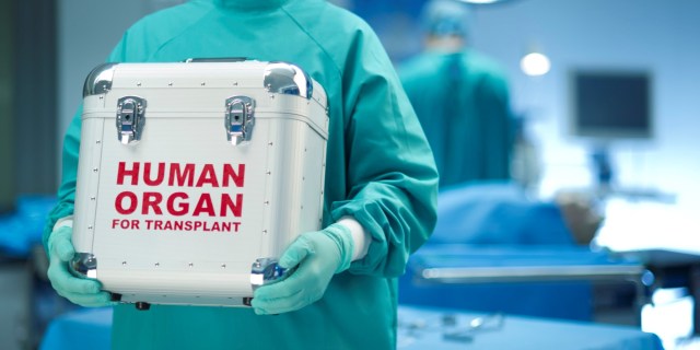
At this moment, there are 115,000 Americans who will die if they don’t get matched with a donated organ. Twenty of them die every day, according to data collected by the United Network for Organ Sharing (UNOS).

Part of the reason that waiting list is so long is because (surprise) organs don’t fare too well without a warm, gooey body keeping them safe. Both a liver and pancreas, for which the UNOS says 14,000 and 900 people respectively are currently waiting, can only be transplanted within twelve hours of donation. Another 4,000 people are waiting for a new heart, but those only last six hours outside the body before they begin to decay.

The 95,000 people waiting on a kidney donation get a little bit more wiggle room — those can last about 30 hours. But considering all of the logistical hurdles and difficulties of matching, donating, transporting, and, transplanting organs, recipients need to be ready for surgery more or less immediately once one is available.

The simplest way to get more organs to the people who need them, it would seem, is to freeze donated organs until they’re needed, like you might do with a casserole.

It’s such a simple idea that scientists and doctors came up with the idea decades ago, but they encountered two major roadblocks that seemed insurmountable — at least, until very recently.

The doctors, cryobiologists, engineers, and physicists at biotech company Arigos Biomedical may have come up with a way to freeze and store organs for as long as necessary. The company, previously funded by two of Peter Thiel’s science-focused foundations, recently raised just under $1 million in a seed round (participants included a venture firm, an angel investor and a family foundation, they note). Arigos co-founder and CEO Tanya Jones tells Futurism that they hope to begin human experiments as soon as 2020; if that goes well the technique could be used in clinics five to seven years after that.

If their technology works, it could shorten or even eliminate some of the organ transplant waitlists in America and around the world.

To understand the science that makes this possible, we need to look at why cryopreservation hasn’t worked in the past.

The first hurdle that cryobiologists need to overcome happens while the organ is on its way down to -120 degrees Celsius, the temperature at which molecular activity stops and organs can be stored indefinitely. People, you may recall, are mostly water, and when water freezes it expands into solid ice. This fact can cause problems as organs freeze, since congealing ice pulls water from nearby cells that need it and can rupture blood vessels as it expands. This is especially a problem since expansion and freezing happen at different times as organs freeze from the outside in.

The second hurdle comes during the warming process: just like an ice cube thrown into a glass of lukewarm water, organs tended to fracture and pop as they thawed. Not too helpful if you’re trying to replace a leaky lung or heart with a non-leaky one.

Scientists solved the first problem, Jones told Futurism, in the 1970s when they discovered a process called vitrification: pumping a cocktail of organic compounds into the organs pulled out most of the water. The remaining solution — water mixed with the added organic molecules — was so full of ­­stuff that it didn’t form ice. Instead, it froze into a type of solid glass that didn’t damage the organs the way ice did. As a result, organs could be frozen without worrying about harmful ice buildup.

But many solutions used in vitrification ended up being toxic. And the second problem, the fracturing issue, still eluded scientists. Over the following decades, most lost hope and gave up.

Jones says that she and Arigos cofounder Stephen Van Sickle found a new solution when Van Sickle took a trip to the library and realized how many old research papers chronicled the attempts to prevent fracturing.

“He went to a library and uncovered a line of old research in the transplant world that was abandoned,” says Jones.

They found a way to flush out all the arteries and veins of the organ and replace it with gas. In the past, scientists had tried replacing the blood and liquid found in a donated heart with oxygen gas, which gave the organs a little bit of extra support and padding as they grew rigid. The field then shifted towards liquids. These liquid perfusions tended to work a little bit better but left the realm of gas cushions as an unexplored loose end.

Jones and Van Sickle decided gas was worth a shot — helium gas in particular, because it wouldn’t be toxic to the donated organs. “You could use any inert noble gas because rule number one: absolutely no dying [it doesn’t kill organs], and rule number two: no blowing up the lab,” says Jones. In helium, they found a way to uniformly cool an organ and also provide its vasculature with some cushioning so that any stress built up during the cooling process wouldn’t cause a fracture. With helium gas in its veins and arteries, an organ is free to shift as it freezes without shattering, just as skyscrapers are engineered to sway rather than topple in the wind.

When they realized that no one else had tried helium, Arigos was born. And it worked really well. Soon their methods became the only way to successfully freeze and thaw organs from any animal larger than a rabbit.

“We have recovered pig kidneys from temperatures of -120 degrees Celsius, which is basically the glass transition temperature,” Jones says. “We tested on pig hearts, and it worked so well and so quickly that we were unexpectedly unprepared to test the recovery.” To be more prepared in the future, the team is working on automating its processes.

Ideally, the technique could create a universal bank of donated organs so that people could be matched with an organ right when they need it, rather than having to wait their turn in line until a suitable match turns up. Or, at least, it could make it much easier to get organs to the people who need them.

A bank of frozen organs could help solve some of the more banal (but very real) hurdles to transplantation. For example, donors and recipients sometimes have different immune systems. It’s more than just their blood types — before a transplant, doctors need to see how many of the six relevant antigens (molecules that can trigger the immune system into rejecting an organ) match up as well.

With a bank of frozen organs at the ready, doctors would be able to not only save more lives with organ transplants, but they could also more carefully match donated organs and recipients to prevent rejection because a better match could be readily available in the freezer.

But it needs FDA approval to make that happen. The company plans to conduct its first pig transplant in 2019, which it hopes will go well enough to convince the FDA to authorize a human trial in 2020. From there, it will take five to seven years for federal approval.

Others in the organ transplantation community are skeptical that Arigos can achieve everything that it claims to do, especially in the timeframe it set out.

“I would predict it would be longer than that, to actual human trials,” David Klassen, Chief Medical Officer at UNOS, tells Futurism. Normally there needs to be a great deal of animal experimentation before human testing is authorized, Klassen says, and these things take time. “That’s guessing on my part, but I would expect it would be a little longer than two years before human trials.”

But still, Klassen says that that the results coming out of organ freezing research are promising, and that he hopes to see continued interest in freezing organs.

“In broad strokes, I do think it is a top priority and should be. I think from the perspective of clinical transplantation, that whole area is a little underappreciated by people who work in the field day to day,” he says. But in the distant future, he also hopes to see the research community move beyond organ banking and develop ways to 3D print or construct organs from scratch, ultimately eliminating the need for donors in the first place.

But not everyone is convinced that cryopreservation is the future of organ transplantation at all, or that Arigos is the company to do it. Robert Kormos, director of the artificial heart program and co-director of the heart transplantation program at the University of Pittsburgh Medical Center, told Futurism that he’s skeptical of organ freezing technology.

“I went to [Arigos Biomedical’s] website and there’s a lot of claims of what they’re purporting to do and what they want to do, but usually what companies like this do is give you a list of publications so you can look at what’s been published in this area,” Kormos says. The lack of published research, completed by either by Arigos staff or other researchers in the field at large, raised a red flag for Kormos, who adds that he has not seen a lot of progress in organ freezing research, a field that he’s seen slow down over the decades.

“This company may have something, but again I’m eager to see what science they’re bringing to the table,” he says. “I think cryopreservation is an interesting concept, but we’re a ways away from that being reality. Again, I haven’t seen the data.”

Until their technique is approved, Arigos is working with some collaborators to improve the way scientists develop new pharmaceuticals. Right now, it’s difficult to bring new drugs to the market because many compounds that work great in animal studies don’t work the same way in humans. As a result, one in five phase I clinical trials, the first of three types of experiments conducted on humans before a drug can be approved, typically fails.

Arigos is planning to sell some slices of frozen, donated human organs to pharmaceutical researchers. That could help researchers determine whether or not a drug is toxic to people before they try it in a full-fledged experiment.

“We’ve got some collaborators set up who are going to explore that with us once we get access to human hearts, which we don’t quite yet,” says Jones.

But even if Arigos’ human trials are successful, the company won’t be able to store every type of organ that someone might need. Because their technique requires filling blood vessels with helium, it doesn’t work so well on organs that don’t have blood vessels, so they’ve only been able to freeze and store organs such as kidneys, livers, hearts, and lungs.

The military would love to be able to store entire frozen limbs, but that’s not on the table right now. If a soldier were to lose a limb after being caught in an explosion, it would be helpful to be able to freeze that limb until it’s ready to be reattached. That would give the soldier time to recover before undergoing surgery.

Freezing technology can’t help there yet. Bone tissue doesn’t take up the cryoprotectant solution well. And there are so many different tissue and cell types in a whole limb, each of which has a different tolerance level to the somewhat-toxic vitrification solution, that the technique wouldn’t really work. Arigos’ technique also wouldn’t work on transplantable corneas since they also have no vasculature, so scientists will need to develop new techniques if we want a universal bank of donated organs.

This also means, as Jones explained, that Arigos’ technology will not allow (presumably very rich) people to totally freeze their bodies and re-emerge in the distant future, as so many sci-fi stories have assured us will be possible. No, Arigos’ focus is strictly on medical uses for frozen organs.

If things go the way Arigos hopes, people worldwide could benefit. “There some countries in the world that don’t even have transplant technology,” says Jones. Aside from the immediate benefits to Americans stuck on an endless waitlist for the next kidney, frozen organs could be shipped or stored anywhere, bringing aid to countries and regions where there’s no waitlist at all.

https://futurism.com/freezing-donated-organs-arigos/

This mad new quantum experiment breaks the idea of ‘Before’ and ‘After,’ creating the ultimate chicken-and-egg situation.

time

by MIKE MCRAE

For around a century it’s been thought that particles don’t have defined properties until we nail them down with a measurement.

That kind of quantum madness opens up a whole world of counter-intuitive paradoxes. Take this one, for example – it’s possible for a single particle to experience two sequences of events at the same time, making it impossible to know which came first.

Physicists from the University of Queensland designed a race course for light that forced a single particle to traverse two pathways at once, making it impossible to say in which order it completed a pair of operations.

In boring old everyday life you could roll a single ball down a ramp and have it ring bell A and then ring bell B. Or, if you’d prefer, you could roll it down another ramp and have it ring B before A.

If you want to get fancy you could even set up a rig so one bell causes the other bell to ring.

None of this is mind blowing, since we’re used to events in the Universe having a set order, where one thing precedes another in such a way that we presume an order of causation.

But nothing is so simple when we accept that reality is a blur of possibility prior to it being measured.

To demonstrate this, the physicists created a physical equivalent of something called a quantum switch, where multiple operations occur while a particle is in a superposition of all its possible locations.

Keeping it simple, the team set up a pathway that split apart and converged again in an interferometer, with access to each fork dependent on the polarisation of the light entering it.

Light waves travelling down each fork in the pathway would then merge and interfere to create a distinctive pattern depending on its properties.

In this particular case, the two light waves were actually the same photon taking both paths at the same time.

Before being measured, a photon can be either vertically or horizontally polarised. Or, more precisely, it’s polarised both vertical and horizontal at the same time until a measurement confirms one over the other.

Since this undefined photon’s polarisation is both vertical and horizontal, it enters both pathways, with the vertically polarised version of the photon barrelling down one channel and the horizontally polarised version heading down the second.

Following the two paths, the team had the quantum equivalent of those bells we mentioned earlier – in the form of lenses that subtly changed the shape of the photon.

The horizontal polarisation would hit ‘bell’ A before striking B, while the vertical polarisation would strike ‘bell’ B, and then A.

An analysis of the interference pattern of the reunited photon revealed signs of this mess of possible sequences.

On one hand, it’s easy to imagine two separate light particles – one horizontally polarised, the other vertically polarised – passing each lens in separate orders.

That’s not what happened, though. This was a single photon with two possible histories, neither of which set in reality until they’re measured.

While the events A and B were independent in this quantum switch, they could be linked to affect one another. A could cause B, or B could cause A … all depending on which history you wanted after the event.

Putting aside daydreams of travelling back in time to undo that big mistake (what were you thinking?!), this does have one possible practical application in the emerging field of quantum communications.

Transmitting photons down a noisy channel could be disastrous for their quantum information, quickly making a mess of their precious superposition. Sending them down channels fitted with a quantum switch, however, could in principle give the quantum information an opportunity to get through.

A paper the team published on the pre-peer review website arxiv.org back in July shows how a quantum switch applied to two noisy channels can allow a superposition to survive.

Whatever weird clockwork is going on in reality’s basement, we won’t pretend to understand it. But the very fact physicists are able to craft it into new technology is truly mindblowing in itself.

This research was published in Physical Review Letters.

https://www.sciencealert.com/quantum-switch-causation-superposition-applied-technology

Machine-learning based model may identify dementia in primary care

machine-learning-model-provides-early-dementia-diagnosis-306451

A machine learning-based model using data routinely gathered in primary care identified patients with dementia in such settings, according to research recently published in BJGP Open.

“Improving dementia care through increased and timely diagnosis is a priority, yet almost half of those living with dementia do not receive a timely diagnosis,” Emmanuel A. Jammeh, PhD, of the science and engineering department at Plymouth University in the United Kingdom, and colleagues wrote.

“A cost-effective tool that can be used by [primary care providers] to identify patients likely to be living with dementia, based only on routine data would be extremely useful. Such a tool could be used to select high-risk patients who could be invited for targeted screening,” they added.

The researchers used Read codes, a set of clinical terms used in the U.K. to summarize data for general practice, to develop a machine learning-based model to identify patients with dementia. The Read codes were selected based on their significant association with patients with dementia, and included codes for risk factors, symptoms and behaviors that are collected in primary care. To test the model, researchers collected Read-encoded data from 26,483 patients living in England aged 65 years and older.

Jammeh and colleagues found that their machine-based model achieved a sensitivity of 84.47% and a specificity of 86.67% for identifying dementia.

“This is the first demonstration of a machine-learning approach to identifying dementia using routinely collected [National Health Service] data, researchers wrote.

“With the expected growth in dementia prevalence, the number of specialist memory clinics may be insufficient to meet the expected demand for diagnosis. Furthermore, although current ‘gold standards’ in dementia diagnosis may be effective, they involve the use of expensive neuroimaging (for example, positron emission tomography scans) and time-consuming neuropsychological assessments which is not ideal for routine screening of dementia,” they continued.

The model will be evaluated with other datasets, and have its validation tested “more extensively” at general practitioner practices in the future, Jammeh and colleagues added. – by Janel Miller

https://www.healio.com/family-medicine/geriatric-medicine/news/online/%7B62392171-6ad7-481a-9289-bd69df49d4a4%7D/machine-learning-based-model-may-identify-dementia-in-primary-care

Traumatic brain injury associated with higher rate of suicide

traumatic-brain-injuries-concussions

1. In this retrospective study, Traumatic Brain Injury (TBI) was associated with a doubling in deaths by suicide, especially within the 6 months post-TBI.

2. The risk of suicide was higher in those with more severe TBI, numerous medical contacts, and longer hospital stays.

Traumatic Brain Injury (TBI) is known to lead to a higher rate of psychiatric symptoms, especially in the early recovery period. While suicide risk has been found to be elevated in some studies, these studies vary widely in the estimated risk and have rarely accounted for important co-variates, such as previous psychiatric history. In this large, retrospective cohort studying including the entire Danish population, the risk of suicide after TBI was found to be twice as high than in patients without TBI, especially within the first 6-months post-TBI. Further, the risk of suicide was greater for those with more severe TBI, numerous medical contacts, and longer hospital stays. As added specificity of the association, the risk of suicide after TBI was found to be elevated compared to those with non-skull fractures. Those with a psychiatric history before TBI were found to be at higher risk of suicide compared to those with TBI alone.

The rigorous study design, exploration of multiple co-variates and sensitivity analyses, and large cohort strongly suggests that TBI leads to higher suicide risk. Future studies determining which specific features of TBI increase suicide risk, such as subsequent disinhibition or depression, will be helpful in determining how TBI and suicide are mechanistically linked.

In-Depth [retrospective cohort]: Using a number of databases covering all those living in Denmark during the study period (1980-2014), 7,418,390 individuals over the age of 10 were tracked for diagnosis of TBI and death by suicide. Covariates in the basic statistical model included sex, age, and calendar year, and a full statistical model included these plus marital status, cohabitation status, socioeconomic status, other injuries, epilepsy, long-term physical disease using the Charlson co-morbidity index, pre-TBI psychiatric disorders, and pre-TBI self-harm behavior. TBI was associated with a 2.64 Incident Rate Ratio (IRR) for suicide in the basic model (CI95 2.55 to 2.74) and 1.90 in the full model (CI95 1.83 to 1.97). Those with severe TBI had a higher rate of suicide than those with mild TBI (IRR 1.32; CI95 1.21 to 1.44). Those with 1 medical contact for TBI had an IRR of 1.75 (CI95 1.68 to 1.83), 2 medical contacts had an IRR of 2.31 (CI95 2.13 to 2.51), and 3 or more medical contacts had an IRR of 2.59 (CI95 2.35 to 2.85). Increasing days in treatment for TBI was associated with higher suicide risk (p < 0.001). The risk of suicide within the first 6 months of TBI was higher than after (IRR 2.10; CI95 1.89 to 2.34). Those with psychological illness after their TBI had higher rates of suicide compared to TBI-only (IRR 4.90; CI95 4.55 to 5.29) and those with psychiatric illness before TBI had higher risks of suicide compared to those with TBI and no psychiatric illness (IRR 2.32; CI95 2.10 to 2.55). Those with TBI had a higher risk of suicide compared to those with non-CNS fractures (IRR 1.73; CI95 1.66 to 1.81).

https://www.psychiatryadvisor.com/neurocognitive-disorders/traumatic-brain-injury-associated-with-higher-rate-of-suicide/article/788955/?utm_source=newsletter&utm_medium=email&utm_campaign=pa-update-dmd-20180825&cpn=psych_md%2cpsych_all&hmSubId=2yAHMYaJqF41&NID=1710903786

Did Scientists Actually Spot Evidence Of Another Universe?

universe
The detailed, all-sky picture of the infant universe created from nine years of WMAP data. The image reveals 13.77 billion year old temperature fluctuations (shown as color differences) that correspond to the seeds that grew to become the galaxies. The signal from our galaxy was subtracted using the multi-frequency data. This image shows a temperature range of ± 200 microKelvin.CREDIT: NASA/WMAP SCIENCE TEAM

by Jesse Shanahan

In a study published earlier this month, a team of theoretical physicists is claiming to have discovered the remnants of previous universes hidden within the leftover radiation from the Big Bang. Our universe is a vast collection of observable matter, like gas, dust, stars, etc., in addition to the ever-elusive dark matter and dark energy. In some sense, this universe is all we know, and even then, we can only directly study about 5% of it, leaving 95% a mystery that scientists are actively working to solve. However, this group of physicists is arguing that our universe isn’t alone; it’s just one in a long line of universes that are born, grow, and die. Among these scientists is mathematical physicist Roger Penrose, who worked closely with Stephen Hawking and currently is the Emeritus Rouse Ball Professor of Mathematics at Oxford University. Penrose and his collaborators follow a cosmological theory called conformal cyclic cosmology (CCC) in which universes, much like human beings, come into existence, expand, and then perish.

As a universe ages, it expands, and the constituent parts grow farther and farther apart from each other. Consequently, the interactions between galaxies that drive star formation and evolution become rarer. Eventually, the stars die out, and the remaining gas and dust is captured by black holes. In one of his most famous theories, Stephen Hawking proposed that this isn’t the end; black holes might have a way to slowly lose mass and energy by radiating certain particles. So, after many eons, the remaining black holes in the universe would disappear, leaving only disparate particles. Seemingly a wasteland, this end-state eventually mirrors the environment of our universe’s birth, and so, the cycle starts anew.

universe 2
Artist’s logarithmic scale conception of the observable universe with the Solar System at the center, inner and outer planets, Kuiper belt, Oort cloud, Alpha Centauri, Perseus Arm, Milky Way galaxy, Andromeda galaxy, nearby galaxies, Cosmic Web, Cosmic microwave radiation and Big Bang’s invisible plasma on the edge.CREDIT: WIKIPEDIA/PABLO CARLOS BUDASSI

When our universe was very young, before any recognizable components like stars, planets, or galaxies formed, it was filled with a dense, hot soup of plasma. As the universe expanded, it cooled, and eventually, particles could combine to form atoms. Eventually, the interaction and fusion of these atoms resulted in all of the matter that we observe today. However, we can still observe the leftover radiation from that initial, dense period in our universe’s history. This leftover glow, called the Cosmic Microwave Background (CMB), is the oldest electromagnetic radiation, and it fills the entirety of our universe. If the CCC theory were true, then there would be hints of previous universes in our universe’s CMB.

At the end of a universe, when those final black holes dissolve, CCC theory states they should leave behind a signature that would survive the death of that universe and persist into the next. Although not definitive proof of previous universes, detecting that signature would be strong evidence in support of CCC theory. In searching for these “Hawking points”, cosmologists face a difficult obstacle as the CMB is faint and varies randomly. However, Penrose is claiming that a comparison between a model CMB with Hawking points and actual data from our CMB has proven that Hawking points actually exist. If true, this would be the first-ever detection of evidence from another universe.

Unfortunately, as groundbreaking as this discovery seems, the scientific community has largely dismissed it. One of the fundamental characteristics of the CMB is that, although it has patterns, the variations are entirely statistically random. In fact, Penrose’s former collaborator, Stephen Hawking, spotted his own initials in the CMB while others have found a deer, a parrot, and numerous other recognizable shapes in the noise. Similarly, the Wilkinson Anisotropy Microscope Probe that mapped the CMB released an interactive image where you can search for familiar shapes and patterns. An avoidable result of both these random fluctuations and the sheer size of the CMB is that if scientists look hard enough, they can find whatever pattern they need, like the existence of Hawking points, perhaps. Another criticism of Penrose’s claim is that if CCC theory holds true, our universe should have tens of thousands of Hawking points in the CMB. Regrettably, Penrose could find only about 20.

Still, the possibility of alternate universes, whether long-dead or existing in parallel to our own, is tantalizing. Many other theories also claim to find traces of other universes hiding in the patterns of the CMB as well. Although it sounds like science fiction, we are left to wonder: is this just the cosmological equivalent of seeing shapes in random clouds or will scientists one day discover that we are one among many infinite universes?

Jesse Shanahan is an astrophysicist, EMT, and science communicator. For more space and language news, follow her on Twitter here.

https://www.forbes.com/sites/jesseshanahan/2018/08/24/did-scientists-actually-spot-evidence-of-another-universe/#2278663f1425