Antarctic Research Center Tries to Mimic Mars Conditions on Earth

Mars exists on Earth…well, at least the closest thing to Mars.

According to CNN, the Concordia research station in Antarctica sits on a plateau that is 3,200 meters above sea level and for about four months every year it is engulfed in complete darkness.

Those who live in the research station live in complete isolation. In fact, CNN reports that the nearest human beings from the station can be found about 372 miles away, making the place more remote than the International Space Station.

And yet, 16 dedicated scientists call the research center home for an entire year.

This is because long time confinement, abnormal day and light cycles, extremely dry air, low oxygen levels, and limited supplies make Mars-like training possible at the research center.

And it will help people get ready for the human race’s eventual voyage to Mars.

“By watching how the human body and mind adapts in Antarctica, we can plan and predict what would happen in space,” Alex Kumar, a doctor with the National Institute for Health Research, told CNN.

http://www.ryot.org/antarctic-research-center-tries-to-mimic-mars-conditions-on-earth/947267

Scientists claim to have determined what Jesus looked like

British scientist Richard Neave used forensic facial reconstruction to reveal what he believes to be a true depiction of the face of Jesus Christ. While the image should not be taken as a definitive model of Jesus, it is a historically accurate representation of how a man born in Jesus’s time and place would have looked.

Of the hundreds of thousands of words written in the Bible, not one gives an accurate description of how Christ looked. And without any firsthand descriptions of Jesus’s appearance to work with either, we’ve long been forced to rely on various artists’ personal interpretations — hence the lean, long-haired Christ so many of us have become familiar with. However, experts have emphasized this depiction is an entirely inaccurate representation of what a man from Jesus’s era may have looked like.

Based on historical records, Jesus Christ was from Galilee, a northern region in what is now modern day Israel. In order to get a better picture of Jesus’s face, Neave, a medical artist who retired from the University of Manchester in England, analyzed three skulls of Galilean Semites from Christ’s era, Popular Mechanics reported. Although the actual image was created three years ago, the picture has recently recirculated the Internet. No doubt, just in time for Christmas.

Neave used the skulls to create a computerized map of the facial structures each man once had. These images were used to create a 3D cast of a typical Galilean skull from the era. Then, using specialized computer programs, Neave recreated muscles and skin out of clay to match the thickness of human facial tissue and to cover the casts of the skull. Once Neave applied simulated skin, a nose, lips, and eyelids to the model, the face began to take shape.

The image’s hair style and coloring was based off drawings from various archeological sites dating back to Christ’s time period. In addition, the image was given a beard, since having one was a popular Jewish tradition at the time. Perhaps one of the most conflicting features of the new image, however, is his hair.

Most images of Christ portray a man with long straight hair. However, not only was this hairstyle uncommon among men at the time, it was even described in the Bible by the apostle Paul as being disrespectful — it’s highly unlikely Paul, being such a devoted follower, would say this about Christ. Most scholars, according to Popular Mechanics, therefore believe Christ to have had short, tight curls instead.

Gone are the lean features of the classic Christ. Instead the image is more muscular and weather-beaten, traits that Neave believes are more fitting of a Jewish carpenter from the first century. What’s more, Neave also suggested Christ was about 5-foot-1 and 110 pounds — the size of the average man from the time period.

While we will likely never know exactly how Christ looked, other scholars agree Neave’s depiction is more historically accurate than those found in Christian children’s books.

http://www.medicaldaily.com/was-jesus-white-forensic-facial-reconstruction-allegedly-shows-what-jesus-really-365668?rel=most_read4

Cancelled wedding converted into large feast for Sacramento homeless

After a California couple called off their wedding, the bride-to-be’s family decided to turn the $35,000 extravagant event into a feast for the homeless.

The bride’s mother, Kari Duane, said Sunday that rather than cancel the reception, they invited Sacramento’s homeless for a once in a lifetime meal Saturday at the Citizen Hotel, one of the city’s finest venues.

Duane said her 27-year-old daughter called her Monday to tell her she and her fiance had decided not go through with the wedding. Soon after, the family decided to share the nonrefundable event with the less fortunate.

“Even though my husband and I were feeling very sad for our daughter, it was heartwarming to see so many people be there and enjoy a meal,” Duane said.

She said they had already paid for a reception that would have hosted 120 guests. About 90 homeless single people, grandparents and whole families with newborns showed up and enjoyed a meal that included appetizers, salad, gnocchi, salmon, and even tri-tip. Some even dressed up for the occasion.

Erika Craycraft arrived with her husband and five children.

“To lose out on something so important to yourself and then give it to someone else is really giving, really kind,” Craycraft told KCRA-TV.

Part of the wedding price tag includes a nonrefundable honeymoon, so on Sunday mother and daughter set off for Belize.

“I hope that when she looks back at this, she knows she was doing something good with a bad situation,” Duane said.

New research finds that evidence of autism shows up in the brain’s blood vessels

by BEC CREW

Evidence of autism can be identified in the composition of blood vessels in the brain, and certain defects or malfunctions in these vessels could serve as a new basis for detection, scientists have found.

While previous research has focussed on the neurological structure and function in a patient’s brain, a team from New York University (NYU) has found evidence of the disorder in the vascular system, suggesting that this could be a new target for medical treatments.

“Our findings show that those afflicted with autism have unstable blood vessels, disrupting proper delivery of blood to the brain,” says lead researcher, Efrain Azmitia.

“In a typical brain, blood vessels are stable, thereby ensuring a stable distribution of blood,” she adds. “Whereas in the autism brain, the cellular structure of blood vessels continually fluctuates, which results in circulation that is fluctuating and, ultimately, neurologically limiting.”

Azmita and her colleagues figured this out by examining the auditory cortex region in human postmortem brain tissue from people with diagnosed autism spectrum disorder (ADS) and an age-matched control group. To mitigate bias, they stripped the samples of all identifiers so they couldn’t tell which was which when examining them at a cellular level.

They found significant increases of two types of protein, called nestin and CD34, in the autistic brain vessels, but not in the control brains, which indicated that the vessels of the autistic patients had a higher level of plasticity. This protein surge was identified in several sections of the autistic brains, including the superior temporal cortex, the fusiform cortex (or face recognition centre), the pons/midbrain, and cerebellum.

This kind of plasticity is characteristic of a process known as angiogenesis, which controls the the production of new blood vessels. Publishing in the Journal of Autism and Developmental Disorders, the researchers suggest that evidence of angiogenesis in autistic brain tissue indicates that these vessels are being formed over and over and are in a state of constant flux. This could mean that inside the brains of people with autism, there’s a significant level of instability in the blood’s delivery mechanism.

“We found that angiogenesis is correlated with more neurogenesis in other brain diseases, therefore there is the possibility that a change in brain vasculature in autism means a change in cell proliferation or maturation, or survival, and brain plasticity in general,” said one of the team, psychiatrist Maura Boldrini. “These changes could potentially affect brain networks.”

So what now? The researchers hope to continue their investigation into how blood vessels in the brain differ in people with and without ADS, and if they can confirm angiogenesis markers as a reliable indication of the disorder, they could have a new detection method on their hands, and perhaps even a new avenue of research for future treatments.

“It’s clear that there are changes in brain vascularisation in autistic individuals from two to 20 years that are not seen in normally developing individuals past the age of two years,” says Azmitia. “Now that we know this, we have new ways of looking at this disorder and, hopefully with this new knowledge, novel and more effective ways to address it.”

http://www.sciencealert.com/evidence-of-autism-can-be-found-in-the-brain-s-blood-vessels-study-finds

Science myths that will not die


False beliefs and wishful thinking about the human experience are common. They are hurting people — and holding back science.

Megan Scudellari

In 1997, physicians in southwest Korea began to offer ultrasound screening for early detection of thyroid cancer. News of the programme spread, and soon physicians around the region began to offer the service. Eventually it went nationwide, piggybacking on a government initiative to screen for other cancers. Hundreds of thousands took the test for just US$30–50.

Across the country, detection of thyroid cancer soared, from 5 cases per 100,000 people in 1999 to 70 per 100,000 in 2011. Two-thirds of those diagnosed had their thyroid glands removed and were placed on lifelong drug regimens, both of which carry risks.

Such a costly and extensive public-health programme might be expected to save lives. But this one did not. Thyroid cancer is now the most common type of cancer diagnosed in South Korea, but the number of people who die from it has remained exactly the same — about 1 per 100,000. Even when some physicians in Korea realized this, and suggested that thyroid screening be stopped in 2014, the Korean Thyroid Association, a professional society of endocrinologists and thyroid surgeons, argued that screening and treatment were basic human rights.

In Korea, as elsewhere, the idea that the early detection of any cancer saves lives had become an unshakeable belief.

This blind faith in cancer screening is an example of how ideas about human biology and behaviour can persist among people — including scientists — even though the scientific evidence shows the concepts to be false. “Scientists think they’re too objective to believe in something as folklore-ish as a myth,” says Nicholas Spitzer, director of the Kavli Institute for Brain and Mind at the University of California, San Diego. Yet they do.

These myths often blossom from a seed of a fact — early detection does save lives for some cancers — and thrive on human desires or anxieties, such as a fear of death. But they can do harm by, for instance, driving people to pursue unnecessary treatment or spend money on unproven products. They can also derail or forestall promising research by distracting scientists or monopolizing funding. And dispelling them is tricky.

Scientists should work to discredit myths, but they also have a responsibility to try to prevent new ones from arising, says Paul Howard-Jones, who studies neuroscience and education at the University of Bristol, UK. “We need to look deeper to understand how they come about in the first place and why they’re so prevalent and persistent.”

Some dangerous myths get plenty of air time: vaccines cause autism, HIV doesn’t cause AIDS. But many others swirl about, too, harming people, sucking up money, muddying the scientific enterprise — or simply getting on scientists’ nerves. Here, Nature looks at the origins and repercussions of five myths that refuse to die.

Myth 1: Screening saves lives for all types of cancer

Regular screening might be beneficial for some groups at risk of certain cancers, such as lung, cervical and colon, but this isn’t the case for all tests. Still, some patients and clinicians defend the ineffective ones fiercely.

The belief that early detection saves lives originated in the early twentieth century, when doctors realized that they got the best outcomes when tumours were identified and treated just after the onset of symptoms. The next logical leap was to assume that the earlier a tumour was found, the better the chance of survival. “We’ve all been taught, since we were at our mother’s knee, the way to deal with cancer is to find it early and cut it out,” says Otis Brawley, chief medical officer for the American Cancer Society.

But evidence from large randomized trials for cancers such as thyroid, prostate and breast has shown that early screening is not the lifesaver it is often advertised as. For example, a Cochrane review of five randomized controlled clinical trials totalling 341,342 participants found that screening did not significantly decrease deaths due to prostate cancer1.

“People seem to imagine the mere fact that you found a cancer so-called early must be a benefit. But that isn’t so at all,” says Anthony Miller at the University of Toronto in Canada. Miller headed the Canadian National Breast Screening Study, a 25-year study of 89,835 women aged 40–59 years old2 that found that annual mammograms did not reduce mortality from breast cancer. That’s because some tumours will lead to death irrespective of when they are detected and treated. Meanwhile, aggressive early screening has a slew of negative health effects. Many cancers grow slowly and will do no harm if left alone, so people end up having unnecessary thyroidectomies, mastectomies and prostatectomies. So on a population level, the benefits (lives saved) do not outweigh the risks (lives lost or interrupted by unnecessary treatment).

Still, individuals who have had a cancer detected and then removed are likely to feel that their life was saved, and these personal experiences help to keep the misconception alive. And oncologists routinely debate what ages and other risk factors would benefit from regular screening.

Focusing so much attention on the current screening tests comes at a cost for cancer research, says Brawley. “In breast cancer, we’ve spent so much time arguing about age 40 versus age 50 and not about the fact that we need a better test,” such as one that could detect fast-growing rather than slow-growing tumours. And existing diagnostics should be rigorously tested to prove that they actually save lives, says epidemiologist John Ioannidis of the Stanford Prevention Research Center in California, who this year reported that very few screening tests for 19 major diseases actually reduced mortality3.

Changing behaviours will be tough. Gilbert Welch at the Dartmouth Institute for Health Policy and Clinical Practice in Lebanon, New Hampshire, says that individuals would rather be told to get a quick test every few years than be told to eat well and exercise to prevent cancer. “Screening has become an easy way for both doctor and patient to think they are doing something good for their health, but their risk of cancer hasn’t changed at all.”

Myth 2: Antioxidants are good and free radicals are bad

In December 1945, chemist Denham Harman’s wife suggested that he read an article in Ladies’ Home Journal entitled ‘Tomorrow You May Be Younger’. It sparked his interest in ageing, and years later, as a research associate at the University of California, Berkeley, Harman had a thought “out of the blue”, as he later recalled. Ageing, he proposed, is caused by free radicals, reactive molecules that build up in the body as by-products of metabolism and lead to cellular damage.

Scientists rallied around the free-radical theory of ageing, including the corollary that antioxidants, molecules that neutralize free radicals, are good for human health. By the 1990s, many people were taking antioxidant supplements, such as vitamin C and β-carotene. It is “one of the few scientific theories to have reached the public: gravity, relativity and that free radicals cause ageing, so one needs to have antioxidants”, says Siegfried Hekimi, a biologist at McGill University in Montreal, Canada.

Yet in the early 2000s, scientists trying to build on the theory encountered bewildering results: mice genetically engineered to overproduce free radicals lived just as long as normal mice4, and those engineered to overproduce antioxidants didn’t live any longer than normal5. It was the first of an onslaught of negative data, which initially proved difficult to publish. The free-radical theory “was like some sort of creature we were trying to kill. We kept firing bullets into it, and it just wouldn’t die,” says David Gems at University College London, who started to publish his own negative results in 2003 (ref. 6). Then, one study in humans7 showed that antioxidant supplements prevent the health-promoting effects of exercise, and another associated them with higher mortality8.

None of those results has slowed the global antioxidant market, which ranges from food and beverages to livestock feed additives. It is projected to grow from US$2.1 billion in 2013 to $3.1 billion in 2020. “It’s a massive racket,” says Gems. “The reason the notion of oxidation and ageing hangs around is because it is perpetuated by people making money out of it.”

Today, most researchers working on ageing agree that free radicals can cause cellular damage, but that this seems to be a normal part of the body’s reaction to stress. Still, the field has wasted time and resources as a result. And the idea still holds back publications on possible benefits of free radicals, says Michael Ristow, a metabolism researcher at the Swiss Federal Institute of Technology in Zurich, Switzerland. “There is a significant body of evidence sitting in drawers and hard drives that supports this concept, but people aren’t putting it out,” he says. “It’s still a major problem.”

Some researchers also question the broader assumption that molecular damage of any kind causes ageing. “There’s a question mark about whether really the whole thing should be chucked out,” says Gems. The trouble, he says, is that “people don’t know where to go now”.

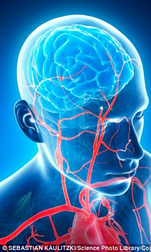
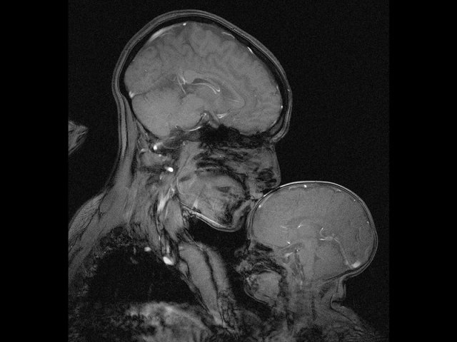
Myth 3: Humans have exceptionally large brains

The human brain — with its remarkable cognition — is often considered to be the pinnacle of brain evolution. That dominance is often attributed to the brain’s exceptionally large size in comparison to the body, as well as its density of neurons and supporting cells, called glia.

None of that, however, is true. “We cherry-pick the numbers that put us on top,” says Lori Marino, a neuroscientist at Emory University in Atlanta, Georgia. Human brains are about seven times larger than one might expect relative to similarly sized animals. But mice and dolphins have about the same proportions, and some birds have a larger ratio.

“Human brains respect the rules of scaling. We have a scaled-up primate brain,” says Chet Sherwood, a biological anthropologist at George Washington University in Washington DC. Even cell counts have been inflated: articles, reviews and textbooks often state that the human brain has 100 billion neurons. More accurate measures suggest that the number is closer to 86 billion. That may sound like a rounding error, but 14 billion neurons is roughly the equivalent of two macaque brains.

Human brains are different from those of other primates in other ways: Homo sapiens evolved an expanded cerebral cortex — the part of the brain involved in functions such as thought and language — and unique changes in neural structure and function in other areas of the brain.

The myth that our brains are unique because of an exceptional number of neurons has done a disservice to neuroscience because other possible differences are rarely investigated, says Sherwood, pointing to the examples of energy metabolism, rates of brain-cell development and long-range connectivity of neurons. “These are all places where you can find human differences, and they seem to be relatively unconnected to total numbers of neurons,” he says.

The field is starting to explore these topics. Projects such as the US National Institutes of Health’s Human Connectome Project and the Swiss Federal Institute of Technology in Lausanne’s Blue Brain Project are now working to understand brain function through wiring patterns rather than size.

Myth 4: Individuals learn best when taught in their preferred learning style

People attribute other mythical qualities to their unexceptionally large brains. One such myth is that individuals learn best when they are taught in the way they prefer to learn. A verbal learner, for example, supposedly learns best through oral instructions, whereas a visual learner absorbs information most effectively through graphics and other diagrams.

There are two truths at the core of this myth: many people have a preference for how they receive information, and evidence suggests that teachers achieve the best educational outcomes when they present information in multiple sensory modes. Couple that with people’s desire to learn and be considered unique, and conditions are ripe for myth-making.

“Learning styles has got it all going for it: a seed of fact, emotional biases and wishful thinking,” says Howard-Jones. Yet just like sugar, pornography and television, “what you prefer is not always good for you or right for you,” says Paul Kirschner, an educational psychologist at the Open University of the Netherlands.

In 2008, four cognitive neuroscientists reviewed the scientific evidence for and against learning styles. Only a few studies had rigorously put the ideas to the test and most of those that did showed that teaching in a person’s preferred style had no beneficial effect on his or her learning. “The contrast between the enormous popularity of the learning-styles approach within education and the lack of credible evidence for its utility is, in our opinion, striking and disturbing,” the authors of one study wrote9.

That hasn’t stopped a lucrative industry from pumping out books and tests for some 71 proposed learning styles. Scientists, too, perpetuate the myth, citing learning styles in more than 360 papers during the past 5 years. “There are groups of researchers who still adhere to the idea, especially folks who developed questionnaires and surveys for categorizing people. They have a strong vested interest,” says Richard Mayer, an educational psychologist at the University of California, Santa Barbara.

In the past few decades, research into educational techniques has started to show that there are interventions that do improve learning, including getting students to summarize or explain concepts to themselves. And it seems almost all individuals, barring those with learning disabilities, learn best from a mixture of words and graphics, rather than either alone.

Yet the learning-styles myth makes it difficult to get these evidence-backed concepts into classrooms. When Howard-Jones speaks to teachers to dispel the learning-styles myth, for example, they often don’t like to hear what he has to say. “They have disillusioned faces. Teachers invested hope, time and effort in these ideas,” he says. “After that, they lose interest in the idea that science can support learning and teaching.”

Myth 5: The human population is growing exponentially (and we’re doomed)

Fears about overpopulation began with Reverend Thomas Malthus in 1798, who predicted that unchecked exponential population growth would lead to famine and poverty.

But the human population has not and is not growing exponentially and is unlikely to do so, says Joel Cohen, a populations researcher at the Rockefeller University in New York City. The world’s population is now growing at just half the rate it was before 1965. Today there are an estimated 7.3 billion people, and that is projected to reach 9.7 billion by 2050. Yet beliefs that the rate of population growth will lead to some doomsday scenario have been continually perpetuated. Celebrated physicist Albert Bartlett, for example, gave more than 1,742 lectures on exponential human population growth and the dire consequences starting in 1969.

The world’s population also has enough to eat. According to the Food and Agriculture Organization of the United Nations, the rate of global food production outstrips the growth of the population. People grow enough calories in cereals alone to feed between 10 billion and 12 billion people. Yet hunger and malnutrition persist worldwide. This is because about 55% of the food grown is divided between feeding cattle, making fuel and other materials or going to waste, says Cohen. And what remains is not evenly distributed — the rich have plenty, the poor have little. Likewise, water is not scarce on a global scale, even though 1.2 billion people live in areas where it is.

“Overpopulation is really not overpopulation. It’s a question about poverty,” says Nicholas Eberstadt, a demographer at the American Enterprise Institute, a conservative think tank based in Washington DC. Yet instead of examining why poverty exists and how to sustainably support a growing population, he says, social scientists and biologists talk past each other, debating definitions and causes of overpopulation.

Cohen adds that “even people who know the facts use it as an excuse not to pay attention to the problems we have right now”, pointing to the example of economic systems that favour the wealthy.

Like others interviewed for this article, Cohen is less than optimistic about the chances of dispelling the idea of overpopulation and other ubiquitous myths (see ‘Myths that persist’), but he agrees that it is worthwhile to try to prevent future misconceptions. Many myths have emerged after one researcher extrapolated beyond the narrow conclusions of another’s work, as was the case for free radicals. That “interpretation creep”, as Spitzer calls it, can lead to misconceptions that are hard to excise. To prevent that, “we can make sure an extrapolation is justified, that we’re not going beyond the data”, suggests Spitzer. Beyond that, it comes down to communication, says Howard-Jones. Scientists need to be effective at communicating ideas and get away from simple, boiled-down messages.

Once a myth is here, it is often here to stay. Psychological studies suggest that the very act of attempting to dispel a myth leads to stronger attachment to it. In one experiment, exposure to pro-vaccination messages reduced parents’ intention to vaccinate their children in the United States. In another, correcting misleading claims from politicians increased false beliefs among those who already held them. “Myths are almost impossible to eradicate,” says Kirschner. “The more you disprove it, often the more hard core it becomes.”

http://www.nature.com/news/the-science-myths-that-will-not-die-1.19022

Nature 528, 322–325 (17 December 2015) doi:10.1038/528322a

1.Ilic, D., Neuberger, M. M., Djulbegovic, M. & Dahm, P. Cochrane Database Syst Rev. 1, CD004720 (2013).
2.Miller, A. B. et al. Br. Med. J. 348, g366 (2014).
3.Saquib, N., Saquib, J. & Ioannidis, J. P. A. Int. J. Epidemiol. 44, 264–277 (2015).
4.Doonan, R. et al. Genes Dev. 22, 3236–3241 (2008).
5.Pérez, V. I. et al. Aging Cell 8, 73–75 (2009).
6.Keaney, M. & Gems, D. Free Radic. Biol. Med. 34, 277–282 (2003).
7.Ristow, M. et al. Proc. Natl Acad. Sci. USA 106, 8665–8670 (2009).
8.Bjelakovic, G., Nikolova, D. & Gluud, C. J. Am. Med. Assoc. 310, 1178–1179 (2013).
9.Pashler, H., McDaniel, M., Rohrer, D. & Bjork, R. Psychol. Sci. Public Interest 9, 105–119 (2008).

Mother-child MRI

While most new moms get their children’s first portrait done at, say, the local mall’s JC Penney Portrait Studio, neuroscientist Rebecca Saxe opted for a slightly different location: the tube of an MRI scanner.

“No one, to my knowledge, had ever made an MR image of a mother and child,” she wrote in a article for Smithsonian magazine.

“We made this one because we wanted to see it.”

A Professor of Cognitive Neuroscience at the Massachusetts Institute of Technology, Saxe told Mic that the inspiration behind the photo had little to do with the typical medical or research-based uses of MRI technology.

“We see brain scan images on TV and in subways advertisements as a proxy for technology and progress… [and] the Madonna is one of the oldest tropes in human art making,” she said of trying to capture the union between science and art in the image.

“These brain scanners are extremely modern technology, only available here and now, to the wealthiest place and time in human history,” she added. “[Yet] the image you see would look the same if it had been made on any continent or in any century, because the biology of human mothers and children you see in the picture has been the same for thousands, probably tens of thousands of years.”

In an interview with Today, Saxe suggested that the image may be indicative of how a child’s brain development is strengthened by a mother’s love. “Some people look at it and see mostly the differences: how thin his skull is, how little space there is between the outside world and his brain. It’s just this very fragile, very thin little shell,” she said. “On the other hand, you can look at it and see how similar it is to his mother’s brain. How close in size — so much closer in size than his hand is.”

Past MRI scans have also suggested that the bond between a child and mother can indeed have a major impact on brain size. Back in 2012, a side-by-side image of two three-year-olds’ brain scans indicated that the size of a neglected child’s brain is significantly smaller than one who was nurtured by his or her mother. Of that particular image, neurology professor Allan Schore told the Telegraph that the development of brain cells is a “consequence of an infant’s interaction with the main caregiver [usually the mother].”

Meanwhile, Saxe believes that the image can also help generate an interest in science. “I hope the main takeaway is that people who don’t normally feel a human connection to science and scientists, have a moment to pause and feel touched, and recognize that the scientific pursuit of self-knowledge is being done for, and by, people like us,” she told Mic.

http://mic.com/articles/130456/this-brain-scan-image-illustrates-the-powerful-bond-between-mother-and-child#.tkqP2UYTE

Zombie nativity scene in Ohio

A “zombie nativity” that spurred complaints and zoning violation notices last year is on display again in a suburban Cincinnati yard, with a change to avoid fines.

Officials have concluded Jasen Dixon’s display complies with local zoning laws because he has removed its roof. Sycamore Township zoning administrator Harry Holbert says the issue was always about the structure and zoning rules, not the zombie figures.

The scene includes a sharp-toothed, grayish figure with black-ringed eyes sitting in the manger where the baby Jesus would be in traditional Christmas nativities

Dixon was threatened with fines last year when officials said the display violated rules on size and placement of yard structures. WCPO-TV (http://bit.ly/1Q06PxD ) reports he removed it before being cited and didn’t have to pay fines.

Women can navigate better when given testosterone, study finds

To investigate whether the differences in how men and women navigate are related to our sex or to cultural conditioning, researchers in Norway measured male and female brain activity while volunteers tried to find their way through a virtual reality maze.

Wearing 3D goggles and using a joystick to make their way through an artificial environment, the participants (18 males and 18 females) had their brain functions continuously recorded by an fMRI scanner as they carried out virtual navigation tasks.

In line with previous findings, the men performed better, using shortcuts, orienting themselves more using cardinal directions, and solving 50 percent more tasks than the women in the study.

“Men’s sense of direction was more effective,” said Carl Pintzka, a neuroscientist at the Norwegian University of Science and Technology (NTNU). “They quite simply got to their destination faster.”

One of the reasons for this is because of the difference in how men and women use their brains when we’re finding our way around. According to the researchers, men use the hippocampus more, whereas women place greater reliance on their brains’ frontal areas.

“That’s in sync with the fact that the hippocampus is necessary to make use of cardinal directions,” said Pintzka. “[M]en usually go in the general direction where [their destination is] located. Women usually orient themselves along a route to get there.”

Generally, the cardinal approach is more efficient, as it depends less on where you start.

But women’s brains make them better at finding objects locally, the researchers say. “In ancient times, men were hunters and women were gatherers. Therefore, our brains probably evolved differently,” said Pintzka. “In simple terms, women are faster at finding things in the house, and men are faster at finding the house.”

What was most remarkable about the study was what happened when the researchers gave women a drop of testosterone to see how it affected their ability to navigate the virtual maze. In a separate experiment, 21 women received a drop of testosterone under their tongues, while 21 got a placebo.

The researchers found that the women receiving testosterone showed improved knowledge of the layout of the maze, and relied on their hippocampus more to find their way around. Having said that, these hormone-derived benefits didn’t enable them to solve more maze tasks in the exercise.

It’s worth bearing in mind that the study used a fairly small sample size in both of the experiments carried out, so the findings need to be read in light of that. Nonetheless, the scientists believe their paper, which is published in Behavioural Brain Research, will help us to better understand the different ways male and female brains work, which could assist in the fight against diseases such as Alzheimer’s.

“Almost all brain-related diseases are different in men and women, either in the number of affected individuals or in severity,” said Pintzka. “Therefore, something is likely protecting or harming people of one sex. Since we know that twice as many women as men are diagnosed with Alzheimer’s disease, there might be something related to sex hormones that is harmful.”

http://www.sciencealert.com/women-can-navigate-better-when-given-testosterone-study-finds

Thanks to Dr. Enrique Leira for bringing this to the It’s Interesting community.

Migraine headaches in the NFL


Jets linebacker Lorenzo Mauldin (55) wears special contact lenses and a helmet shade to prevent migraines.

By ZACH SCHONBRUN

FWhen he woke last Sunday morning, Jeremy Kerley sensed trouble already coming on. Fitful sleep is often his trigger, he said. The migraine eventually hit him like an anvil late in last week’s game against the Giants.

His eyes grew blurry and he felt what he described as a “sharp, shooting, throbbing pain.” He wanted to sit down. He wanted to lie down. He knew he needed to leave the field.

Kerley, the Jets’ punt returner, departed to the locker room and did not return. As the Jets came from behind to beat the Giants in overtime, he was receiving intravenous fluids and oxygen to help relieve the anguish from a struggle that has afflicted him since high school.

For Kerley, migraines are the silent menace that constantly lurks. They ambush him almost once a month, even though he rarely talks about it. He knew his grandfather got them; only recently, he discovered that his dad did, too. He just never knows when they will affect him.

Though Kerley is one of approximately 38 million Americans who suffer from them, migraines are not something that is openly discussed in N.F.L. locker rooms. They are far more common in women, and often minimized as simply a headache, a stigma that Kerley acknowledged could make it difficult to pull himself out of a game.

But those who do struggle with migraines — which the Migraine Research Foundation considers a neurological disease, like epilepsy — understand the plight. When Kerley felt a severe headache coming on last season after a game at Minnesota, his teammate Percy Harvin patted him on the back.

“I know how you feel,” Harvin said quietly. He has struggled with migraines throughout his career.

Kerley did the same thing earlier this season, after linebacker Lorenzo Mauldin revealed that he had had migraines since adolescence. Kerley gave him recommendations about nutritional supplements that he found helpful, like fish oil and magnesium. Mauldin also now takes prescription medication to both relieve and prevent severe headaches.

He said that light could often trigger his migraine episodes, so Mauldin wears special contact lenses and a protective shade on his helmet.

“It hurts because it’s pulsating and you can’t really stop it,” Mauldin said. “With a bruise or something, you can put alcohol or peroxide over it and it’ll be fine. Or if you’ve hurt a muscle, you can ice it. But you can’t put ice over a migraine.”

In September, a migraine forced Ohio State quarterback Cardale Jones to the emergency room, something that is not uncommon, said Dr. Melissa Leber, the director of emergency department sports medicine at the Icahn School of Medicine at Mount Sinai. She typically treats patients intravenously. But that often cannot relieve the crippling symptoms right away.

“Some people can’t even get out of bed,” Leber said. “Others can function just while not feeling well. It really runs the gamut for how debilitating it can be.”

Migraines are thought to be related to the brain’s trigeminal nerve, which can grow hypersensitive and cause pain signals to fire throughout the brain, typically concentrated around the eyes or temples. Though migraines are strongly hereditary, showing up in people who have had no sports history, they are often clinically similar to post-traumatic headaches, like the headaches that arise after a concussion, according to Dr. Tad Seifert, a neurologist at Norton Healthcare in Louisville, Ky.

During the summer, Seifert led a study of 74 high school football players in the Louisville area and found that 33.8 percent of them suffered from migraines, a rate twice that of the normal population. The rate rose to 37.5 percent in players who reported having sustained a concussion once in their lives, and 40.7 percent in those who reported multiple concussions.

“The elephant in the room is whether there is some influence of contact sports and the development of frequent or chronic headache later in life,” Seifert said. “And if so, how much?”

Seifert, who also chairs an N.C.A.A. task force on headaches, said that he expected to publish a similar report involving 834 Division I athletes in the spring. Though he would not go into detail about the results, he said that it looked to be “very similar to what we’ve found in this sample of high school players.” Mauldin, it should be noted, sustained a concussion earlier this season.

There is no cure for migraines, and sufferers often go the rest of their lives “controlling” the issue, Seifert said, comparing it to those dealing with high blood pressure or diabetes. What concerns him, though, are the studies that have shown that people with migraines are more susceptible to concussions, and when they do sustain one, it takes them longer to recover.

“We know that the migraine brain is just wired differently,” Seifert said. “And we know that it’s a brain that is hypersensitive to external injury. And those pain receptors that are in overdrive — it takes that much longer to calm down and return to baseline.”

In the time it takes for the receptors to settle, though, the pain can bring a linebacker to his knees.

“When they pop up out of nowhere, you start to feel a sensation like in between the middle of your forehead,” Mauldin said. “But it’s in the back of your head as well. It’s like somebody’s punching you in the side of the head.”

Kerley said he had yet to receive a migraine disease diagnosis, but he thinks it could be related to difficulties he regularly has with sleeping, being someone who has sleep apnea. When he feels a headache coming on, he has a nasal spray that he said often cured his symptoms within a half-hour. But last Sunday, it was too late.

“If you don’t catch it while it’s early, it could get pretty bad,” Kerley said. “Mine got there.”

Former Space X vet seeks to disrupt the stiletto status quo with ‘comfort’ heels

By Carolyne Zinko

If humans have been wearing shoes for at least 40,000 years, and modern styles for the past century, does a new line of women’s high heels really require space-age engineering to make them more fit for the foot?

Silicon Valley native Dolly Singh thinks so.

The former employee of Elon Musk’s Space X is aiming to send shock waves through the $40 billion-a-year shoe industry with her line of Thesis Couture stilettos. The high-performance heels have been created by a team of designers, engineers, doctors — and a rocket scientist and astronaut, no less — and promise to be more comfortable than other high heels on the market.

Singh was motivated by walking the shiny white floors of Space X in Los Angeles in heels for five years and realizing she had two choices: “I could wear uglier shoes, or wear pretty shoes and wind up with ugly deformed feet.”

The shoes, which initially will feature a 4-inch heel, are three years in the making. They’ve been built around an entirely new foot mold — or last, in footwear industry parlance — rather than designed around existing lasts used in mass manufacturing.

“In most cases, people take the existing architecture — the shank, the sole as it is — and they’ll try to add some cushion,” Singh said during a recent San Francisco visit. “They’ll add air pockets, they’ll do this or that. And those are all evolutionary, small improvements, whereas what we wanted to do was a revolutionary improvement.”

Armed with the new last, her team has used structural engineering principles to redistribute the load on the ball and toes of the feet, and space age plastics and rubber for the shoe’s frame and heel. Combined, the advances will offer better shock absorption and arch support. Most other high heeled shoes, in contrast, contain metal rods for strength.

The load on the ball of the foot is reduced from 80 percent to 50 percent, and the shoe’s platform reduces shock by 50 percent, Singh said. Arch support is hidden inside the frame, and the heel is cleverly designed to be larger, to provide stability, without appearing visually clunky.

“From a physics standpoint, high heels as they’re designed today are not at all thoughtful,” Singh said. “High heels are probably the only product you can find where there’s $40 billion a year of commerce, and where for almost a century no innovation has taken place. That’s insane.”

The line is scheduled to launch in March, with a debut style produced in a limited edition of 1,500 pairs, each pair numbered and signed, in much the same way Musk launched his Tesla Model S series.

The debut edition will be priced at $925, with daytime looks to follow at $350 a pair, evening looks at $600 or so, and red carpet styles at about $1,000 a pair.

In January, Thesis Couture will conduct trunk shows with a prototype in New York, Los Angeles and San Francisco at which customers can try on the shoes and place orders for the debut collection. Trunk show dates have not been finalized.

The name Thesis Couture, by the way, derives from her belief that her thesis about the new shoe design is a defensible theory. Take that, Stone Age foot wraps.

http://www.sfchronicle.com/style/article/Former-Space-X-vet-seeks-to-disrupt-the-stiletto-6690389.php