Researchers find new way to stimulate the natural cellular recycling process, which may help treat patients with neurodegenerative disease.

Brown University researchers studying the biology of aging have demonstrated a new strategy for stimulating autophagy, the process by which cells rebuild themselves by recycling their own worn-out parts.

In a study published in the journal Cell Reports, the researchers show that the approach increased the lifespans of worms and flies, and experiments in human cells hint that the strategy could be useful in future treatments for Alzheimer’s disease, ALS and other age-related neurodegenerative conditions.

“Autophagy dysfunction is present across a range of age-related diseases including neurodegeneration,” said Louis Lapierre, an assistant professor of molecular biology, cell biology and biochemistry at Brown who led the work. “We and others think that by learning how to influence this process pharmacologically, we might be able to affect the progression of these diseases. What we’ve shown here is a new and conserved entry point for stimulating autophagy.”

Autophagy has become a hot topic in recent years, earning its discoverer the Nobel Prize in Physiology and Medicine in 2016. The process involves the rounding up of misfolded proteins and obsolete organelles within a cell into vesicles called autophagosomes. The autophagosomes then fuse with a lysosome, an enzyme-containing organelle that breaks down those cellular macromolecules and converts it into components the cell can re-use.

Lapierre and his colleagues wanted to see if they could increase autophagy by manipulating a transcription factor (a protein that turns gene expression on and off) that regulates autophagic activity. In order for the transcription factor to switch autophagic activity on, it needs to be localized in the nucleus of a cell. So Lapierre and his team screened for genes that enhance the level of the autophagy transcription factor, known as TFEB, within nuclei.

Using the nematode C. elegans, the screen found that reducing the expression of a protein called XPO1, which transports proteins out of the nucleus, leads to nuclear accumulation of the nematode version of TFEB. That accumulation was associated with an increase in markers of autophagy, including increased autophagosome, autolysosomes as well as increased lysosome biogenesis. There was also a marked increase in lifespan among the treated nematodes of between about 15 and 45 percent.

“What we showed was that by blocking the escape of this transcription factor from the nucleus, we could not only influence autophagy but we could get an increase in lifespan as well,” Lapierre said.

The next step was to see if there were drugs that could mimic the effect of the gene inhibition used in the screening experiment. The researchers found that selective inhibitors of nuclear export (SINE), originally developed to inhibit XPO1 to treat cancers, had a similar effect — increasing markers of autophagy and significantly increasing lifespan in nematodes.

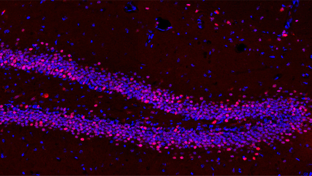
The researchers then tested SINE on a genetically modified fruit fly that serves as a model organism for the neurodegenerative disease ALS. Those experiments showed a small but significant increase in the lifespans of the treated flies. “Our data suggests that these compounds can alleviate some of the neurodegeneration in these flies,” Lapierre said.

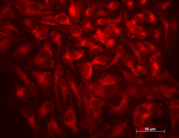
As a final step, the researchers set out to see if XPO1 inhibition had similar effects on autophagy in human cells as it had in the nematodes. After treating a culture of human HeLa cells with SINE, the researchers found that, indeed, TFEB concentrations in nuclei increased, as did markers of autophagic activity and lysosomal biogenesis.

“Our study tells us that the regulation of the intracellular partitioning of TFEB is conserved from nematodes to humans and that SINE could stimulate autophagy in humans,” Lapierre said. “SINE have been recently shown in clinical trials for cancer to be tolerated, so the potential for using SINE to treat other age-related diseases is there.”

Future research, Lapierre said, will focus on testing these drugs in more clinically relevant models of neurodegenerative diseases. But this initial research is a proof of concept for this strategy as a means to increase autophagy and potentially treat age-related diseases.

Lapierre is a faculty member in the newly approved Center on the Biology of Aging within the Brown Institute for Translational Science. This center, led by Professor of Biology John Sedivy, studies the biological mechanisms of aging. The center’s mission is to expand biomedical research and education programs in the emerging discipline of biogerontology, and to bring forth scientific discoveries related to aging and associated disorders.

Drug target for curing the common cold

UK scientists believe they may have found a way to combat the common cold.

Rather than attacking the virus itself, which comes in hundreds of versions, the treatment targets the human host.

It blocks a key protein in the body’s cells that cold viruses normally hijack to self-replicate and spread.

This should stop any cold virus in its tracks if given early enough, lab studies suggest. Safety trials in people could start within two years.

The Imperial College London researchers are working on making a form of the drug that can be inhaled, to reduce the chance of side-effects.

In the lab, it worked within minutes of being applied to human lung cells, targeting a human protein called NMT, Nature Chemistry journal reports.

All strains of cold virus need this human protein to make new copies of themselves.

Researcher Prof Ed Tate said: “The idea is that we could give it to someone when they first become infected and it would stop the virus being able to replicate and spread.

“Even if the cold has taken hold, it still might help lessen the symptoms.

“This could be really helpful for people with health conditions like asthma, who can get quite ill when they catch a cold.”

He said targeting the host rather than the infection was “a bit radical” but made sense because the viral target was such a tricky one.

Cold viruses are not only plentiful and diverse, they also evolve rapidly, meaning they can quickly develop resistance to drugs.

The test drug completely blocked several strains of cold virus without appearing to harm the human cells in the lab. Further studies are needed to make sure it is not toxic in the body though.

Dr Peter Barlow of the British Society for Immunology said: “While this study was conducted entirely in vitro – using cells to model Rhinovirus infection in the laboratory – it shows great promise in terms of eventually developing a drug treatment to combat the effects of this virus in patients.”

Fighting a cold
Colds spread very easily from person to person. And the viruses that cause the infections can live on hands and surfaces for 24 hours.

Painkillers and cold remedies might help ease the symptoms. But currently there is nothing that will halt the infection.

You can catch a cold by:

– inhaling tiny droplets of fluid that contain the cold virus – these are launched into the air when an infected person coughs or sneezes
– touching an object or surface contaminated by infected droplets and then touching your mouth, nose or eyes
– touching the skin of someone who has the infected droplets on their skin and then touching your mouth, nose or eyes

Symptoms – a runny or blocked nose, sneezing and sore throat – usually come on quickly and peak after a couple of days. Most people will feel better after a week or so. But a mild cough can persist for a few weeks.

http://www.bbc.com/news/health-44107481

Deep brain stimulation may help children with Rett syndrome


The dentate gyrus of a mouse that received deep brain stimulation, with cell nuclei in blue and expression of the gene c-Fos in red.

By Shawna Williams

Even as patients with Parkinson’s disease, obsessive-compulsive disorder, and other conditions turn to deep brain stimulation (DBS) to keep their symptoms in check, it’s been unclear to scientists why the therapy works. Now, researchers in Texas report that in mice, the treatment dials the activity of hundreds of genes up or down in brain cells. Their results, published in eLife March 23, hint that DBS’s use could be expanded to include improving learning and memory in people with intellectual disabilities.

“The paper is very well done. . . . It’s really a rigorous study,” says Zhaolan “Joe” Zhou, a neuroscientist at the University of Pennsylvania’s Perelman School of Medicine who reviewed the paper for eLife. Now that the genes and pathways DBS affects are known, researchers can home in on ways to improve the treatment, or perhaps combine the therapy with pharmacological approaches to boost its effect, he says.

In DBS, two electrodes are surgically implanted in a patient’s brain (the area depends on the disorder being treated), and connected to generators that are placed in the chest. Gentle pulses of electricity are then passed continuously through the electrodes. The treatment reduces motor symptoms in many people with Parkinson’s, and allows some patients to reduce their use of medications, but it does not eliminate symptoms or slow the disease’s progression.

In addition to its use in movement disorders, DBS is being explored as a potential therapy for a range of other brain-related disorders. For instance, as a way to boost learning and memory in people with Alzheimer’s disease, researchers are looking into stimulating the fimbria-fornix, a brain region thought to regulate the activity of the memory-storing hippocampus.

Such studies made Huda Zoghbi, a neurogeneticist at Baylor College of Medicine, wonder what effect DBS might have on learning- and memory-related disorders that strike earlier in life. “We rationalized that maybe in Alzheimer’s, many of the neurons are already gone, but perhaps in a healthier brain, like that of a Rett syndrome model, we can test the idea if stimulation of the fornix can improve learning and memory,” she explains. Rett syndrome, a genetic disease that almost exclusively strikes girls, includes intellectual disability, autism-like deficits in social interactions, and a loss of motor function. Several years ago, Zoghbi and colleagues tried zapping the fimbria-fornix, a C-shape bundle of nerves adjacent to the hippocampus in the brain, in mouse models of Rett syndrome. Published in 2015, their results showed that after two weeks of daily, one-hour DBS sessions, the mice with an intellectual disability performed like their peers without the disorder on a range of hippocampus-dependent tasks.

“We were struck that everything became indistinguishable after deep brain stimulation from a baseline normal,” Zoghbi says. This prompted her team to ask, “How does it work at a molecular level?” The answer, she thought, could determine whether DBS of the fimbria-fornix has the potential to serve as a multipurpose tool, treating not just Rett syndrome but other childhood-onset intellectual disabilities with a variety of causes. “It’s going to be really tough, perhaps, to solve these diseases one gene at a time, so that learning can be corrected,” she says. “You could eventually consider an intervention that can be broadly applicable, irrespective of the molecular cause of the defect.”

For the latest study, the research team analyzed baseline differences in gene activity between mice with and without the Rett syndrome–like condition in a part of the hippocampus called the dentate gyrus. They also treated the mice with the intellectual disability once with 45 minutes of DBS. Of the many genes with marked differences in initial activity between the two groups of mice, one-quarter (39 genes) became normal in the Rett mice after treatment, they report.

Zoghbi’s group also tested the effects of DBS in normal mice; in addition to changing the activity levels of thousands of genes, the researchers found, the treatment prompted alternative splicing of the RNA copies of other genes, which would result in differences in the resulting proteins. Many of the genes affected by the alternative splicing are known to be involved in the growth of new neurons or in maintaining the synapses through which brain cells communicate. In the 2015 study, the group had found that DBS enhances some hippocampus-related abilities in wildtype mice, such as spatial learning.

For hints as to whether DBS might have the potential to treat intellectual disabilities other than Rett syndrome, the researchers compared their list of genes whose activity levels changed after DBS in normal mice with existing data on genes known to have abnormal expression levels in mouse models of several such disorders. As with Rett syndrome, DBS in wildtype mice altered the activity levels of about one-quarter of the genes involved in each of the disorders.

The fact that a short period of stimulation had such profound effects on gene expression is interesting, says Svjetlana Miocinovic, a movement-disorders neurologist at Emory University who was not involved in the study. Most research on the mechanism of DBS has focused on changes it induces in the electrical or physical properties of the brain, she tells The Scientist. “I think this kind of study, where they actually look at the molecular environment in these neurons that are exposed to stimulation . . . is really the way to figure out what exactly is going on and how is that neural plasticity accomplished.”

Now that they have a way to measure such molecular effects, Zoghbi and her collaborators plan to optimize DBS for models of intellectual disabilities—figuring out how long the current needs to be on, for example, and how often. Another question they’d like to address is whether stimulating other brain areas in addition to the fimbria-fornix could add to the benefits seen in the mice.

Zoghbi emphasizes that even if DBS turns out to be safe and effective for children with Rett syndrome, it won’t be a silver bullet, because patients will have missed out on some important developmental milestones. “To really get the full benefit,” she says, “we’re going to have to combine any intervention with intensive physical and behavioral therapy.”

A. Pohodish et al., “Forniceal deep brain stimulation induces gene expression and splicing changes that promote neurogenesis and plasticity,” eLife, doi:10.7554/eLife.34031, 2018.

https://www.the-scientist.com/?articles.view/articleNo/54581/title/Deep-Brain-Stimulation-Affects-the-Activity-of-Hundreds-of-Genes/&utm_campaign=TS_DAILY%20NEWSLETTER_2018&utm_source=hs_email&utm_medium=email&utm_content=62919749&_hsenc=p2ANqtz-_Wc4T-kVW-WjClgnxcSv-OaWmpr8r7DtQJQRRztFWWATkUzGmgHVi5RLo74kNAO-J2872OymH-_uX0WKTVquRF4v7QAw&_hsmi=62919749

Scientists transfer memory from one snail to another

By Ashley Yeager

Researchers have transferred a memory from one snail to another via RNA, they report today (May 14) in eNeuro. If confirmed in other species, the finding may lead to a shift in scientists’ thinking about how memories are made—rather than cemented in nerve-cell connections, they may be spurred on by RNA-induced epigenetic changes.

“The study suggests that RNA populations are the missing link in the search for memory,” Bridget Queenan, a neuroscientist at the University of California, Santa Barbara, who was not involved in the study, writes in an email to The Scientist. “If circulating neural RNAs can transfer behavioral states and tendencies, orchestrating both the transient feeling and the more permanent memory, it suggests that human memory—just like mood—will only be explained by exploring the interplay between bodies and brains.”

For decades, researchers have tried to pinpoint how, when, and where memories form. In the 1940s, Canadian psychologist Donald Hebb proposed memories are made in the connections between neurons, called synapses, and stored as those connections grow stronger and more abundant. Experiments in the 1960s, however, suggested RNA could play a role in making memories, though the work was largely written off as irreproducible.

Study coauthor David Glanzman of the University of California, Los Angeles, has been working on the cell biology of learning and memory for nearly 40 years, and says for the majority of that time he believed memory was stored at synapses. Several years ago, though, he and his colleagues began replicating memory-erasing research done in rodents in California sea hares (Aplysia californica), a type of marine snail also called a sea slug. The team found that the snail synapses built to “store” a memory weren’t necessarily the synapses that were removed from the neural circuits in the memory-erasing experiments.

“It was completely arbitrary which synaptic connections got erased,” Glanzman says. “That suggested maybe the memory wasn’t stored at the synapse but somewhere else.”

Glanzman turned his attention to RNA because of those earlier hints it was related to memory, and also because of recent experiments suggesting long-term memory was stored in the cell bodies of neurons, not synapses. He picked Aplysia because it has been a longtime model organism for memory studies. Like all mollusks, these snails have groups of neurons called ganglia, rather than brains. Their nervous systems comprise about 20,000 neurons, and the cells are some of the biggest and easily identifiable among nerve cells in all animals. In the snail’s gut, for example, are specific sensory and motor neurons that control the withdrawal of a fleshy, spout-like organ on the snail’s back called a siphon and the contraction of a caterpillar-looking gill, which the animal uses to breathe.

When touched lightly on the siphon, the neurons fire, retract the tissue, and contract the gill within the body cavity for a few seconds to protect it against attack. Sticking electrodes in the snail’s tail and shocking it makes this defensive response last longer, tens of seconds, and sometimes up to almost a minute. By repeatedly shocking the snail’s tail, the animal learns to stay in that defensive position when touched on the siphon, even weeks after the shocks end.

In his team’s latest experiments, Glanzman and his colleagues zapped snails’ tails, then pulled the abdominal neurons from the shocked snails, extracted their RNA, dissolved the RNA into deionized water, and injected the solution into the necks of snails that had never been shocked. (For a control, the team also took RNA from non-shocked snails and injected into naive snails.) When tapped on the siphon 24 hours later, snails that got RNA from shocked snails withdrew their siphon and gill for significantly longer (almost 40 seconds) than did snails that got RNA from non-shocked animals (less than 10 seconds).

DNA methylation appeared to be essential for the transfer of the memory among snails. When Glanzman and his colleagues blocked DNA methylation in snails getting RNA from shocked ones, the injected snails withdrew their siphons for only a few seconds when tapped on the siphon.

Glanzman wanted to know if the RNA from shocked snails actually affected the neuronal connections of the snails receiving the injections any differently than RNA from nonshocked snails. So, in a third test, he and his team removed sensory neurons from nonshocked snails, cultured the cells in a dish, and then exposed the cells to RNA from shocked snails. Zapping the culture with a bit of current excited the sensory neurons much more than neurons treated with RNA from nonshocked snails. RNA from shocked snails also enhanced a subset of synapses between sensory and motor neurons in vitro, suggesting it was indeed the RNA that transported the memory, Glanzman explains.

The idea “seems quite radical as we don’t have a specific mechanism for how it works in a non-synaptic manner,” Bong-Kiun Kaang, a neuroscientist at Seoul National University who was not involved in the study, writes in an email to The Scientist. Kaang notes there are “many critical questions that need to be addressed to further validate the author’s argument,” such as what kinds of noncoding RNAs are specifically involved, how are the RNAs transferred among neurons, and how much do RNAs at the synapse play a role? The experiments should also be replicated in organisms other than snails, he says.

Glanzman says that in his next experiments he will attempt to identify the RNAs involved, and he has an idea for the mechanism, too. The memory is not stored in the RNA itself, he speculates—instead, noncoding RNA produces epigenetic changes in the nucleus of neurons, thereby storing the memory.

“This idea is probably going to strike most of my colleagues as extremely improbable,” Glanzman says. “But if we’re right, we’re just at the beginning of understanding how memory works.”

A. Bédécarrats et al., “RNA from trained Aplysia can induce an epigenetic engram for long-term sensitization in untrained Aplysia,” eNeuro, doi.org/10.1523/ENEURO.0038-18.2018, 2018.

https://www.the-scientist.com/?articles.view/articleNo/54565/title/RNA-Moves-a-Memory-From-One-Snail-to-Another/

New peptide treatment for obese patients suffering from insatiable hunger

In a new study researchers from the Institute for Experimental Pediatric Endocrinology of the Charité – Universitätsmedizin Berlin have successfully treat patients whose obesity is caused by a genetic defect. Aside from its beneficial effects on the patients, the researchers also provided insights into the fundamental signaling pathways regulating satiety of the new drug. The results of this research have been published in Nature Medicine*.

A mutation in the gene encoding the leptin receptor (LEPR) can cause extreme hunger starting with the first months of life. As a result, affected individuals develop extreme obesity during childhood. Increased exercise and reduced caloric intake are usually insufficient to stabilize body-weight. In many cases, obesity surgery fails to deliver any benefits, meaning that a drug-based treatment approach becomes increasingly important.

Two years ago, Dr. Peter Kühnen and the working group successfully demonstrated that treatment with a peptide, which activates the melanocortin 4 receptor (MC4R) could play a central role in the body’s energy metabolism and body weight regulation. Leptin, which is also known as the satiety (or starvation) hormone, normally binds to the LEPR, triggering a series of steps that leads to the production of melanocyte-stimulating hormone (MSH). The act of MSH by binding to its receptor, the melanocortin 4 receptor (MC4R) which transduce the satiety signal to the body. However, if the LEPR is defective, the signaling cascade is interrupted. The patient’s hunger remains unabated, placing them at greater risk of becoming obese. As part of this current study, researchers used a peptide that binds to the MC4R in the brain, and this activation trigger the normal satiety signal. Working in cooperation with the Clinical Research Unit at the Berlin Institute of Health (BIH), the researchers were able to record significant weight loss in patients with genetic defects affecting the LEPR.

“We also wanted to determine why the used peptide was so effective and why, in contrast to other preparations with a similar mode of action, it did not produce any severe side effects,” explains Dr. Kühnen. “We were able to demonstrate that this treatment leads to the activation of a specific and important signaling pathway, whose significance had previously been underestimated.” Dr. Kühnen’s team is planning to conduct further research to determine whether other patients might benefit from this drug: “It is possible that other groups of patients with dysfunctions affecting the same signaling pathway might be suitable candidates for this treatment.”

*Clément K, et al., MC4R agonism promotes durable weight loss in patients with leptin receptor deficiency, Nature Medicine (2018), doi:10.1038/s41591-018-0015-9.

https://www.charite.de/en/service/press_reports/artikel/detail/den_unstillbaren_hunger_abschalten/

Scientists at Indiana University discover the first evidence that non-human animals can replay episodic memory in their heads, which could help find new treatments for patients with Alzheimer’s disease or normal memory loss with aging

Neuroscientists at Indiana University have reported the first evidence that non-human animals can mentally replay past events from memory. The discovery could help advance the development of new drugs to treat Alzheimer’s disease.

The study, led by IU professor Jonathon Crystal, appears today in the journal Current Biology.

“The reason we’re interested in animal memory isn’t only to understand animals, but rather to develop new models of memory that match up with the types of memory impaired in human diseases such as Alzheimer’s disease,” said Crystal, a professor in the IU Bloomington College of Arts and Sciences’ Department of Psychological and Brain Sciences and director of the IU Bloomington Program in Neuroscience.

Under the current paradigm, Crystal said most preclinical studies on potential new Alzheimer’s drugs examine how these compounds affect spatial memory, one of the easiest types of memory to assess in animals. But spatial memory is not the type of memory whose loss causes the most debilitating effects of Alzheimer’s disease.

“If your grandmother is suffering from Alzheimer’s, one of the most heartbreaking aspects of the disease is that she can’t remember what you told her about what’s happening in your life the last time you saw her,” said Danielle Panoz-Brown, an IU Ph.D. student who is the first author on the study. “We’re interested in episodic memory — and episodic memory replay — because it declines in Alzheimer’s disease, and in aging in general.”

Episodic memory is the ability to remember specific events. For example, if a person loses their car keys, they might try to recall every single step — or “episode” — in their trip from the car to their current location. The ability to replay these events in order is known as “episodic memory replay.” People wouldn’t be able to make sense of most scenarios if they couldn’t remember the order in which they occurred, Crystal said.

To assess animals’ ability to replay past events from memory, Crystal’s lab spent nearly a year working with 13 rats, which they trained to memorize a list of up to 12 different odors. The rats were placed inside an “arena” with different odors and rewarded when they identified the second-to-last odor or fourth-to-last odor in the list.

The team changed the number of odors in the list before each test to confirm the odors were identified based upon their position in the list, not by scent alone, proving the animals were relying on their ability to recall the whole list in order. Arenas with different patterns were used to communicate to the rats which of the two options was sought.

After their training, Crystal said, the animals successfully completed their task about 87 percent of the time across all trials. The results are strong evidence the animals were employing episodic memory replay.

Additional experiments confirmed the rats’ memories were long-lasting and resistant to “interference” from other memories, both hallmarks of episodic memory. They also ran tests that temporarily suppressed activity in the hippocampus — the site of episodic memory — to confirm the rats were using this part of their brain to perform their tasks.

Crystal said the need to find reliable ways to test episodic memory replay in rats is urgent since new genetic tools are enabling scientists to create rats with neurological conditions similar to Alzheimer’s disease. Until recently, only mice were available with the genetic modifications needed to study the effect of new drugs on these symptoms.

“We’re really trying push the boundaries of animal models of memory to something that’s increasingly similar to how these memories work in people,” he said. “If we want to eliminate Alzheimer’s disease, we really need to make sure we’re trying to protect the right type of memory.”

https://news.iu.edu/stories/2018/05/iub/releases/10-scientists-find-first-evidence-animals-can-mentally-replay-past-events.html

Organic printing inks may restore sight to blind people


An array of semitransparent organic pixels on top of a ultrathin sheet of gold. The thickness of both the organic islands and the underlying gold is more than one-hundred times thinner than a single neuron.

SUMMARY: A simple retinal prosthesis is under development. Fabricated using cheap and widely-available organic pigments used in printing inks and cosmetics, it consists of tiny pixels like a digital camera sensor on a nanometric scale. Researchers hope that it can restore sight to blind people.

Researchers led by Eric Glowacki, principal investigator of the organic nanocrystals subgroup in the Laboratory of Organic Electronics, Linköping University, have developed a tiny, simple photoactive film that converts light impulses into electrical signals. These signals in turn stimulate neurons (nerve cells). The research group has chosen to focus on a particularly pressing application, artificial retinas that may in the future restore sight to blind people. The Swedish team, specializing in nanomaterials and electronic devices, worked together with researchers in Israel, Italy and Austria to optimise the technology. Experiments in vision restoration were carried out by the group of Yael Hanein at Tel Aviv University in Israel. Yael Hanein’s group is a world-leader in the interface between electronics and the nervous system.

The results have recently been published in the scientific journal Advanced Materials.

The retina consists of several thin layers of cells. Light-sensitive neurons in the back of the eye convert incident light to electric signals, while other cells process the nerve impulses and transmit them onwards along the optic nerve to an area of the brain known as the “visual cortex.” An artificial retina may be surgically implanted into the eye if a person’s sight has been lost as a consequence of the light-sensitive cells becoming degraded, thus failing to convert light into electric pulses.

The artificial retina consists of a thin circular film of photoactive material, and is similar to an individual pixel in a digital camera sensor. Each pixel is truly microscopic — it is about 100 times thinner than a single cell and has a diameter smaller than the diameter of a human hair. It consists of a pigment of semi-conducting nanocrystals. Such pigments are cheap and non-toxic, and are commonly used in commercial cosmetics and tattooing ink.

“We have optimised the photoactive film for near-infrared light, since biological tissues, such as bone, blood and skin, are most transparent at these wavelengths. This raises the possibility of other applications in humans in the future,” says Eric Glowacki.

He describes the artificial retina as a microscopic doughnut, with the crystal-containing pigment in the middle and a tiny metal ring around it. It acts without any external connectors, and the nerve cells are activated without a delay.

“The response time must be short if we are to gain control of the stimulation of nerve cells,” says David Rand, postdoctoral researcher at Tel Aviv University. “Here, the nerve cells are activated directly. We have shown that our device can be used to stimulate not only neurons in the brain but also neurons in non-functioning retinas.”

https://www.sciencedaily.com/releases/2018/05/180502104043.htm

Targeting astrocytes, the brain cells that support neurons, in the brain might help alleviate symptoms of Alzheiemer’s disease

A study by scientists of the German Center for Neurodegenerative Diseases (DZNE) points to a novel potential approach against Alzheimer’s disease. In studies in mice, the researchers were able to show that blocking a particular receptor located on astrocytes normalized brain function and improved memory performance. Astrocytes are star-shaped, non-neuronal cells involved in the regulation of brain activity and blood flow. The findings are published in the Journal of Experimental Medicine (JEM).

Alzheimer’s disease is a common and currently incurable brain disorder leading to dementia, whose mechanisms remain incompletely understood. The disease appears to be sustained by a combination of factors that include pathological changes in blood flow, neuroinflammation and detrimental changes in brain cell activity.

“The brain contains different types of cells including neurons and astrocytes”, explains Dr. Nicole Reichenbach, a postdoc researcher at the DZNE and first author of the paper published in JEM. “Astrocytes support brain function and shape the communication between neurons, called synaptic transmission, by releasing a variety of messenger proteins. They also provide metabolic and structural support and contribute to the regulation of blood flow in the brain.”

Glitches in network activity

Similar to neurons, astrocytes are organized into functional networks that may involve thousands of cells. “For normal brain function, it is crucial that networks of brain cells coordinate their firing rates. It’s like in a symphony orchestra where the instruments have to be correctly tuned and the musicians have to stay in synchrony in order to play the right melody”, says Professor Gabor Petzold, a research group leader at the DZNE and supervisor of the current study. “Interestingly, one of the main jobs of astrocytes is very similar to this: to keep neurons healthy and to help maintain neuronal network function. However, in Alzheimer’s disease, there is aberrant activity of these networks. Many cells are hyperactive, including neurons and astrocytes. Hence, understanding the role of astrocytes, and targeting such network dysfunctions, holds a strong potential for treating Alzheimer’s.”

Astrocyte-targeted treatment alleviated memory impairment

Petzold and colleagues tested this approach in an experimental study involving mice. Due to a genetic disposition, these rodents exhibited certain symptoms of Alzheimer’s similar to those that manifest in humans with the disease. In the brain, this included pathological deposits of proteins known as “Amyloid-beta plaques” and aberrant network activity. In addition, the mice showed impaired learning ability and memory.

In their study, the DZNE scientists targeted a cell membrane receptor called P2Y1R, which is predominately expressed by astrocytes. Previous experiments by Petzold and colleagues had revealed that activation of this receptor triggers cellular hyperactivity in mouse models of Alzheimer’s. Therefore, the researchers treated groups of mice with different P2Y1R antagonists. These chemical compounds can bind to the receptor, thus switching it off. The treatment lasted for several weeks.

“We found that long-term treatment with these drugs normalized the brain’s network activity. Furthermore, the mice’s learning ability and memory greatly improved”, Petzold says. On the other hand, in a control group of wild type mice this treatment had no significant effect on astrocyte activity. “This indicates that P2Y1R inhibition acts quite specifically. It does not dampen network activity when pathological hyperactivity is absent.”

New approaches for research and therapies?

Petzold summarizes: “This is an experimental study that is currently not directly applicable to human patients. However, our results suggest that astrocytes, as important safeguards of neuronal health and normal network function, may hold the potential for novel treatment options in Alzheimer’s disease.” In future studies, the scientists intend to identify additional novel pathways in astrocytes and other cells as potential drug targets.

Reference:
Reichenbach, N., Delekate, A., Breithausen, B., Keppler, K., Poll, S., Schulte, T., . . . Petzold, G. C. (2018). P2Y1 receptor blockade normalizes network dysfunction and cognition in an Alzheimer’s disease model. The Journal of Experimental Medicine. doi:10.1084/jem.20171487

https://www.dzne.de/en/news/public-relations/press-releases/press/detail/the-brains-rising-stars-new-options-against-alzheimers/

Using science to study whether there should be two or only one space between types sentences

In the beginning, the rules of the space bar were simple. Two spaces after each period. Every time. Easy.

That made sense in the age of the typewriter. Letters of uniform width looked cramped without extra space after the period. Typists learned not to do it.

But then, at the end of the 20th century, the typewriter gave way to the word processor, and the computer, and modern variable-width fonts. And the world divided.

Some insisted on keeping the two-space rule. They couldn’t get used to seeing just one space after a period. It simply looked wrong.

Some said this was blasphemy. The designers of modern fonts had built the perfect amount of spacing, they said. Anything more than a single space between sentences was too much.

And so the rules of typography fell into chaos. “Typing two spaces after a period is totally, completely, utterly, and inarguably wrong,” Farhad Manjoo wrote in Slate in 2011. “You can have my double space when you pry it from my cold, dead hands,” Megan McArdle wrote in the Atlantic the same year. (And yes, she double-spaced it.)

This schism has actually existed throughout most of typed history, the writer and type enthusiast James Felici once observed (in a single-spaced essay).

The rules of spacing have been wildly inconsistent going back to the invention of the printing press. The original printing of the U.S. Declaration of Independence used extra long spaces between sentences. John Baskerville’s 1763 Bible used a single space. WhoevenknowswhateffectPietroBembowasgoingforhere.Single spaces. Double spaces. Em spaces. Trends went back and forth between continents and eras for hundreds of years, Felici wrote.It’s not a good look.

And that’s just English. Somewrittenlanguageshavenospacesatall and o thers re quire a space be tween ev e ry syl la ble.

Ob viously, thereneed to be standards. Unless you’re doing avant – garde po e try, or something , you can’tjustspacew ords ho w e v e r y o u want. That would be insanity. Or at least, obnoxious.

Enter three psychology researchers from Skidmore College, who decided it’s time for modern science to sort this out once and for all.

“Professionals and amateurs in a variety of fields have passionately argued for either one or two spaces following this punctuation mark,” they wrote in a paper published last week in the journal Attention, Perception, & Psychophysics.

They cite dozens of theories and previous research, arguing for one space or two. A 2005 study that found two spaces reduced lateral interference in the eye and helped reading. A 2015 study that found the opposite. A 1998 experiment that suggested it didn’t matter.

“However,” they wrote, “to date, there has been no direct empirical evidence in support of these claims, nor in favor of the one-space convention.”

So the researchers, Rebecca L. Johnson, Becky Bui and Lindsay L. Schmitt, rounded up 60 students and some eye tracking equipment, and set out to heal the divide.

First, they put the students in front of computers and dictated a short paragraph, to see how many spaces they naturally used. Turns out, 21 of the 60 were “two-spacers,” and the rest typed with close-spaced sentences that would have horrified the Founding Fathers.

The researchers then clamped each student’s head into place, and used an Eyelink 1000 to record where they looked as they silently read 20 paragraphs. The paragraphs were written in various styles: one-spaced, two-spaced, and strange combinations like two spaces after commas, but only one after periods. And vice versa, too.

And the verdict was: two spaces after the period is better. It makes reading slightly easier.

Reading speed only improved marginally, the paper found, and only for the 21 “two-spacers,” who naturally typed with two spaces between sentences. The majority of one-spacers, on the other hand, read at pretty much the same speed either way. And reading comprehension was unaffected for everyone, regardless of how many spaces followed a period.

The major reason to use two spaces, the researchers wrote, was to make the reading process smoother, not faster. Everyone tended to spend fewer milliseconds staring at periods when a little extra blank space followed it.

(Putting two spaces after a comma, if you’re wondering, slowed down reading speed, so don’t do that.)

The study’s authors concluded that two-spacers in the digital age actually have science on their side, and more research should be done to “investigate why reading is facilitated when periods are followed by two spaces.”

But no sooner did the paper publish than the researchers discovered that science doesn’t necessarily govern matters of the space bar.

Johnson and her co-authors submitted the paper with two spaces after each period — as was proper. And the journal deleted all the    extra spaces anyway.

https://www.washingtonpost.com/news/speaking-of-science/wp/2018/05/04/one-space-between-each-sentence-they-said-science-just-proved-them-wrong-2/?noredirect=on&utm_term=.857b06f6cb87

Clock Nucleus and Aggression Centres Linked, Implication For Alzheimer’s Treatment

A research team at University of Copenhagen including a researcher from the Faculty of Health and Medical Sciences has discovered a circuit in the brains of mice connecting circadian rhythm to aggressive behaviour. The discovery is particularly interesting to Alzheimer’s patients who experience increased aggression at night. The researchers have developed special protein tools capable of turning off the cells in the brain causing the behaviour.

Each year around 8,000 Danes are diagnosed with a form of dementia. Alzheimer’s disease is one of them. The disease manifests itself in memory difficulties in particular, but can also result in personality changes and mood swings.

When the sun sets 20 per cent of all Alzheimer’s patients experience increased bewilderment, anxiety, unease, disorientation, irritation and aggression. This phenomenon is called ‘sundowning’ or sundown syndrome. At worst, the condition can mean that the patient must be left in professional care, as it can be difficult for family members to handle. The cause of the condition is unknown, but previous research has suggested that it is connected to the circadian rhythm.

A research team including a researcher from the Department of Drug Design and Pharmacology at the University of Copenhagen is now able to confirm this connection. The researchers have identified and mapped a circuit between the part of the brain containing the circadian clock or circadian rhythm and a part of the brain controlling aggression.

’We have shown that the circadian clock in mice is closely linked to an aggression centre in the mouse brain by a cell circuit. The human brain has those same groups of cells that the circuit goes through. With this knowledge, we are now enabled to target this circuit pharmacologically and target cells that make people aggressive at the end of the day’, says Assistant Professor Timothy Lynagh from the Department of Drug Design and Pharmacology at the University of Copenhagen.

Turn off the Aggression
The inner clock or circadian rhythm is located in the part of the brain called suprachiasmatic nucleus. One of the parts of the brain that control aggressive behaviour is called the ventromedial hypothalamus. Researchers have previously observed a connection between the two parts of the brain, though none have had knowledge of the specific circuit connecting them.

Using electrophysiology and microscopy, the researchers measured the activity of the brain cells at main author Clifford Saper’s laboratory in Boston. They also turned off parts of the cell circuit in the brains of mice to map the circuit and to identify the cells connecting the two parts of the brain. To map circuits in the brain you need a protein tool that can turn off the various cells to determine their function. Assistant Professor Timothy Lynagh has designed precisely such a tool.

‘We take a receptor and mutate it, so that it is not sensitive to anything in the brain, but very sensitive to a particular drug. The tool works like an on/off switch. When you put the protein tool in the mouse brain, under normal circumstances, nothing will happen. But when you give the animal the drug, the cells that have the receptor on them will be turned off’, Timothy Lynagh explains.

Using this tool, the researchers can thus in theory turn off the cells that cause people suffering from sundown syndrome to become more aggressive at night.

May Be Used on Humans 20 Years into the Future
The tool can also be used in other contexts than sundown syndrome. In other studies, Tim Lynagh’s tool has been used to turn off cells in rats linked to anxiety and fear.

‘If you can start understanding which cells in the brain lead to which problems, you can then put this tool into any of those parts of the brain. The person who takes the drug will then have the cells causing the problem turned off’, Timothy Lynagh says.

Even though the study was conducted on mice, the tool and the knowledge the research has generated can potentially be used in the treatment of humans.

‘Because of the huge advances that are coming along with CRISPR, I would be tempted to say that based on a recent demonstration of gene therapy for brain disease, potentially, it could be used in the human brain in 20 years’ time. Of course it needs a lot more research’, he says.

Reference:
Todd, W. D., Fenselau, H., Wang, J. L., Zhang, R., Machado, N. L., Venner, A., … & Lowell, B. B. (2018). A hypothalamic circuit for the circadian control of aggression. Nature neuroscience, 1.

http://healthsciences.ku.dk/news/2018/05/researchers-discover-connection-between-circadian-rhythm-and-aggression/