Signals long thought to be “noise” appear to represent a distinct form of brain activity.
By Tanya Lewis
Every few seconds a wave of electrical activity travels through the brain, like a large swell moving through the ocean. Scientists first detected these ultraslow undulations decades ago in functional magnetic resonance imaging (fMRI) scans of people and other animals at rest—but the phenomenon was thought to be either electrical “noise” or the sum of much faster brain signals and was largely ignored.
Now a study that measured these “infraslow” (less than 0.1 hertz) brain waves in mice suggests they are a distinct type of brain activity that depends on an animal’s conscious state. But big questions remain about these waves’ origin and function.
An fMRI scan detects changes in blood flow that are assumed to be linked to neural activity. “When you put someone in a scanner, if you just look at the signal when you don’t ask the subject to do anything, it looks pretty noisy,” says Marcus Raichle, a professor of radiology and neurology at Washington University School of Medicine in St. Louis and senior author of the new study, published in April in Neuron. “All this resting-state activity brought to the forefront: What is this fMRI signal all about?”
To find out what was going on in the brain, Raichle’s team employed a combination of calcium/hemoglobin imaging, which uses fluorescent molecules to detect the activity of neurons at the cellular level, and electrophysiology, which can record signals from cells in different brain layers. They performed both measurements in awake and anesthetized mice; the awake mice were resting in tiny hammocks in a dark room.
The team found that infraslow waves traveled through the cortical layers of the awake rodents’ brains—and changed direction when the animals were anesthetized. The researchers say these waves are distinct from so-called delta waves (between 1 and 4 Hz) and other higher-frequency brain activity.
These superslow waves may be critical to how the brain functions, Raichle says. “Think of, say, waves on the water of Puget Sound. You can have very rough days where you have these big groundswells and then have whitecaps sitting on top of them,” he says. These “swells” make it easier for brain areas to become active—for “whitecaps” to form, in other words.
Other researchers praised the study’s general approach but were skeptical that it shows the infraslow waves are totally distinct from other brain activity. “I would caution against jumping to a conclusion that resting-state fMRI is measuring some other property of the brain that’s got nothing to do with the higher-frequency fluctuations between areas of the cortex,” says Elizabeth Hillman, a professor of biomedical engineering at Columbia University’s Zuckerman Institute, who was not involved in the work. Hillman published a study in 2016 finding that resting-state fMRI signals represent neural activity across a range of frequencies, not just low ones.
More studies are needed to tease apart how these different types of brain signals are related. “These kinds of patterns are very new,” Hillman notes. “We haven’t got much of a clue what they are, and figuring out what they are is really, really difficult.”
Dr. Thomas Hainmüller and Prof. Dr. Marlene Bartos of the Institute of Physiology of the University of Freiburg have established a new model to explain how the brain stores memories of tangible events. The model is based on an experiment that involved mice seeking a place where they received rewards in a virtual environment. The scientific journal “Nature” has published the study.
In the world of the mouse’s video game, the walls that depict a corridor four meters long are made up of green and blue patterned blocks. The floor is marked with turquoise dots. A short distance away, there’s a brown disc on the floor that looks like a cookie. That’s the symbol for the reward location. The mouse heads for it, gets there, and the symbol disappears. The next cookie promptly appears a bit further down the corridor. The mouse is surrounded by monitors and is standing on a styrofoam ball that is floating on compressed air and turns beneath the mouse when it runs. The ball makes it possible to transfer of the mouse’s movements to the virtual environment. If the mouse reaches the reward symbol, a straw is used to give it a drop of soy milk and stimulate it to form memories of its experiences in the virtual world. The mouse learns when, and at which location, it will receive a reward. It also learns how to locate itself and discriminate between different corridors in the video game.
Viewing the brain with a special microscope
“As the mouse is getting to know its environment, we use a special microscope to look from the outside into its brain and we record the activities of its nerve cells on video,” explains Thomas Hainmüller, a physician and doctoral candidate in the MD/PhD program of the Spemann Graduate School of Biology and Medicine (SGBM) of the University of Freiburg. He says that works because, in reality, the head of the mouse remains relatively still under the microscope as it runs through the virtual world of the video game. On the recordings, the mice’s genetically-manipulated nerve cells flash as soon as they become active. Hainmüller and Marlene Bartos, a Professor of Systemic and Cellular Neurobiology are using this method to investigate how memories are sorted and retrieved. “We repeatedly place the mouse in the virtual world on consecutive days,” says Hainmüller. “In that way, we can observe and compare the activity of the nerve cells in different stages of memory formation,” he explains.
Nerve cells encode places
The region of the brain called the hippocampus plays a decisive role in the formation of memory episodes – or memories of tangible experiences. Hainmüller and Bartos published that the nerve cells in the hippocampus create a map of the virtual world in which single neurons code for actual places in the video game. Earlier studies done at the Freiburg University Medical Center showed that nerve cells in the human hippocampus code video games in the same way. The cells become activated and flash when the mouse is at the respective place, otherwise they remain dark. “To our surprise, we found very different maps inside the hippocampus,” reports Hainmüller. In part, they provide an approximate overview of the position of the mouse in the corridor, yet they also consider time and context factors, and above all, information about in which of the corridors the mouse is located. The maps are also updated during the days of the experiment and as a result can be recognized as a learning process.
Better understanding of memory formation
The research team summarizes, saying that their observations provide a model that explains how activity of the nerve cells in the hippocampus can map the space, time and and context of memory episodes. The findings allow for better understanding of the biological processes that effect the formation of memory in the brain. Hainmüller says, “In the long term, we would like to use our results to contribute to the development of treatments to help people with neurological and psychiatric illnesses.”
Original publication
Thomas Hainmüller and Marlene Bartos (2018): Parallel emergence of stable and dynamic memory engrams in the hippocampus. In: Nature. doi: 10.1038/s41586-018-0191-2
Leading theories propose that sleep presents an opportune time for important, new memories to become stabilized. And it’s long been known which brain waves are produced during sleep. But in a new study, researchers set out to better understand the brain mechanisms that secure memory storage.
The team from Northwestern and Princeton universities set out to find more direct and precisely timed evidence for the involvement of one particular sleep wave — known as the “sleep spindle.”
In the study, sleep spindles, described as bursts of brain activity typically lasting around one second, were linked to memory reactivation. The paper, “Sleep spindle refractoriness segregates periods of memory reactivation,” published today in the journal Current Biology.
“The most novel aspect of our study is that we found these spindles occur rhythmically — about every three to six seconds — and this rhythm is related to memory,” said James W. Antony, first author of the study and a postdoctoral fellow in Princeton’s Computational Memory Lab.
Three experiments explored how recent memories are reactivated during sleep. While volunteers took an afternoon nap, sound cues were surreptitiously played. Each was linked to a specific memory. The researchers’ final experiment showed that if cues were presented at opportune times such that spindles could follow them, the linked memories were more likely to be retained. If they were presented when a spindle was unlikely to follow, the linked memories were more likely to be forgotten.
“One particularly remarkable aspect of the study was that we were able to monitor spindles moment by moment while people slept,” said Ken A. Paller, senior author of the study and professor of psychology at Northwestern’s Weinberg College of Arts and Sciences. “Therefore, we could know when the brain was most ready for us to prompt memory reactivation.”
If the researchers reminded people of a recently learned fact, a spindle would likely be evident in the cerebral cortex, and memory for that information would be improved, added Paller, also director of Northwestern’s Cognitive Neuroscience Program.
“In memory research, we know it’s important to segregate experiences while you’re awake so that everything doesn’t just blend together,” said Antony, who worked in Paller’s lab at Northwestern as a doctoral student. “If that happens, you may have difficulty retrieving information because so many things will come to mind at once. We believe the spindle rhythmicity shown here might play a role in segregating successive memory reactivations from each other, preventing overlap that might cause later interference between memories.”
Ultimately, the researchers’ goal is to understand how sleep affects memory under natural conditions and how aging or disease can impact these functions.
“With that goal in mind, we’ve helped elucidate the importance of sleep spindles more generally,” Antony said.
Paller said they are on the trail of the physiology of memory reactivation.
“Future work will be needed to see how spindles fit together with other aspects of the physiology of memory and will involve other types of memory testing and other species,” Paller said.
In addition to Antony and Paller, co-authors are Luis Piloto, Margaret Wang, Paula Pacheco and Kenneth A. Norman, all of Princeton.
Many people tend to look back on the past with rose-colored glasses, remembering the good times and the good feelings…while forgetting the bad.
But a new study suggests that heavy marijuana users may have some trouble letting go of negative emotions tied to memories — a phenomenon that’s also seen in people with depression. Earlier research has also linked marijuana use with depression.
Although the new results are very preliminary, the findings, presented here on Friday (May 25) at the annual meeting of the Association for Psychological Science, may offer clues about the link between marijuana use and depression.
Rose-colored memories
The study explored a psychological phenomenon called “fading affect bias,” in which people tend to hold on to positive feelings tied to their memories more than they hold on to negative feelings. In other words, negative feelings related to our memories fade faster than positive ones.
Psychologists have hypothesized that this phenomenon, which is generally seen in people without mental health conditions, may serve as a sort of “psychological immune system,” said study lead author Daniel Pillersdorf, a graduate student in psychology at the University of Windsor in Ontario. This may be “so that we think more pleasantly in general, and don’t have that cognitive burden of holding on to negative emotions associated with memories,” Pillersdorf said.
Some previous studies have suggested that this fading affect bias may be different for people who use drugs, but no studies have looked at whether marijuana use could affect this phenomenon.
In the new study, the researchers analyzed information from 46 heavy marijuana users — most of whom used the drug at least four times a week — and 51 people who didn’t use marijuana. Participants were asked to recall, and provide written descriptions of, three pleasant memories and three unpleasant memories from the past year. The participants were then asked to rate the intensity of emotion tied to those memories, on a scale of negative 10, meaning extremely unpleasant, to positive 10, or extremely pleasant. They rated their emotions both at the time the memory was made, and at the current time. (Marijuana users were not under the influence at the time the researchers asked them the questions.)
The researchers found that both marijuana users and non-users showed fading affect bias, but for marijuana users, the fading was a lot less.
“They were hanging on to that unpleasant affect over time, much more” than non-users, Pillersdorf told Live Science. “They were less able … to shed that unpleasantness associated with their memories.”
The study also found that marijuana users tended to recall life events in more general terms than specific ones. For example, when asked about a happy event in the past year, marijuana users were more likely to respond with general or broad answers such as “I went on vacation,” rather than recalling a specific event or day, such as “I attended my college graduation.” This phenomenon is known as over-general autobiographical memory, and it’s also linked with depression, Pillersdorf said.
It’s important to note that the new study found only an association and cannot determine why marijuana users show less fading affect bias, and more overgeneral memory, than non-users.
Link with depression?
Even so, the new findings agree with previous research that has found a link between heavy marijuana use and depression. However, researchers don’t know why marijuana and depression are linked — it could be that marijuana use plays a role in developing depression, or that people who are already depressed are more likely to use the drug. [7 Ways Marijuana May Affect the Brain]
Based on the new findings, one hypothesis is that the decreased “fading” of negative memories in marijuana users could be contributing to the development or continuing of depression, Pillersdorf said. “It may be that, chronic or frequent cannabis use is putting [a person] more at risk for the development or continuing of depression,” he said. However, Pillersdorf stressed that this is just a hypothesis that would need to be investigated with future research.
To further investigate the link, researchers will need to study marijuana users and non-users over long periods of time. For example, researchers could start with people in their late teens or early 20s, who don’t have depression, and see if those who use marijuana frequently are more likely to eventually develop depression than non-users.
Additional studies could also investigate whether other substances have an effect on fading affect bias, Pillersdorf said.
The study has not yet been published in a peer-reviewed journal.
An auditory illusion that’s making the rounds online seems to have divided people into passionate camps depending on whether they hear the word “Yanny” or “Laurel” when listening to a recording.
If you hear one, you don’t hear the other, and you’ll be convinced the audio clip could only be saying … “Laurel” (in my case). Are you #teamyanny or #teamlaurel?
There’s some science to suggest that depending on how you look at the explanation, either both teams are correct or neither are. That’s because no “true” word has been recorded, Andrew Oxenham, a professor in the Departments of Psychology and Otolaryngology at the University of Minnesota, told Live Science.
The illusion first popped up on Reddit a few days ago. It is being likened to the famous dress debate of 2015, in which some people swore the garment was black and blue and others said it was white and gold. According to a study of that illusion, people saw the different colors because of assumptions the brain made about the illumination of the dress under different lighting conditions.
Filling in missing information
This latest “illusion,” although based on auditory perception and not vision, also likely boils down to the brain’s wackiness. One idea is that, if there is any ambiguity about a sound or word, the brain will lock onto one word or sound and deem that the correct interpretation. When there is a “perceptually ambiguous stimulus,” the University of Sydney’s David Alais told The Guardian, “the brain locks on to a single perceptual interpretation. Here, the Yanny/Laurel sound is meant to be ambiguous because each sound has a similar timing and energy content — so, in principle, it’s confusable.”
Alais, who studies audiovisual perception, added, “All of this goes to highlight just how much the brain is an active interpreter of sensory input, and thus that the external world is less objective than we like to believe.”
Researchers are saying it’s the auditory version of the so-called Rubin’s vase, an image that is visually ambiguous and can be interpreted in one of two ways: as the profiles of two people, or a vase, according to various news reports on the illusion.
Because your brain plays tricks on you here, your expectations about what you’ll hear, or even your past experiences, could shape whether you feel strongly about Team Yanny or Team Laurel, The Guardian reported.
In addition to sending vital auditory clues to your brain, your ears play a role in this maddening Yanny/Laurel interpretation. Each sound is made up of several frequencies, and those that create “Yanny” are higher than those for “Laurel,” said Lars Riecke, a cognitive neuroscientist at Maastricht University in the Netherlands, as reported by The Verge. The speakers you’re using may change the frequency, leading to the different interpretations, he added.
But your ear shape and your age could also play roles. Turns out, as people age, they start to lose the ability to hear the higher sounds, so they may be more likely to hear “Laurel,” which was the case for Alais, who is 52.
Sound frequencies
“Basically, there is no ‘true’ word and the stimulus has ‘clues’ based on the formant frequencies that point to either one or the other word,” Oxenham said. A formant refers to the frequencies that carry the most energy when a sound is made, and they depend on the different parts of a person’s vocal tract.
The shape of the tract and the resulting frequencies that come out when a person speaks are due to the placement of the tongue, according to psycholinguist Suzy Styles of the Nanyang Technological University, who tweeted about the Yanny/Laurel puzzle.
It seems like a speech synthesizer must have created the clip, according to Oxram and Styles. In normal speech, Styles tweeted, there are three formants that a person produces, but in this clip, there are more than three.
“So unless this speaker had two completely separate tongues, this ambiguous speech has been carefully crafted to fool the ears. Shall we call it an Ear-llusion?,” Styles tweeted.
Reportedly, if you mess with the sound on your speakers to remove the high frequencies, you’ll hear “Laurel” and vice versa when you remove the lower frequencies.
Why Laurel or Yanny?
As for what makes a person sway one way or the other after listening to this audio clip, that’s anyone’s guess for now.
“I’m not sure that anyone knows why some people hear it one way and other people hear it another way, but that’s often the way with these visual and auditory illusions — our brains ‘fill in’ missing information, and how that happens seems to vary a lot from one person to the next,” Oxenham said.
Bharath Chandrasekaran, an associate professor in the Department of Communication Sciences and Disorders at the University of Texas at Austin, said he doesn’t know either, but he’s planning to find out. He told The Verge that he is going to look for volunteers in both camps and then run tests in which he looks at their brain waves while they listen to the audio clip.
Neuroscientists at Indiana University have reported the first evidence that non-human animals can mentally replay past events from memory. The discovery could help advance the development of new drugs to treat Alzheimer’s disease.
The study, led by IU professor Jonathon Crystal, appears today in the journal Current Biology.
“The reason we’re interested in animal memory isn’t only to understand animals, but rather to develop new models of memory that match up with the types of memory impaired in human diseases such as Alzheimer’s disease,” said Crystal, a professor in the IU Bloomington College of Arts and Sciences’ Department of Psychological and Brain Sciences and director of the IU Bloomington Program in Neuroscience.
Under the current paradigm, Crystal said most preclinical studies on potential new Alzheimer’s drugs examine how these compounds affect spatial memory, one of the easiest types of memory to assess in animals. But spatial memory is not the type of memory whose loss causes the most debilitating effects of Alzheimer’s disease.
“If your grandmother is suffering from Alzheimer’s, one of the most heartbreaking aspects of the disease is that she can’t remember what you told her about what’s happening in your life the last time you saw her,” said Danielle Panoz-Brown, an IU Ph.D. student who is the first author on the study. “We’re interested in episodic memory — and episodic memory replay — because it declines in Alzheimer’s disease, and in aging in general.”
Episodic memory is the ability to remember specific events. For example, if a person loses their car keys, they might try to recall every single step — or “episode” — in their trip from the car to their current location. The ability to replay these events in order is known as “episodic memory replay.” People wouldn’t be able to make sense of most scenarios if they couldn’t remember the order in which they occurred, Crystal said.
To assess animals’ ability to replay past events from memory, Crystal’s lab spent nearly a year working with 13 rats, which they trained to memorize a list of up to 12 different odors. The rats were placed inside an “arena” with different odors and rewarded when they identified the second-to-last odor or fourth-to-last odor in the list.
The team changed the number of odors in the list before each test to confirm the odors were identified based upon their position in the list, not by scent alone, proving the animals were relying on their ability to recall the whole list in order. Arenas with different patterns were used to communicate to the rats which of the two options was sought.
After their training, Crystal said, the animals successfully completed their task about 87 percent of the time across all trials. The results are strong evidence the animals were employing episodic memory replay.
Additional experiments confirmed the rats’ memories were long-lasting and resistant to “interference” from other memories, both hallmarks of episodic memory. They also ran tests that temporarily suppressed activity in the hippocampus — the site of episodic memory — to confirm the rats were using this part of their brain to perform their tasks.
Crystal said the need to find reliable ways to test episodic memory replay in rats is urgent since new genetic tools are enabling scientists to create rats with neurological conditions similar to Alzheimer’s disease. Until recently, only mice were available with the genetic modifications needed to study the effect of new drugs on these symptoms.
“We’re really trying push the boundaries of animal models of memory to something that’s increasingly similar to how these memories work in people,” he said. “If we want to eliminate Alzheimer’s disease, we really need to make sure we’re trying to protect the right type of memory.”
Synesthetes can taste sounds, smell colors or see scents, and research proves these people experience reality differently.
By Laura Moss
I know that the number four is yellow, but I have a friend who insists four is red.
She also says four has a motherly personality, but my four has no personality — none of my numbers do. But all of my numbers have colors, and so do my letters, days and months.
My friend and I both have synesthesia, a perceptual condition in which the stimulation of one sense triggers an automatic, involuntary experience in another sense.
Synesthesia can occur between just about any combination of senses or cognitive pathways.
Synesthetes — or people who have synesthesia — may see sounds, taste words or feel a sensation on their skin when they smell certain scents. They may also see abstract concepts like time projected in the space around them, like the image on the right.
Many synesthetes experience more than one form of the condition. For example, my friend and I both have grapheme-color synesthesia — numbers and letters trigger a color experience, even though my experience differs from hers.
Because her numbers have personalities, she also has a form of synesthesia known as ordinal-linguistic personification.
Scientists used to think synesthesia was quite rare, but they now think up to 4 percent of the population has some form of the condition.
What’s it like?
David Eagleman, a neuroscientist and director of the Laboratory for Perception and Action at the Baylor College of Medicine, isn’t a synesthete, but he often uses this analogy to explain the phenomenon.
When you see this photo, you likely think “President Barack Obama” even though those words aren’t written anywhere on the picture. Your brain automatically and involuntarily makes that connection, much like my brain makes a connection between the number four and the color yellow.
“It’s not the same as a hallucination,” Eagleman explains in the documentary “Red Mondays and Gemstone Jalapenos.” “It’s not actually interfering with their ability to see, so in that same way, you could picture a giant orange pumpkin sitting in front of you, but that doesn’t prevent you from seeing through that and past that.”
Synesthesia is a sensory phenomenon that’s unrelated to memory, so if you’re not a synesthete, you could teach yourself to associate a color with a certain number for example, but your brain wouldn’t respond the same way a synesthete’s would.
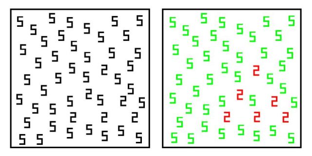
For instance, someone without grapheme-color synesthesia would have a more difficult time picking out the black twos from the black fives in the image on the left.
However, if your numbers have colors, you’ll see the triangle of twos almost instantly.
But this task may be even easier for some grapheme-color synesthetes.
Daniel Smilek, a psychology professor at the University of Waterloo, has identified two groups of synesthetes among those who associate colors with letters and numbers. There are projectors, those whose colors fill the printed letter in front of them, and associators who see the colors in their mind’s eye, like I do.
What about people who can hear silent videos?
Synesthesia doesn’t just apply to people who associate certain colors with images. Some people have the ability to hear sounds in videos when there is actually no sound being played.
Psychologist Chris Fassnidge calls this phenomenon “visually evoked auditory response” (vEAR). While it’s technically not synesthesia, Fassnidge believes it’s a new form that warrants further study. “Some people describe it as a buzzing sound in their head,” Fassnidge told Vox. “For other people, it’s kind of like a white noise. And then other people say it varies depending on what it is they are looking at.”
A 2008 study suggests that vEAR is fairly common — affecting 20 to 30 percent of people — and many people may not realize they are associating faint sounds with imagery.
“A lot of people don’t realize they have this thing until you start testing for it in the laboratory,” Fassnidge said. “Maybe because they co-occur so frequently you either aren’t aware of the mental sound until you strip away everything else.”
What causes it?
About 40 percent of synesthetes have a first-degree relative with synesthesia, and many synesthetes recall having synesthesia as long as they can remember.
“I was definitely playing with it when I was 5 or 6 years old because I remember raiding my parents’ record cabinet, searching for records that I liked to listen to for colors,” said Sean Day, a synesthete who associates colors with both sounds and tastes.
A 2018 study conducted by scientists from the Max Planck Institute for Psycholinguistics and the University of Cambridge analyzed DNA samples from several families who have multiple generations of synesthetes. They concluded that while the families differed in DNA variations, there was one commonality. There was an enhancement of genes involved in cell migration and axonogenesis – a process that enables brain cells to wire up to their correct partners.
“This research is revealing how genetic variation can modify our sensory experiences, potentially via altered connectivity in the brain,” Professor Simon Baron-Cohen stated in the study. “Synesthesia is a clear example of neurodiversity which we should respect and celebrate.”
Other experts believe that everyone may be born with the ability to experience synesthesia.
Daphne Maurer, a psychologist at McMaster University, has speculated that all of us may be born with the neural connections that allow synesthesia, but that most of us lose those connections as we grow.
Eagleman acknowledges there may be synesthetic correspondences in the brains of non-synesthetes, but that people are unaware of them until they’re teased out.
He points to something called the bouba/kiki effect as an example. When asked to choose which of two shapes on the right is named “bouba” and which is “kiki,” most people choose kiki for the angular shape and bouba for the rounded one.
Research also shows that people are likely to say that louder tones are brighter than soft ones and that darker liquids smell stronger than lighter ones.
In his book, “Wednesday Is Indigo Blue,” Eagleman says these examples prove that these analogies are actually “pre-existing relationships.”
“In this way, synesthetic associations our ancestors established long ago grew into the more abstract expressions we know today — and this is why metaphors make sense,” he writes.
However, synesthesia differs from these examples in that the sensory experience triggered is automatic and unlearned, making it different from metaphorical thinking.
“It’s a genuine phenomenon, and people who have it are actually experiencing the world differently,” Eagleman said.
How is it tested?
Consistency is one of the best ways to test for synesthesia.
“If you tell me that your letter ‘J’ is a very particular shade of powder blue … I can test you on that and have you identify exactly the shade that best matches,” Eagleman said. “If you’re just being poetic or metaphorical or making something up, then you can’t capture those colors again. But if you’re really synesthetic, then you’ll be able to pick exactly those colors out years later.”
Researchers also look at synesthetes’ brains. Using positron-emission tomography and functional magnetic resonance imaging, they’ve found that people who report seeing colors in music, for example, have increased activation in the visual areas of the brain in response to sound.
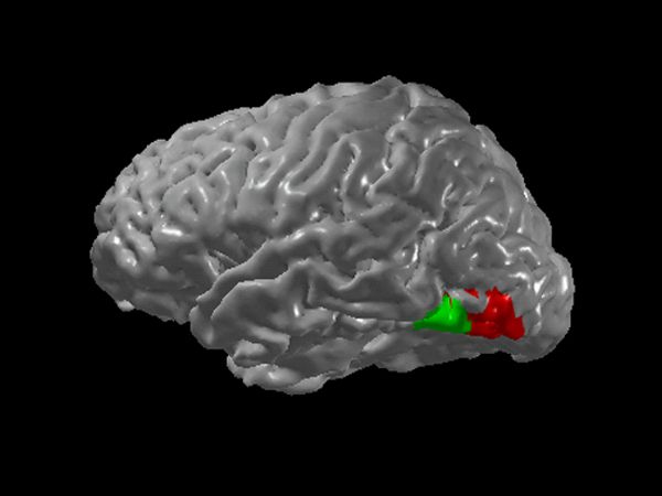
Pictured right are the regions of the brain that are thought to be cross-activated in grapheme-color synesthesia.
The pros and cons
Some synesthetes say their condition can be uncomfortable at times. For example, seeing words printed in the wrong color can be strange, or certain names may taste bad to a synesthete. Others report suffering sensory overload or feeling embarrassed at a young age when they describe experiences they didn’t know were atypical.
However, most synesthetes think of their abilities as a gift and wouldn’t want to lose them.
“You’ve experienced extremely unpleasant odors,” Day points out. “Do you want to permanently lose your sense of smell?”
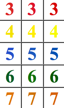
There may also be some benefits to being a synesthete, such as an ability to discern similar colors and easily memorize information. For example, I might not remember a digit in a phone number, but I’ll have an impression of green and therefore know the mystery number is six. (Some of my numbers are pictured above, as depicted by the Synesthesia Battery.)
In 2005, Daniel Tammet set the European record for pi memorization by memorizing 22,514 digits in five hours. He attributed the feat to his ability to see numbers with color, texture and sound.
There’s also evidence that synesthesia may enhance creativity. A 2004 study at the University of California had a group of students take the Torrance Tests of Creative Thinking. The synesthetes who took the test scored more than twice as high in every category.
In some instances, the neurological condition has even led to unique job opportunities. Some car manufacturers, for example, are hiring synesthetes to help designers create cars that are more pleasing to potential drivers.
And synesthetes keep good company. The list of known synesthetes is long and includes Vladimir Nabokov, Vincent Van Gogh, Marilyn Monroe, Billy Joel and Mary J. Blige.
Musician Pharrell Williams associates music with colors and says he can’t imagine life without this “gift.”
“If it was taken from me suddenly I’m not sure that I could make music,” he told Psychology Today. “I wouldn’t be able to keep up with it. I wouldn’t have a measure to understand.”
You don’t remember it, but you woke up at least 100 times last night. These spontaneous arousals, lasting less than 15 seconds each, occur roughly every five minutes and don’t seem to affect how well-rested you feel. They are unrelated to waking up from a bad dream or your partner tossing and turning. Instead, they seem to be linked to some internal biological mechanism.
Frequently waking up throughout the night may have protected early humans from predators by increasing their awareness of their surroundings during sleep. “The likelihood someone would notice an animal is higher [if they] wake up more often,” says Ronny Bartsch, a senior lecturer in the Department of Physics at Bar-Ilan University in Israel. “When you wake up, you’re more prone to hear things. In deep sleep, you’re completely isolated.”
Sleep scientists, however, have been stumped as to what triggers these nocturnal disruptions. In a new Science Advances paper Bartsch proposes an innovative hypothesis that spontaneous arousals are due to random electrical activity in a specific set of neurons in the brain—aptly named the wake-promoting neurons.
Even when you are asleep your brain cells continuously buzz with a low level of electrical activity akin to white noise on the radio. Occasionally, this electrical clamor reaches a threshold that triggers the firing of neurons. The new paper suggests that when random firing occurs in the wake-promoting neurons, a person briefly jerks awake. But this is countered by a suite of sleep-promoting neurons that helps one quickly fall back to sleep.
Low-level electrical activity in neurons increases in colder temperatures whereas warmer temperatures flatten it. As a result, there should be fewer spontaneous arousals in hot weather. To test this theory, the researchers created computer models that mapped how neuronal noise should act at different temperatures and how the varying electrical activity could affect spontaneous arousals. They also measured sleep in zebra fish, which have similar day/night cycles to humans but are ectothermic, meaning their body temperature is controlled by the environment rather than by internal processes.
The researchers compared the fish’s sleep rates at four different water temperatures: 77, 82 (ideal for zebra fish), 84 and 93 degrees Fahrenheit. Across the board, the colder the water the more often the zebra fish woke up and the longer they stayed awake. The data from the zebra fish and the models of temperature, neuronal noise and arousal matched perfectly. “I think their theory is a perfectly good one and may even be correct,” says Clifford Saper, a neuroscientist at Harvard Medical School’s Division of Sleep Medicine and head of Neurology at Beth Israel Deaconess Medical Center who was not involved with the study. “But the experiment they did doesn’t test that hypothesis.”
The zebra fish experiment shows the fish wake up more frequently and stay awake for longer in colder temperatures but reveals nothing about these animals’ neuronal noise—or humans’, for that matter. Bartsch says that, so far, no studies have figured out how to measure neuronal noise in a sleeping animal.
The idea that warm temperatures cause fewer nocturnal disruptions also seemingly flies in the face of conventional wisdom that a colder bedroom leads to better sleep. But waking up because you are hot and uncomfortable is different from these brief spontaneous arousals. In fact, our bodies are pretty good at regulating their core brain and body temperatures, so the difference of a few degrees outside would not alter neuronal activity. In contrast, zebra fish’s temperature varies quite a bit. Saper says because of this zebra fish “are probably the last animal that I would use to try to make this point.”
Bartsch emphasizes the study is not trying to make a claim about thermoregulation in adults but he says it may have implications for newborn babies. “Because very young infants are more ectothermic than endothermic, their arousability could scale similarly to fish for different ambient temperatures.”
Infants are not as good at regulating their own temperature and so are more vulnerable to changes in the environment. (This is why premature babies have to be kept in incubators.) Consequently, the researchers think newborns may be more susceptible to heat-related fluctuations in neuronal noise.
The theory may have important implications for infant sleep. Although they may be disruptive to parents, spontaneous arousals could help save a baby’s life. Sudden infant death syndrome (SIDS) has been a leading cause of mortality in children between one month and one year of age and yet largely remains a mystery. One idea is that SIDS is caused by a stoppage in breathing, often through accidental suffocation. Waking up during the night can prompt babies to shift or cry out, helping to ensure that they do not have anything obstructing their airways and are still breathing. “We came up again with a theory that the babies with SIDS have low neuronal noise and therefore they have lower arousals,” says Hila Dvir, a physicist at Bar-Ilan. “Because they have low arousals, they are less protected from any hypoxic event—a shortage of oxygen.”
Not everyone is convinced, though. “Over the years, people have come up with ideas to explain SIDS, like a single explanation for it, and they just keep hitting dead ends with it because it’s probably a complex, heterogeneous situation,” says Rafael Pelayo, a clinical professor at the Stanford Center for Sleep Sciences and Medicine “It is a cool idea that this neuronal noise is explaining the arousals. I just think they jumped a little bit when they got into SIDS. It has to be more complicated than that.”
A research team at University of Copenhagen including a researcher from the Faculty of Health and Medical Sciences has discovered a circuit in the brains of mice connecting circadian rhythm to aggressive behaviour. The discovery is particularly interesting to Alzheimer’s patients who experience increased aggression at night. The researchers have developed special protein tools capable of turning off the cells in the brain causing the behaviour.
Each year around 8,000 Danes are diagnosed with a form of dementia. Alzheimer’s disease is one of them. The disease manifests itself in memory difficulties in particular, but can also result in personality changes and mood swings.
When the sun sets 20 per cent of all Alzheimer’s patients experience increased bewilderment, anxiety, unease, disorientation, irritation and aggression. This phenomenon is called ‘sundowning’ or sundown syndrome. At worst, the condition can mean that the patient must be left in professional care, as it can be difficult for family members to handle. The cause of the condition is unknown, but previous research has suggested that it is connected to the circadian rhythm.
A research team including a researcher from the Department of Drug Design and Pharmacology at the University of Copenhagen is now able to confirm this connection. The researchers have identified and mapped a circuit between the part of the brain containing the circadian clock or circadian rhythm and a part of the brain controlling aggression.
’We have shown that the circadian clock in mice is closely linked to an aggression centre in the mouse brain by a cell circuit. The human brain has those same groups of cells that the circuit goes through. With this knowledge, we are now enabled to target this circuit pharmacologically and target cells that make people aggressive at the end of the day’, says Assistant Professor Timothy Lynagh from the Department of Drug Design and Pharmacology at the University of Copenhagen.
Turn off the Aggression
The inner clock or circadian rhythm is located in the part of the brain called suprachiasmatic nucleus. One of the parts of the brain that control aggressive behaviour is called the ventromedial hypothalamus. Researchers have previously observed a connection between the two parts of the brain, though none have had knowledge of the specific circuit connecting them.
Using electrophysiology and microscopy, the researchers measured the activity of the brain cells at main author Clifford Saper’s laboratory in Boston. They also turned off parts of the cell circuit in the brains of mice to map the circuit and to identify the cells connecting the two parts of the brain. To map circuits in the brain you need a protein tool that can turn off the various cells to determine their function. Assistant Professor Timothy Lynagh has designed precisely such a tool.
‘We take a receptor and mutate it, so that it is not sensitive to anything in the brain, but very sensitive to a particular drug. The tool works like an on/off switch. When you put the protein tool in the mouse brain, under normal circumstances, nothing will happen. But when you give the animal the drug, the cells that have the receptor on them will be turned off’, Timothy Lynagh explains.
Using this tool, the researchers can thus in theory turn off the cells that cause people suffering from sundown syndrome to become more aggressive at night.
May Be Used on Humans 20 Years into the Future
The tool can also be used in other contexts than sundown syndrome. In other studies, Tim Lynagh’s tool has been used to turn off cells in rats linked to anxiety and fear.
‘If you can start understanding which cells in the brain lead to which problems, you can then put this tool into any of those parts of the brain. The person who takes the drug will then have the cells causing the problem turned off’, Timothy Lynagh says.
Even though the study was conducted on mice, the tool and the knowledge the research has generated can potentially be used in the treatment of humans.
‘Because of the huge advances that are coming along with CRISPR, I would be tempted to say that based on a recent demonstration of gene therapy for brain disease, potentially, it could be used in the human brain in 20 years’ time. Of course it needs a lot more research’, he says.
Reference:
Todd, W. D., Fenselau, H., Wang, J. L., Zhang, R., Machado, N. L., Venner, A., … & Lowell, B. B. (2018). A hypothalamic circuit for the circadian control of aggression. Nature neuroscience, 1.
What if we could edit the sensations we feel; paste in our brain pictures that we never saw, cut out unwanted pain or insert non-existent scents into memory?
UC Berkeley neuroscientists are building the equipment to do just that, using holographic projection into the brain to activate or suppress dozens and ultimately thousands of neurons at once, hundreds of times each second, copying real patterns of brain activity to fool the brain into thinking it has felt, seen or sensed something.
The goal is to read neural activity constantly and decide, based on the activity, which sets of neurons to activate to simulate the pattern and rhythm of an actual brain response, so as to replace lost sensations after peripheral nerve damage, for example, or control a prosthetic limb.
“This has great potential for neural prostheses, since it has the precision needed for the brain to interpret the pattern of activation. If you can read and write the language of the brain, you can speak to it in its own language and it can interpret the message much better,” said Alan Mardinly, a postdoctoral fellow in the UC Berkeley lab of Hillel Adesnik, an assistant professor of molecular and cell biology. “This is one of the first steps in a long road to develop a technology that could be a virtual brain implant with additional senses or enhanced senses.”
Mardinly is one of three first authors of a paper appearing online April 30 in advance of publication in the journal Nature Neuroscience that describes the holographic brain modulator, which can activate up to 50 neurons at once in a three-dimensional chunk of brain containing several thousand neurons, and repeat that up to 300 times a second with different sets of 50 neurons.
“The ability to talk to the brain has the incredible potential to help compensate for neurological damage caused by degenerative diseases or injury,” said Ehud Isacoff, a UC Berkeley professor of molecular and cell biology and director of the Helen Wills Neuroscience Institute, who was not involved in the research project. “By encoding perceptions into the human cortex, you could allow the blind to see or the paralyzed to feel touch.”
Holographic projection
Each of the 2,000 to 3,000 neurons in the chunk of brain was outfitted with a protein that, when hit by a flash of light, turns the cell on to create a brief spike of activity. One of the key breakthroughs was finding a way to target each cell individually without hitting all at once.
To focus the light onto just the cell body — a target smaller than the width of a human hair — of nearly all cells in a chunk of brain, they turned to computer generated holography, a method of bending and focusing light to form a three-dimensional spatial pattern. The effect is as if a 3D image were floating in space.
In this case, the holographic image was projected into a thin layer of brain tissue at the surface of the cortex, about a tenth of a millimeter thick, though a clear window into the brain.
“The major advance is the ability to control neurons precisely in space and time,” said postdoc Nicolas Pégard, another first author who works both in Adesnik’s lab and the lab of co-author Laura Waller, an associate professor of electrical engineering and computer sciences. “In other words, to shoot the very specific sets of neurons you want to activate and do it at the characteristic scale and the speed at which they normally work.”
The researchers have already tested the prototype in the touch, vision and motor areas of the brains of mice as they walk on a treadmill with their heads immobilized. While they have not noted any behavior changes in the mice when their brain is stimulated, Mardinly said that their brain activity — which is measured in real-time with two-photon imaging of calcium levels in the neurons — shows patterns similar to a response to a sensory stimulus. They’re now training mice so they can detect behavior changes after stimulation.
Prosthetics and brain implants
The area of the brain covered — now a slice one-half millimeter square and one-tenth of a millimeter thick — can be scaled up to read from and write to more neurons in the brain’s outer layer, or cortex, Pégard said. And the laser holography setup could eventually be miniaturized to fit in a backpack a person could haul around.
Mardinly, Pégard and the other first author, postdoc Ian Oldenburg, constructed the holographic brain modulator by making technological advances in a number of areas. Mardinly and Oldenburg, together with Savitha Sridharan, a research associate in the lab, developed better optogenetic switches to insert into cells to turn them on and off. The switches — light-activated ion channels on the cell surface that open briefly when triggered — turn on strongly and then quickly shut off, all in about 3 milliseconds, so they’re ready to be re-stimulated up to 50 or more times per second, consistent with normal firing rates in the cortex.
Pégard developed the holographic projection system using a liquid crystal screen that acts like a holographic negative to sculpt the light from 40W lasers into the desired 3D pattern. The lasers are pulsed in 300 femtosecond-long bursts every microsecond. He, Mardinly, Oldenburg and their colleagues published a paper last year describing the device, which they call 3D-SHOT, for three-dimensional scanless holographic optogenetics with temporal focusing.
“This is the culmination of technologies that researchers have been working on for a while, but have been impossible to put together,” Mardinly said. “We solved numerous technical problems at the same time to bring it all together and finally realize the potential of this technology.”
As they improve their technology, they plan to start capturing real patterns of activity in the cortex in order to learn how to reproduce sensations and perceptions to play back through their holographic system.
Reference:
Mardinly, A. R., Oldenburg, I. A., Pégard, N. C., Sridharan, S., Lyall, E. H., Chesnov, K., . . . Adesnik, H. (2018). Precise multimodal optical control of neural ensemble activity. Nature Neuroscience. doi:10.1038/s41593-018-0139-8