When artificial intelligence was allowed to learn how to move through a virtual environment, it spontaneously generated patterns of activity found in grid neurons of the human brain.

Futuristic cityscape maze.

By Diana Kwon

A computer program can learn to navigate through space and spontaneously mimics the electrical activity of grid cells, neurons that help animals navigate their environments, according to a study published May 9 in Nature.

“This paper came out of the blue, like a shot, and it’s very exciting,” Edvard Moser, a neuroscientist at the Kavli Institute for Systems Neuroscience in Norway who was not involved in the work, tells Nature in an accompanying news story. “It is striking that the computer model, coming from a totally different perspective, ended up with the grid pattern we know from biology.” Moser shared a Nobel Prize for the discovery of grid cells with neuroscientists May-Britt Moser and John O’Keefe in 2014.

When scientists trained an artificial neural network to navigate in the form of virtual rats through a simulated environment, they found that the algorithm produced patterns of activity similar to that found in the grid cells of the human brain. “We wanted to see whether we could set up an artificial network with an appropriate task so that it would actually develop grid cells,” study coauthor Caswell Barry of University College London, tells Quanta. “What was surprising was how well it worked.”

The team then tested the program in a more-complex, maze-like environment, and found that not only did the virtual rats make their way to the end, they were also able to outperform a human expert at the task.

“It is doing the kinds of things that animals do and that is to take direct routes wherever possible and shortcuts when they are available,” coauthor Dharshan Kumaran, a senior researcher at Google’s AI company DeepMind, tells The Guardian.

DeepMind researchers hope to use these types of artificial neural networks to study other parts of the brain, such as those involved in understanding sound and controlling limbs, according to Wired. “This has proven to be extremely hard with traditional neuroscience so, in the future, if we could improve these artificial models, we could potentially use them to understand other brain functionalities,” study coauthor Andrea Banino, a research scientist at DeepMind, tells Wired. “This would be a giant step toward the future of brain understanding.”

https://www.the-scientist.com/?articles.view/articleNo/54534/title/Artificial-Intelligence-Mimics-Navigation-Cells-in-the-Brain/&utm_campaign=TS_DAILY%20NEWSLETTER_2018&utm_source=hs_email&utm_medium=email&utm_content=62845247&_hsenc=p2ANqtz-_1eI9gR1hZiJ5AMHakKnqqytoBx4h3r-AG5kHqEt0f3qMz5KQh5XeBQGeWxvqyvET-l70AGfikSD0n3SiVYETaAbpvtA&_hsmi=62845247

Startup Nectome is pitching a mind-uploading service that is “100 percent fatal”

by Antonio Regalado

The startup accelerator Y Combinator is known for supporting audacious companies in its popular three-month boot camp.

There’s never been anything quite like Nectome, though.

Next week, at YC’s “demo days,” Nectome’s cofounder, Robert McIntyre, is going to describe his technology for exquisitely preserving brains in microscopic detail using a high-tech embalming process. Then the MIT graduate will make his business pitch. As it says on his website: “What if we told you we could back up your mind?”

So yeah. Nectome is a preserve-your-brain-and-upload-it company. Its chemical solution can keep a body intact for hundreds of years, maybe thousands, as a statue of frozen glass. The idea is that someday in the future scientists will scan your bricked brain and turn it into a computer simulation. That way, someone a lot like you, though not exactly you, will smell the flowers again in a data server somewhere.

This story has a grisly twist, though. For Nectome’s procedure to work, it’s essential that the brain be fresh. The company says its plan is to connect people with terminal illnesses to a heart-lung machine in order to pump its mix of scientific embalming chemicals into the big carotid arteries in their necks while they are still alive (though under general anesthesia).

The company has consulted with lawyers familiar with California’s two-year-old End of Life Option Act, which permits doctor-assisted suicide for terminal patients, and believes its service will be legal. The product is “100 percent fatal,” says McIntyre. “That is why we are uniquely situated among the Y Combinator companies.”

There’s a waiting list

Brain uploading will be familiar to readers of Ray Kurzweil’s books or other futurist literature. You may already be convinced that immortality as a computer program is definitely going to be a thing. Or you may think transhumanism, the umbrella term for such ideas, is just high-tech religion preying on people’s fear of death.

Either way, you should pay attention to Nectome. The company has won a large federal grant and is collaborating with Edward Boyden, a top neuroscientist at MIT, and its technique just claimed an $80,000 science prize for preserving a pig’s brain so well that every synapse inside it could be seen with an electron microscope.

McIntyre, a computer scientist, and his cofounder Michael McCanna have been following the tech entrepreneur’s handbook with ghoulish alacrity. “The user experience will be identical to physician-assisted suicide,” he says. “Product-market fit is people believing that it works.”

Nectome’s storage service is not yet for sale and may not be for several years. Also still lacking is evidence that memories can be found in dead tissue. But the company has found a way to test the market. Following the example of electric-vehicle maker Tesla, it is sizing up demand by inviting prospective customers to join a waiting list for a deposit of $10,000, fully refundable if you change your mind.

So far, 25 people have done so. One of them is Sam Altman, a 32-year-old investor who is one of the creators of the Y Combinator program. Altman tells MIT Technology Review he’s pretty sure minds will be digitized in his lifetime. “I assume my brain will be uploaded to the cloud,” he says.

Old idea, new approach

The brain storage business is not new. In Arizona, the Alcor Life Extension Foundation holds more than 150 bodies and heads in liquid nitrogen, including those of baseball great Ted Williams. But there’s dispute over whether such cryonic techniques damage the brain, perhaps beyond repair.

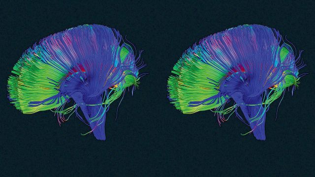
So starting several years ago, McIntyre, then working with cryobiologist Greg Fahy at a company named 21st Century Medicine, developed a different method, which combines embalming with cryonics. It proved effective at preserving an entire brain to the nanometer level, including the connectome—the web of synapses that connect neurons.

A connectome map could be the basis for re-creating a particular person’s consciousness, believes Ken Hayworth, a neuroscientist who is president of the Brain Preservation Foundation—the organization that, on March 13, recognized McIntyre and Fahy’s work with the prize for preserving the pig brain.

There’s no expectation here that the preserved tissue can be actually brought back to life, as is the hope with Alcor-style cryonics. Instead, the idea is to retrieve information that’s present in the brain’s anatomical layout and molecular details.

“If the brain is dead, it’s like your computer is off, but that doesn’t mean the information isn’t there,” says Hayworth.

A brain connectome is inconceivably complex; a single nerve can connect to 8,000 others, and the brain contains millions of cells. Today, imaging the connections in even a square millimeter of mouse brain is an overwhelming task. “But it may be possible in 100 years,” says Hayworth. “Speaking personally, if I were a facing a terminal illness I would likely choose euthanasia by [this method].”

A human brain

The Nectome team demonstrated the seriousness of its intentions starting this January, when McIntyre, McCanna, and a pathologist they’d hired spent several weeks camped out at an Airbnb in Portland, Oregon, waiting to purchase a freshly deceased body.

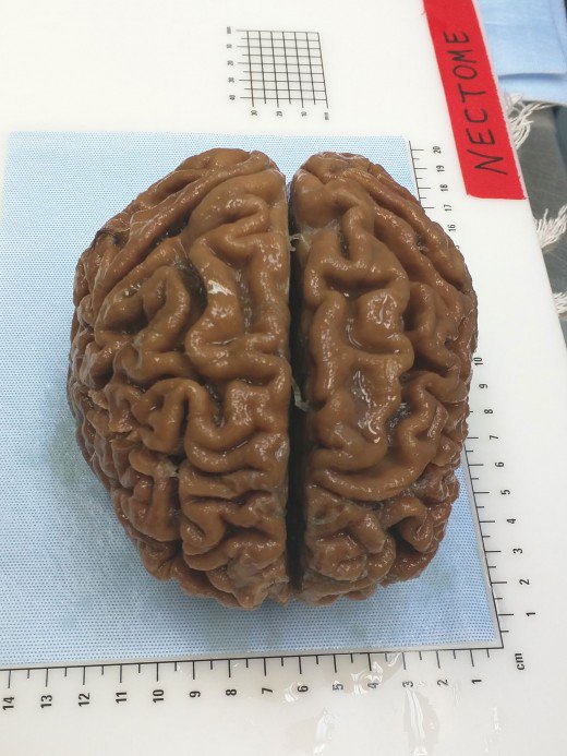
In February, they obtained the corpse of an elderly woman and were able to begin preserving her brain just 2.5 hours after her death. It was the first demonstration of their technique, called aldehyde-stabilized cryopreservation, on a human brain.

Fineas Lupeiu, founder of Aeternitas, a company that arranges for people to donate their bodies to science, confirmed that he provided Nectome with the body. He did not disclose the woman’s age or cause of death, or say how much he charged.

The preservation procedure, which takes about six hours, was carried out at a mortuary. “You can think of what we do as a fancy form of embalming that preserves not just the outer details but the inner details,” says McIntyre. He says the woman’s brain is “one of the best-preserved ever,” although her being dead for even a couple of hours damaged it. Her brain is not being stored indefinitely but is being sliced into paper-thin sheets and imaged with an electron microscope.

McIntyre says the undertaking was a trial run for what the company’s preservation service could look like. He says they are seeking to try it in the near future on a person planning doctor-assisted suicide because of a terminal illness.

Hayworth told me he’s quite anxious that Nectome refrain from offering its service commercially before the planned protocol is published in a medical journal. That’s so “the medical and ethics community can have a complete round of discussion.”

“If you are like me, and think that mind uploading is going to happen, it’s not that controversial,” he says. “But it could look like you are enticing someone to commit suicide to preserve their brain.” He thinks McIntyre is walking “a very fine line” by asking people to pay to join a waiting list. Indeed, he “may have already crossed it.”

Crazy or not ?

Some scientists say brain storage and reanimation is an essentially fraudulent proposition. Writing in our pages in 2015, the McGill University neuroscientist Michael Hendricks decried the “abjectly false hope” peddled by transhumanists promising resurrection in ways that technology can probably never deliver.

“Burdening future generations with our brain banks is just comically arrogant. Aren’t we leaving them with enough problems?” Hendricks told me this week after reviewing Nectome’s website. “I hope future people are appalled that in the 21st century, the richest and most comfortable people in history spent their money and resources trying to live forever on the backs of their descendants. I mean, it’s a joke, right? They are cartoon bad guys.”

Nectome has received substantial support for its technology, however. It has raised $1 million in funding so far, including the $120,000 that Y Combinator provides to all the companies it accepts. It has also won a $960,000 federal grant from the U.S. National Institute of Mental Health for “whole-brain nanoscale preservation and imaging,” the text of which foresees a “commercial opportunity in offering brain preservation” for purposes including drug research.

About a third of the grant funds are being spent in the MIT laboratory of Edward Boyden, a well-known neuroscientist. Boyden says he’s seeking to combine McIntyre’s preservation procedure with a technique MIT invented, expansion microscopy, which causes brain tissue to swell to 10 or 20 times its normal size, and which facilitates some types of measurements.

I asked Boyden what he thinks of brain preservation as a service. “I think that as long as they are up-front about what we do know and what we don’t know, the preservation of information in the brain might be a very useful thing,” he replied in an e-mail.

The unknowns, of course, are substantial. Not only does no one know what consciousness is (so it will be hard to tell if an eventual simulation has any), but it’s also unclear what brain structures and molecular details need to be retained to preserve a memory or a personality. Is it just the synapses, or is it every fleeting molecule? “Ultimately, to answer this question, data is needed,” Boyden says.

Demo day

Nectome has been honing its pitch for Y Combinator’s demo days, trying to create a sharp two-minute summary of its ideas to present to a group of elite investors. The team was leaning against showing an image of the elderly woman’s brain. Some people thought it was unpleasant. The company had also walked back its corporate slogan, changing it from “We archive your mind” to “Committed to the goal of archiving your mind,” which seemed less like an overpromise.

McIntyre sees his company in the tradition of “hard science” startups working on tough problems like quantum computing. “Those companies also can’t sell anything now, but there is a lot of interest in technologies that could be revolutionary if they are made to work,” he says. “I do think that brain preservation has amazing commercial potential.”

He also keeps in mind the dictum that entrepreneurs should develop products they want to use themselves. He sees good reasons to save a copy of himself somewhere, and copies of other people, too.

“There is a lot of philosophical debate, but to me a simulation is close enough that it’s worth something,” McIntyre told me. “And there is a much larger humanitarian aspect to the whole thing. Right now, when a generation of people die, we lose all their collective wisdom. You can transmit knowledge to the next generation, but it’s harder to transmit wisdom, which is learned. Your children have to learn from the same mistakes.”

“That was fine for a while, but we get more powerful every generation. The sheer immense potential of what we can do increases, but the wisdom does not.”

https://www.technologyreview.com/s/610456/a-startup-is-pitching-a-mind-uploading-service-that-is-100-percent-fatal/

A metabolic shift in neurons may provide insight into neurodegenerative diseases


A key metabolic pathway must be switched off during neuron development or fewer neurons (green, on the right) survive.

by Jennifer Hicks

Researchers at the Salk Institute of Biological Studies released a study in the July 12 issue of eLife, which identifies the point at which there’s a dramatic metabolic shift in developing neurons. This discovery of the path a neuron takes during development could help provide insight into neurodegenerative diseases such as Alzheimer’s and Parkinson’s disease.

In a press release, Tony Hunter, American Cancer Society Professor, Salk Molecular and Cell Biology Laboratory said there’s relatively little understanding about how neuron metabolism is first established.

Oxidative stress leads to disruptions in neural cells which are key players in neurodegenerative diseases like Parkinson’s or ALS. The brain needs oxygen to survive but by knowing when and how neuron metabolism goes off track and mitochondria fail to function properly in these diseases, researchers can begin to devise ways to re-route metabolic processes to prevent degeneration.

“Aside from enabling us to understand this process during neuronal development, the work also allows us to better understand neurodegenerative disease,” added Hunter.

What the researchers found in the study was that while neurons shut off the aerobic glycolysis to survive during the metabolic process at the same time neurons also had to kick-start oxidative phosphorylation in order to survive. When the researchers stopped that metabolic process from happening, the neurons died. A neuron dysfunction of any kind can potentially lead to neurodegenerative disease for a number of reasons.

http://www.forbes.com/sites/jenniferhicks/2016/07/31/a-look-at-the-metabolic-shift-in-neurons-for-insight-into-neurodegenerative-disease/#14296174e07b

Mystery of what sleep does to our brains may finally be solved

By Clare Wilson

It is one of life’s great enigmas: why do we sleep? Now we have the best evidence yet of what sleep is for – allowing housekeeping processes to take place that stop our brains becoming overloaded with new memories.

All animals studied so far have been found to sleep, but the reason for their slumber has eluded us. When lab rats are deprived of sleep, they die within a month, and when people go for a few days without sleeping, they start to hallucinate and may have epileptic seizures.

One idea is that sleep helps us consolidate new memories, as people do better in tests if they get a chance to sleep after learning. We know that, while awake, fresh memories are recorded by reinforcing connections between brain cells, but the memory processes that take place while we sleep have remained unclear.

Support is growing for a theory that sleep evolved so that connections in the brain can be pruned down during slumber, making room for fresh memories to form the next day. “Sleep is the price we pay for learning,” says Giulio Tononi of the University of Wisconsin-Madison, who developed the idea.

Now we have the most direct evidence yet that he’s right. Tononi’s team measured the size of these connections or synapses in brain slices taken from mice. The synapses in samples taken at the end of a period of sleep were 18 per cent smaller than those in samples taken from before sleep, showing that the synapses between neurons are weakened during slumber.

A good night’s sleep

Tononi announced these findings at the Federation of European Neuroscience Societies meeting in Copenhagen, Denmark, last week. “The data was very solid and well documented,” says Maiken Nedergaard of the University of Rochester, who attended the conference.

“It’s an extremely elegant idea,” says Vladyslav Vyazovskiy of the University of Oxford

If the housekeeping theory is right, it would explain why, when we miss a night’s sleep, the next day we find it harder to concentrate and learn new information – we may have less capacity to encode new experiences. The finding suggests that, as well as it being important to get a good night’s sleep after learning something, we should also try to sleep well the night before.

It could also explain why, if our sleep is interrupted, we feel less refreshed the next day. There is some indirect evidence that deep, slow-wave sleep is best for pruning back synapses, and it takes time for our brains to reach this level of unconsciousness.

Waking refreshed

Previous evidence has also supported the housekeeping theory. For instance, EEG recordings show that the human brain is less electrically responsive at the start of the day – after a good night’s sleep – than at the end, suggesting that the connections may be weaker. And in rats, the levels of a molecule called the AMPA receptor – which is involved in the functioning of synapses – are lower at the start of their wake periods.

The latest brain-slice findings that synapses get smaller is the most direct evidence yet that the housekeeping theory is right, says Vyazovskiy. “Structural evidence is very important,” he says. “That’s much less affected by other confounding factors.”

Protecting what matters

Getting this data was a Herculean task, says Tononi. They collected tiny chunks of brain tissue, sliced it into ultrathin sections and used these to create 3D models of the brain tissue to identify the synapses. As there were nearly 7000 synapses, it took seven researchers four years.

The team did not know which mouse was which until last month, says Tononi, when they broke the identification code, and found their theory stood up.

“People had been working for years to count these things. You start having stress about whether it’s really possible for all these synapses to start getting fatter and then thin again,” says Tononi.

The team also discovered that some synapses seem to be protected – the biggest fifth stayed the same size. It’s as if the brain is preserving its most important memories, says Tononi. “You keep what matters.”

https://www.newscientist.com/article/2096921-mystery-of-what-sleep-does-to-our-brains-may-finally-be-solved/

Toxoplasma infection might trigger neurodegenerative disease


Infection with the common parasite Toxoplasma gondii promotes accumulation of a neurotransmitter in the brain called glutamate, triggering neurodegenerative diseases in individuals predisposed to such conditions.

Written by Honor Whiteman

This is the finding of a new study conducted by researchers from the University of California-Riverside (UC-Riverside), recently published in PLOS Pathogens.

T. gondii is a single-celled parasite that can cause a disease known as toxoplasmosis.

Infection with the parasite most commonly occurs through eating undercooked, contaminated meat or drinking contaminated water.

It may also occur through accidentally swallowing the parasite after coming into contact with cat feces – by cleaning a litter tray, for example.

Though more than 60 million people in the United States are believed to be infected with T. gondii, few people become ill from it; a healthy immune system can normally stave it off.

As such, most people who become infected with the parasite are unaware of it.

Those who do become ill from T. gondii infection may experience flu-like symptoms – such as swollen lymph glands or muscle aches – that last for at least a month.

In severe cases, toxoplasmosis can cause damage to the eyes, brain, and other organs, though such complications usually only arise in people with weakened immune systems.

The new study, however, suggests there may be another dark side to T. gondii infection: it may lead to development of neurodegenerative disease in people who are predisposed to it.

To reach their findings, lead author Emma Wilson – an associate professor in the Division of Biomedical Sciences at the UC-Riverside School of Medicine – and colleagues focused on how T. gondii infection in mice affects glutamate production

How a build-up of glutamate can damage the brain

Glutamate is an amino acid released by nerve cells, or neurons. It is one of the brain’s most abundant excitatory neurotransmitters, aiding communication between neurons.

However, previous studies have shown that too much glutamate may cause harm; a build-up of glutamate is often found in individuals with traumatic brain injury (TBI) and people with certain neurodegenerative diseases, such as multiple sclerosis (MS) and amyotrophic lateral sclerosis (ALS).

The researchers explain that excess glutamate accumulates outside of neurons, and this build-up is regulated by astrocytes – cells in the central nervous system (CNS).

Astrocytes use a glutamate transporter called GLT-1 in an attempt to remove excess glutamate from outside of neurons and convert it into a less harmful substance called glutamine, which cells use for energy.

“When a neuron fires, it releases glutamate into the space between itself and a nearby neuron,” explains Wilson. “The nearby neuron detects this glutamate, which triggers a firing of the neuron. If the glutamate isn’t cleared by GLT-1 then the neurons can’t fire properly the next time and they start to die.”


T. gondii increases glutamate by inhibiting GLT-1

n mice infected with T. gondii, the researchers identified an increase in glutamate levels.

They found that the parasite causes astrocytes to swell, which impairs their ability to regulate glutamate accumulation outside of neurons.

Furthermore, the parasite prevents GLT-1 from being properly expressed, which causes an accumulation of glutamate and misfiring of neurons. This may lead to neuronal death, and ultimately, neurodegenerative disease.

“These results suggest that in contrast to assuming chronic Toxoplasma infection as quiescent and benign, we should be aware of the potential risk to normal neurological pathways and changes in brain chemistry.” – Emma Wilson

Next, the researchers gave the infected mice an antibiotic called ceftriaxone, which has shown benefits in mouse models of ALS and a variety of CNS injuries.

They found the antibiotic increased expression of GLT-1, which led to a reduction in glutamate build-up and restored neuronal function.

Wilson says their study represents the first time that T. gondii has been shown to directly disrupt a key neurotransmitter in the brain.

“More direct and mechanistic research needs to be performed to understand the realities of this very common pathogen,” she adds.

While their findings indicate a link between T. gondii infection and neurodegenerative disease, Wilson says they should not be cause for panic.

“We have been living with this parasite for a long time,” she says. “It does not want to kill its host and lose its home. The best way to prevent infection is to cook your meat and wash your hands and vegetables. And if you are pregnant, don’t change the cat litter.”

The team now plans to further investigate what causes the reduced expression of GLT-1 in T. gondii infection.

http://www.medicalnewstoday.com/articles/310865.php

The interesting way that your brain makes space to build new and stronger connections so you can learn more

There’s an old saying in neuroscience: neurons that fire together wire together. This means the more you run a neuro-circuit in your brain, the stronger that circuit becomes. This is why, to quote another old saw, practice makes perfect. The more you practice piano, or speaking a language, or juggling, the stronger those circuits get.

For years this has been the focus for learning new things. But as it turns out, the ability to learn is about more than building and strengthening neural connections. Even more important is our ability to break down the old ones. It’s called “synaptic pruning.” Here’s how it works.

Imagine your brain is a garden, except instead of growing flowers, fruits, and vegetables, you grow synaptic connections between neurons. These are the connections that neurotransmitters like dopamine, seratonin, and others travel across.

“Glial cells” are the gardeners of your brain—they act to speed up signals between certain neurons. But other glial cells are the waste removers, pulling up weeds, killing pests, raking up dead leaves. Your brain’s pruning gardeners are called “microglial cells.” They prune your synaptic connections. The question is, how do they know which ones to prune?

Researchers are just starting to unravel this mystery, but what they do know is the synaptic connections that get used less get marked by a protein, C1q (as well as others). When the microglial cells detect that mark, they bond to the protein and destroy—or prune—the synapse.

This is how your brain makes the physical space for you to build new and stronger connections so you can learn more.

Have you ever felt like your brain is full? Maybe when starting a new job, or deep in a project. You’re not sleeping enough, even though you’re constantly taking in new information. Well, in a way, your brain actually is full

When you learn lots of new things, your brain builds connections, but they’re inefficient, ad hoc connections. Your brain needs to prune a lot of those connections away and build more streamlined, efficient pathways. It does that when we sleep.

Your brain cleans itself out when you sleep—your brain cells shrinking by up to 60% to create space for your glial gardeners to come in take away the waste and prune the synapses.

Have you ever woken up from a good night’s rest and been able to think clearly and quickly? That’s because all the pruning and pathway-efficiency that took place overnight has left you with lots of room to take in and synthesize new information—in other words, to learn.

This is the same reason naps are so beneficial to your cognitive abilities. A 10- or 20-minute nap gives your microglial gardeners the chance to come in, clear away some unused connections, and leave space to grow new ones.

Thinking with a sleep-deprived brain is like hacking your way through a dense jungle with a machete. It’s overgrown, slow-going, exhausting. The paths overlap, and light can’t get through. Thinking on a well-rested brain is like wandering happily through Central Park; the paths are clear and connect to one another at distinct spots, the trees are in place, you can see far ahead of you. It’s invigorating.

And in fact, you actually have some control over what your brain decides to delete while you sleep. It’s the synaptic connections you don’t use that get marked for recycling. The ones you do use are the ones that get watered and oxygenated. So be mindful of what you’re thinking about.

If you spend too much time reading theories about the end of Game of Thrones and very little on your job, guess which synapses are going to get marked for recycling?

If you’re in a fight with someone at work and devote your time to thinking about how to get even with them, and not about that big project, you’re going to wind up a synaptic superstar at revenge plots but a poor innovator.

To take advantage of your brain’s natural gardening system, simply think about the things that are important to you. Your gardeners will strengthen those connections and prune the ones that you care about less. It’s how you help the garden of your brain flower.

http://www.fastcompany.com/3059634/your-most-productive-self/your-brain-has-a-delete-button-heres-how-to-use-it

Exploring the Biology of Eating Disorders

With the pressure for a certain body type prevalent in the media, eating disorders are on the rise. But these diseases are not completely socially driven; researchers have uncovered important genetic and biological components as well and are now beginning to tease out the genes and pathways responsible for eating disorder predisposition and pathology.

As we enter the holiday season, shoppers will once again rush into crowded department stores searching for the perfect gift. They will be jostled and bumped, yet for the most part, remain cheerful because of the crisp air, lights, decorations, and the sound of Karen Carpenter’s contralto voice ringing out familiar carols.

While Carpenter is mainly remembered for her musical talents, unfortunately, she is also known for introducing the world to anorexia nervosa (AN), a severe life-threatening mental illness characterized by altered body image and stringent eating patterns that claimed her life just before her 33rd birthday in 1983.

Even though eating disorders (ED) carry one of the highest mortality rates of any mental illness, many researchers and clinicians still view them as socially reinforced behaviors and diagnose them based on criteria such as “inability to maintain body weight,” “undue influence of body weight or shape on self-evaluation,” and “denial of the seriousness of low body weight” (1). This way of thinking was prevalent when Michael Lutter, then an MD/PhD student at the University of Texas Southwestern Medical Center, began his psychiatry residency in an eating disorders unit. “I just remember the intense fear of eating that many patients exhibited and thought that it had to be biologically driven,” he said.

Lutter carried this impression with him when he established his own research laboratory at the University of Iowa. Although clear evidence supports the idea that EDs are biologically driven—they predominantly affect women and significantly alter energy homeostasis—a lack of well-defined animal models combined with the view that they are mainly behavioral abnormalities have hindered studies of the neurobiology of EDs. Still, Lutter is determined to find the biological roots of the disease and tease out the relationship between the psychiatric illness and metabolic disturbance using biochemistry, neuroscience, and human genetics approaches.

We’ve Only Just Begun

Like many diseases, EDs result from complex interactions between genes and environmental risk factors. They tend to run in families, but of course, for many family members, genetics and environment are similar enough that teasing apart the influences of nature and nurture is not easy. Researchers estimate that 50-80% of the predisposition for developing an ED is genetic, but preliminary genome-wide analyses and candidate gene studies failed to identify specific genes that contribute to the risk.

According to Lutter, finding ED study participants can be difficult. “People are either reluctant to participate, or they don’t see that they have a problem,” he reported. Set on finding the genetic underpinnings of EDs, his team began recruiting volunteers and found 2 families, 1 with 20 members, 10 of whom had an ED and another with 5 out of 8 members affected. Rather than doing large-scale linkage and association studies, the team decided to characterize rare single-gene mutations in these families, which led them to identify mutations in the first two genes, estrogen-related receptor α (ESRRA) and histone deacetylase 4 (HDAC4), that clearly associated with ED predisposition in 2013 (1).

“We have larger genetic studies on-going, including the collection of more families. We just happened to publish these two families first because we were able to collect enough individuals and because there is a biological connection between the two genes that we identified,” Lutter explained.

ESRRA appears to be a transcription factor upregulated by exercise and calorie restriction that plays a role in energy balance and metabolism. HDAC4, on the other hand, is a well-described histone deacteylase that has previously been implicated in locomotor activity, body weight homeostasis, and neuronal plasticity.

Using immunoprecipitation, the researchers found that ESRRA interacts with HDAC4, in both the wild type and mutant forms, and transcription assays showed that HDAC4 represses ESRRA activity. When Lutter’s team repeated the transcription assays using mutant forms of the proteins, they found that the ESRRA mutation seen in one family significantly reduced the induction of target gene transcription compared to wild type, and that the mutation in HDAC4 found in the other family increased transcriptional repression for ESRRA target genes.

“ESRRA is a well known regulator of mitochondrial function, and there is an emerging view that mitochondria in the synapse are critical for neurotransmission,” Lutter said. “We are working on identifying target pathways now.”

Bless the Beasts and the Children

Finding genes associated with EDs provides the groundwork for molecular studies, but EDs cannot be completely explained by the actions of altered transcription factors. Individuals suffering these disorders often experience intense anxiety, intrusive thoughts, hyperactivity, and poor coping strategies that lead to rigid and ritualized behaviors and severe crippling perfectionism. They are less aware of their emotions and often try to avoid emotion altogether. To study these complex behaviors, researchers need animal models.

Until recently, scientists relied on mice with access to a running wheel and restricted access to food. Under these conditions, the animals quickly increase their locomotor activity and reduce eating, frequently resulting in death. While some characteristics of EDs—excessive exercise and avoiding food—can be studied in these mice, the model doesn’t allow researchers to explore how the disease actually develops. However, Lutter’s team has now introduced a promising new model (3).

Based on their previous success with identifying the involvement of ESRRA and HDAC4 in EDs, the researchers wondered if mice lacking ESRRA might make suitable models for studies on ED development. To find out, they first performed immunohistochemistry to understand more about the potential cognitive role of ESRRA.

“ESRRA is not expressed very abundantly in areas of the brain typically implicated in the regulation of food intake, which surprised us,” Lutter said. “It is expressed in many cortical regions that have been implicated in the etiology of EDs by brain imaging like the prefrontal cortex, orbitofrontal cortex, and insula. We think that it probably affects the activity of neurons that modulate food intake instead of directly affecting a core feeding circuit.”

With these data, the team next tried providing only 60% of the normal daily calories to their mice for 10 days and looked again at ESRRA expression. Interestingly, ESRRA levels increased significantly when the mice were insufficiently fed, indicating that the protein might be involved in the response to energy balance.

Lutter now believes that upregulation of ESRRA helps organisms adapt to calorie restriction, an effect possibly not happening in those with ESRRA or HDAC4 mutations. “This makes sense for the clinical situation where most individuals will be doing fine until they are challenged by something like a diet or heavy exercise for a sporting event. Once they start losing weight, they don’t adapt their behaviors to increase calorie intake and rapidly spiral into a cycle of greater and greater weight loss.”

When Lutter’s team obtained mice lacking ESRRA, they found that these animals were 15% smaller than their wild type littermates and put forth less effort to obtain food both when fed restricted calorie diets and when they had free access to food. These phenotypes were more pronounced in female mice than male mice, likely due to the role of estrogen signaling. Loss of ESRRA increased grooming behavior, obsessive marble burying, and made mice slower to abandon an escape hole after its relocation, indicating behavioral rigidity. And the mice demonstrated impaired social functioning and reduced locomotion.

Some people with AN exercise extensively, but this isn’t seen in all cases. “I would say it is controversial whether or not hyperactivity is due to a genetic predisposition (trait), secondary to starvations (state), or simply a ritual that develops to counter the anxiety of weight related obsessions. Our data would suggest that it is not due to genetic predisposition,” Lutter explained. “But I would caution against over-interpretation of mouse behavior. The locomotor activity of mice is very different from people and it’s not clear that you can directly translate the results.”

For All We Know

Going forward, Lutter’s group plans to drill down into the behavioral phenotypes seen in their ESRRA null mice. They are currently deleting ESRRA from different neuronal cell types to pair individual neurons with the behaviors they mediate in the hope of working out the neural circuits involved in ED development and pathology.

In addition, the team has created a mouse line carrying one of the HDAC4 mutations previously identified in their genetic study. So far, this mouse “has interesting parallels to the ESRRA-null mouse line,” Lutter reported.

The team continues to recruit volunteers for larger-scale genetic studies. Eventually, they plan to perform RNA-seq to identify the targets of ESRRA and HDAC4 and look into their roles in mitochondrial biogenesis in neurons. Lutter suspects that this process is a key target of ESRRA and could shed light on the cognitive differences, such as altered body image, seen in EDs. In the end, a better understanding of the cells and pathways involved with EDs could create new treatment options, reduce suffering, and maybe even avoid the premature loss of talented individuals to the effects of these disorders.

References

1. Lutter M, Croghan AE, Cui H. Escaping the Golden Cage: Animal Models of Eating Disorders in the Post-Diagnostic and Statistical Manual Era. Biol Psychiatry. 2015 Feb 12.

2. Cui H, Moore J, Ashimi SS, Mason BL, Drawbridge JN, Han S, Hing B, Matthews A, McAdams CJ, Darbro BW, Pieper AA, Waller DA, Xing C, Lutter M. Eating disorder predisposition is associated with ESRRA and HDAC4 mutations. J Clin Invest. 2013 Nov;123(11):4706-13.

3. Cui H, Lu Y, Khan MZ, Anderson RM, McDaniel L, Wilson HE, Yin TC, Radley JJ, Pieper AA, Lutter M. Behavioral disturbances in estrogen-related receptor alpha-null mice. Cell Rep. 2015 Apr 21;11(3):344-50.

http://www.biotechniques.com/news/Exploring-the-Biology-of-Eating-Disorders/biotechniques-361522.html

Scientists achieve implantation of memory into the brains of mice while they sleep

Sleeping minds: prepare to be hacked. For the first time, conscious memories have been implanted into the minds of mice while they sleep. The same technique could one day be used to alter memories in people who have undergone traumatic events.

When we sleep, our brain replays the day’s activities. The pattern of brain activity exhibited by mice when they explore a new area during the day, for example, will reappear, speeded up, while the animal sleeps. This is thought to be the brain practising an activity – an essential part of learning. People who miss out on sleep do not learn as well as those who get a good night’s rest, and when the replay process is disrupted in mice, so too is their ability to remember what they learned the previous day.

Karim Benchenane and his colleagues at the Industrial Physics and Chemistry Higher Educational Institution in Paris, France, hijacked this process to create new memories in sleeping mice. The team targeted the rodents’ place cells – neurons that fire in response to being in or thinking about a specific place. These cells are thought to help us form internal maps, and their discoverers won a Nobel prize last year.

Benchenane’s team used electrodes to monitor the activity of mice’s place cells as the animals explored an enclosed arena, and in each mouse they identified a cell that fired only in a certain arena location. Later, when the mice were sleeping, the researchers monitored the animals’ brain activity as they replayed the day’s experiences. A computer recognised when the specific place cell fired; each time it did, a separate electrode would stimulate brain areas associated with reward.

When the mice awoke, they made a beeline for the location represented by the place cell that had been linked to a rewarding feeling in their sleep. A brand new memory – linking a place with reward – had been formed.

It is the first time a conscious memory has been created in animals during sleep. In recent years, researchers have been able to form subconscious associations in sleeping minds – smokers keen to quit can learn to associate cigarettes with the smells of rotten eggs and fish in their sleep, for example.

Previous work suggested that if this kind of subconscious learning had occurred in Benchenane’s mice, they would have explored the arena in a random manner, perhaps stopping at the reward-associated location. But these mice headed straight for the location, suggesting a conscious memory. “The mouse develops a goal-directed behaviour to go towards the place,” says Benchenane. “It proves that it’s not an automatic behaviour. What we create is an association between a particular place and a reward that can be consciously accessed by the mouse.”

“The mouse is remembering enough abstract information to think ‘I want to go to a certain place’, and go there when it wakes up,” says neuroscientist Neil Burgess at University College London. “It’s a bigger breakthrough [than previous studies] because it really does show what the man in the street would call a memory – the ability to bring to mind abstract knowledge which can guide behaviour in a directed way.”

Benchenane doesn’t think the technique can be used to implant many other types of memories, such as skills – at least for the time being. Spatial memories are easier to modify because they are among the best understood.

His team’s findings also provide some of the strongest evidence for the way in which place cells work. It is almost impossible to test whether place cells function as an internal map while animals are awake, says Benchenane, because these animals also use external cues, such as landmarks, to navigate. By specifically targeting place cells while the mouse is asleep, the team were able to directly test theories that specific cells represent specific places.

“Even when those place cells fire in sleep, they still convey spatial information,” says Benchenane. “That provides evidence that when you’ve got activation of place cells during the consolidation of memories in sleep, you’ve got consolidation of the spatial information.”

Benchenane hopes that his technique could be developed to help alter people’s memories, perhaps of traumatic events (see “Now it’s our turn”, below).

Loren Frank at the University of California, San Francisco, agrees. “I think this is a really important step towards helping people with memory impairments or depression,” he says. “It is surprising to me how many neurological and psychiatric illnesses have something to do with memory, including schizophrenia and obsessive compulsive disorder.”

“In principle, you could selectively change brain processing during sleep to soften memories or change their emotional content,” he adds.

Journal reference: Nature Neuroscience, doi:10.1038/nn.3970

http://www.newscientist.com/article/dn27115-new-memories-implanted-in-mice-while-they-sleep.html#.VP_L9uOVquD

Thanks to Kebmodee for bringing this to the attention of the It’s Interesting community.

New research suggests that memories may not be stored by synaptic connections between nerve cells

New research suggests that memories may not be stored by synaptic connections between neurons in the brain, but rather synapses may allow the expression of memories that are stored elsewhere in the neuron.

The revolutionary study by academics at the University of California has suggested for the first time that memories are not stored in synapses as previously thought. It is synapses, the connections between brain cells, that are destroyed by Alzheimer’s.

The breakthrough, reported in the highly regarded online journal eLife, could mean that it becomes possible to restore lost memories.

“Long-term memory is not stored at the synapse,” said David Glanzman, the study’s co-author and professor of integrative biology and physiology and of neurobiology at UCLA. “That’s a radical idea, but that’s where the evidence leads. The nervous system appears to be able to regenerate lost synaptic connections. If you can restore the synaptic connections, the memory will come back. It won’t be easy, but I believe it’s possible.”

Professor Glanzman’s team studied the marine snail Aplysia to understand the animal’s learning and memory functions. Glanzman was particularly interested in the Aplysia’s defensive reactions and the sensory and motor neurons responsible for its withdrawal response.

“If you train an animal on a task, inhibit its ability to produce proteins immediately after training, and then test it 24 hours later, the animal doesn’t remember the training,” said Prof. Glanzman. “However, if you train an animal, wait 24 hours, and then inject a protein synthesis inhibitor in its brain, the animal shows perfectly good memory 24 hours later. In other words, once memories are formed, if you temporarily disrupt protein synthesis, it doesn’t affect long-term memory. That’s true in the Aplysia and in human’s brains.”

As part of the test, the snails were given a number of electric shocks, which in themselves would not usually produce long-term memories. The team found that the memories they thought had been completely erased earlier in the experiment had returned, suggesting that synaptic connections that had previously been lost were apparently restored.

“That suggests that the memory is not in the synapses but somewhere else,” said Glanzman. “We think it’s in the nucleus of the neurons. We haven’t proved that, though.”

He added that the research could be a major breakthrough for Alzheimer’s sufferers as even though the disease destroys synapses in the brain, memories might not necessarily destroyed.

“As long as the neurons are still alive, the memory will still be there, which means you may be able to recover some of the lost memories in the early stages of Alzheimer’s,” said Prof Glanzman.

http://www.telegraph.co.uk/news/science/11307411/Cure-for-memory-loss-could-be-on-the-horizon.html

New research may help explain why curiosity promotes better memory

Everyone knows it’s easier to learn about a topic you’re curious about. Now, a new study reveals what’s going on in the brain during that process, revealing that such curiosity may give a person a memory boost.

When participants in the study were feeling curious, they were better at remembering information even about unrelated topics, and brain scans showed activity in areas linked to reward and memory.

The results, detailed October 2 in the journal Neuron, hint at ways to improve learning and memory in both healthy people and those with neurological disorders, the researchers said.

“Curiosity may put the brain in a state that allows it to learn and retain any kind of information, like a vortex that sucks in what you are motivated to learn, and also everything around it,” Matthias Gruber, a memory researcher at the University of California, Davis, said in a statement. “These findings suggest ways to enhance learning in the classroom and other settings.”

Gruber and his colleagues put people in a magnetic resonance imaging (MRI) scanner and showed them a series of trivia questions, asking them to rate their curiosity about the answers to those questions. Later, the participants were shown selected trivia questions, then a picture of a neutral face during a 14-second delay, followed by the answer. Afterward, the participants were given a surprise memory test of the faces, and then a memory test of the trivia answers.

Not surprisingly, the study researchers found that people remembered more information about the trivia when they were curious about the trivia answers. But unexpectedly, when the participants were curious, they were also better at remembering the faces, an entirely unrelated task. Participants who were curious were also more likley than others to remember both the trivia information and unrelated faces a day later, the researchers found.

The brain scans showed that, compared with when their curiosity wasn’t piqued, when people were curious, they showed more activation of brain circuits in the nucleus accumbens, an area involved in reward. These same circuits, mediated by the neurochemical messenger dopamine, are involved in forms of external motivation, such as food, sex or drug addiction.

Finally, being curious while learning seemed to produce a spike of activity in the hippocampus, an area involved in forming new memories, and strengthened the link between memory and reward brain circuits.

The study’s findings not only highlight the importance of curiosity for learning in healthy people, but could also give insight into neurological conditions. For example, as people age, their dopamine circuits tend to deteriorate, so understanding how curiosity affects these circuits could help scientists develop treatments for patients with memory disorders, the researchers said.

http://www.livescience.com/48121-curiosity-boosts-memory-learning.html