Opioids might worsen chronic pain, study finds

Written by Honor Whiteman

Anew study has questioned the benefits of opioid painkillers, after finding the drugs might worsen chronic pain rather than ease it.

Study co-leader Prof. Peter Grace, of the University of Colorado at Boulder (CU-Boulder), and colleagues recently published their findings in the Proceedings of the National Academy of Sciences.

Opioids are among the most commonly used painkillers in the United States; almost 250 million opioid prescriptions were written in 2013 – the equivalent to one bottle of pills for every American adult.

Previous studies have suggested opioids – such as codeine, oxycodone, morphine, and fentanyl – are effective pain relievers. They bind to proteins in the brain, spinal cord, and gastrointestinal tract called opioid receptors, reducing pain perception.

Increasing use and abuse of opioids, however, has become a major public health concern in the U.S.; opioid overdoses are responsible for 78 deaths in the country every day.

Now, Prof. Grace and colleagues have questioned whether opioids really work for pain relief, after finding the opioid morphine worsened chronic pain in rats.

Just 5 days of morphine treatment increased chronic pain in rats
According to Prof. Grace, previous studies assessing morphine use have focused on how the drug affects pain in the short term.

With this in mind, the researchers set out to investigate the longer-term effects of morphine use for chronic pain.

For their study, the team assessed two groups of rats with chronic nerve pain. One group was treated with morphine, while the other was not.

Compared with the non-treatment group, the team found that the chronic pain of the morphine group worsened with just 5 days of treatment. What is more, this effect persisted for several months.

“We are showing for the first time that even a brief exposure to opioids can have long-term negative effects on pain,” says Prof. Grace. “We found the treatment was contributing to the problem.”

Another ‘ugly side’ to opioids
According to the authors, the combination of morphine and nerve injury triggered a “cascade” of glial cell signaling, which increased chronic pain.

Glial cells are the “immune cells” of the central nervous system, which support and insulate nerve cells and aid nerve injury recovery.

They found that this cascade activated signaling from a protein called interleukin-1beta (IL-1b), which led to overactivity of nerve cells in the brain and spinal cord that respond to pain. This process can increase and prolong pain.

The researchers say their findings have important implications for individuals with chronic pain – a condition that is estimated to affect around 100 million Americans.

“The implications for people taking opioids like morphine, oxycodone and methadone are great, since we show the short-term decision to take such opioids can have devastating consequences of making pain worse and longer lasting. This is a very ugly side to opioids that had not been recognized before.”

Study co-leader Prof. Linda Watkins, CU-Boulder

It is not all bad news, however. The researchers found they were able to reverse morphine’s pain-increasing effect using a technique called “designer receptor exclusively activated by designer drugs” (DREADD), which involves the use of a targeted drug that stops glial cell receptors from recognizing opioids.

“Importantly, we’ve also been able to block the two main receptors involved in this immune response, including Toll-Like receptor 4 (TLR4) and another one called P2X7R, which have both been separately implicated in chronic pain before,” notes Prof. Grace.

“By blocking these receptors, we’re preventing the immune response from kicking in, enabling the painkilling benefits of morphine to be delivered without resulting in further chronic pain.”

He adds that drugs that can block such receptors are currently in development, but it is likely to be at least another 5 years before they are available for clinical use.

http://www.medicalnewstoday.com/articles/310645.php

Thanks to Kebmnodee for bringing this to the attention of the It’s Interesting community.

That New Superbug Was Found in a UTI and That’s Key

BR3GWM bacteria streaked and grows on an agar plate in the lab

by SARAH ZHANG

THE WOMAN HARBORING E. coli resistant to colistin did not know it, and it’s only luck that we do. Her doctor would have never prescribed that last-resort antibiotic for a routine urinary tract infection—it can cause serious kidney damage. But her doctor did take a urine sample, which ended up at the Walter Reed National Military Medical Center, where researchers had recently started testing for colistin resistance. The test came back positive. Then the came scary headlines about a new superbug in the US.

Superbugs are bacteria with genetic mutations that let them survive humanity’s harshest weapons in germ warfare: antibiotics. The gene behind this E. coli’s colistin resistance is called mcr-1. It first emerged last year when Chinese researchers found it in samples from hospital patients and raw pork. Why pork? Colistin’s serious side effects mean it’s no longer used as a human antibiotic in many countries. But in China, farmers have been adding it by the pound into feed to fatten animals up.

Once epidemiologists knew to look for mcr-1, they found it in Malaysia, England and then the rest of Europe. It was only a matter of time before colistin resistance turned up in the US. On the same day news came out about this woman’s colistin-resistant UTI, the Department of Health and Human Services also announced it found mcr-1 in a sample from a pig intestine.

Colistin is not used in animal feed in the US, so it’s unclear how colistin-resistant bacteria ended up infecting that woman—or that pig. But food and people move freely across borders. And more even seriously, US animal farmers do use other antibiotics—even human ones—on chicken, pigs, and cows. A growing body of research has linked antibiotic use in food animals to drug-resistant bouts of food poisoning from salmonella, campylobacter, and MRSA. Even more interesting is a possible link between antibiotics on meat and urinary tract infections, which science journalist Maryn McKenna has covered extensively. The Food and Drug Administration issued a guidance last year for farms to phase out medically important antibiotics, though only voluntarily.


The Rise of the Drug-Resistant UTI

Urinary tract infections are damn common—annoyingly common if you ask many women. And antibiotic resistant UTIs are on the rise, too: From 2000 to 2010, the number of UTIs resistant to the antibiotic Cipro went from 3 percent to 17.1 percent. Because UTIs afflict so many people, they’re fairly representative antibiotic resistance out there in people community—especially compared to the resistant infections that epidemiologists tend to study most intensely, like ones that kill already sick hospital patients. “UTIs are a good picture of what people are being exposed to on a daily basis” says Amee Manges, an epidemiologist at the University of British Columbia. Case in point: That colistin-resistant bacteria in the woman from Philadelphia.

Manges has spent the past fifteen years studying the link between antibiotic use in meat production, especially poultry, and UTIs. Back when she was working on her doctoral thesis at the University of California, Berkeley, she kept seeing young, otherwise healthy students with UTIs. Originally, she thought she was going to track sexual transmission of the E. coli that caused such infections. With that kind of sporadic sexual transmission, she should have seen many different strains. But when she DNA fingerprinted the bacteria, she found they were all the same strain—the same pattern you’d see from a single source, like if the campus cafeteria gave everyone food poisoning. She was never able to trace those UTI cases back to the original source, but she’s been working on the question ever since.

UTIs are so hard to trace because the infection might not set in until long after a patient first acquired to bacteria. Say a woman eats some undercooked chicken. “The bacteria just hangs out in your intestine for months or possibly years,” says Manges. Then you get risk factor for UTI—sex or a catheter insertion—and that bacteria makes its way from, ahem, the end of your gut to the urethra. But getting people to remember what they ate a week ago is hard. Getting people to remember what they ate a year ago? Hahaha.

The Surveillance Net
Nevertheless, Manges and others have found that strains on meat match strains found in UTIs. Because of the difficulty in tracing UTIs, that evidence is not as ironclad as the evidence for antibiotics use and antibiotic-resistant food poisoning. With routine surveillance of UTIs though, epidemiologists could get a better handle of not only resistant bacteria that come from meat—but also other sources like drinking water or travel or family members being in the hospital. But that surveillance doesn’t happen. “There’s no organized infrastructure to get a good handle about resistance rates across communities,” says Kalpana Gupta, an infectious disease specialist at Boston University.

When patients walk in with UTIs, doctors will often hand out antibiotics without doing a urine culture. Growing the bacteria takes two days—testing for antibiotic-resistance a third—and by that time the patient is usually on the mend already. The fact that the women in Philadelphia got tested was unusual. The fact that her sample was tested against colistin even more so. As Gupta says, “Colistin is not something we would even use to treat UTIs.” (Resistance to another class of antibiotics triggered that extra test in this case.)

The Centers for Disease Control and Prevention is now following up with the woman in Philadelphia to find out she ended up with that colistin-strain of E. coli, which has never been found in the US before. Her infection was fortunately not resistant to all antibiotics. But what makes the colistin-resistance gene mcr-1 so worrisome is that it’s on a small loop of DNA that different bacteria easily swap back and forth. Someday, another bacteria already immune to all other antibiotics will pick up mcr-1, too. It’s only a matter of time.

The wider the surveillance net though, the more quickly we’ll find it.

In Search For Cures, Scientists Create Embryos That Are Both Animal And Human

A handful of scientists around the United States are trying to do something that some people find disturbing: make embryos that are part human, part animal.

The researchers hope these embryos, known as chimeras, could eventually help save the lives of people with a wide range of diseases.

One way would be to use chimera embryos to create better animal models to study how human diseases happen and how they progress.

Perhaps the boldest hope is to create farm animals that have human organs that could be transplanted into terminally ill patients.

But some scientists and bioethicists worry the creation of these interspecies embryos crosses the line. “You’re getting into unsettling ground that I think is damaging to our sense of humanity,” says Stuart Newman, a professor of cell biology and anatomy at the New York Medical College.

The experiments are so sensitive that the National Institutes of Health has imposed a moratorium on funding them while officials explore the ethical issues they raise.

Nevertheless, a small number of researchers are pursuing the work with alternative funding. They hope the results will persuade the NIH to lift the moratorium.

“We’re not trying to make a chimera just because we want to see some kind of monstrous creature,” says Pablo Ross, a reproductive biologist at the University of California, Davis. “We’re doing this for a biomedical purpose.”

The NIH is expected to announce soon how it plans to handle requests for funding.

Recently, Ross agreed to let me visit his lab for an unusual look at his research. During the visit, Ross demonstrated how he is trying to create a pancreas that theoretically could be transplanted into a patient with diabetes.

The first step involves using new gene-editing techniques to remove the gene that pig embryos need to make a pancreas.

Working under an elaborate microscope, Ross makes a small hole in the embryo’s outer membrane with a laser. Next, he injects a molecule synthesized in the laboratory to home in on and delete the pancreas gene inside. (In separate experiments, he has done this to sheep embryos, too.)

After the embryos have had their DNA edited this way, Ross creates another hole in the membrane so he can inject human induced pluripotent stem cells, or iPS for short, into the pig embryos.

Like human embryonic stem cells, iPS cells can turn into any kind of cell or tissue in the body. The researchers’ hope is that the human stem cells will take advantage of the void in the embryo to start forming a human pancreas.

Because iPS cells can be made from any adult’s skin cells, any organs they form would match the patient who needs the transplant, vastly reducing the risk that the body would reject the new organ.

But for the embryo to develop and produce an organ, Ross has to put the chimera embryos into the wombs of adult pigs. That involves a surgical procedure, which is performed in a large operating room across the street from Ross’s lab.

The day Ross opened his lab to me, a surgical team was anesthetizing an adult female pig so surgeons could make an incision to get access to its uterus.

Ross then rushed over with a special syringe filled with chimera embryos. He injected 25 embryos into each side of the animal’s uterus. The procedure took about an hour. He repeated the process on a second pig.

Every time Ross does this, he then waits a few weeks to allow the embryos to develop to their 28th day — a time when primitive structures such as organs start to form.

Ross then retrieves the chimeric embryos to dissect them so he can see what the human stem cells are doing inside. He examines whether the human stem cells have started to form a pancreas, and whether they have begun making any other types of tissues.

The uncertainty is part of what makes the work so controversial. Ross and other scientists conducting these experiments can’t know exactly where the human stem cells will go. Ross hopes they’ll only grow a human pancreas. But they could go elsewhere, such as to the brain.

“If you have pigs with partly human brains you would have animals that might actually have consciousness like a human,” Newman says. “It might have human-type needs. We don’t really know.”

That possibility raises new questions about the morality of using the animals for experimentation. Another concern is that the stem cells could form human sperm and human eggs in the chimeras.

“If a male chimeric pig mated with a female chimeric pig, the result could be a human fetus developing in the uterus of that female chimera,” Newman says. Another possibility is the animals could give birth to some kind of part-human, part-pig creature.

“One of the concerns that a lot of people have is that there’s something sacrosanct about what it means to be human expressed in our DNA,” says Jason Robert, a bioethicist at Arizona State University. “And that by inserting that into other animals and giving those other animals potentially some of the capacities of humans that this could be a kind of violation — a kind of, maybe, even a playing God.”

Ross defends what his work. “I don’t consider that we’re playing God or even close to that,” Ross says. “We’re just trying to use the technologies that we have developed to improve peoples’ life.”

Still, Ross acknowledges the concerns. So he’s moving very carefully, he says. For example, he’s only letting the chimera embryos develop for 28 days. At that point, he removes the embryos and dissects them.

If he discovers the stem cells are going to the wrong places in the embryos, he says he can take steps to stop that from happening. In addition, he’d make sure adult chimeras are never allowed to mate, he says.

“We’re very aware and sensitive to the ethical concerns,” he says. “One of the reasons we’re doing this research the way we’re doing it is because we want to provide scientific information to inform those concerns.”

Ross is working with Juan Carlos Izpisua Belmonte from the Salk Intitute for Biological Studies in La Jolla, Calif., and Hiromitsu Nakauchi at Stanford University. Daniel Garry of the University of Minnesota and colleagues are conducting similar work. The research is funded in part by the Defense Department and the California Institute for Regenerative Medicine (CIRM).

http://www.npr.org/sections/health-shots/2016/05/18/478212837/in-search-for-cures-scientists-create-embryos-that-are-both-animal-and-human

Thanks to Kebmodee for bringing this to the attention of the It’s Interesting community.

Viral and Bacterial Links to the Brain’s Decline


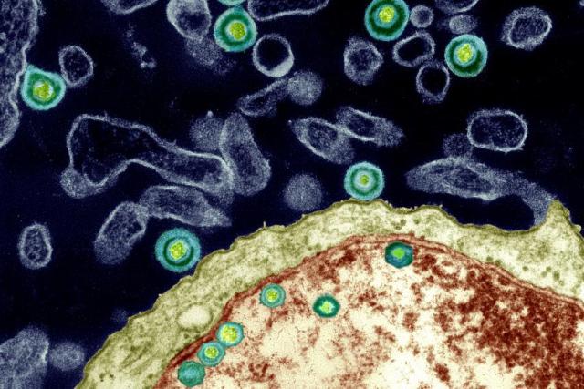
Herpes simplex viruses pass through the outer protein coat of a nucleus, magnified 40,000 times. Dr. Ruth Itzhak’s research published in 1997 revealed a potential link to the presence of HSV-1 (one specific variety of Herpes simplex) and the onset of Alzheimer’s in 60 percent of the cases they studied. However, she has only been able to study a low number of cases since the work has received only a cursory nod from the greater research world and little funding.

By Ed Cara

As recently as the 1970s, doctors stubbornly treated complaints of festering open sores in the stomach as a failing of diet or an inability to manage stress. Though we had long accepted the basic premise of Louis Pasteur’s germ theory—that flittering short bursts of disease and death are often caused by microscopic beings that could be stopped by sanitary food, water and specially crafted drugs—many researchers ardently resisted the idea that they could also trigger more complicated, chronic illnesses.

When it came to ulcers, no one believed that any microorganisms could endure in the acidic cauldron of our digestive system. It took the gumshoe work of Australian doctors and medical researchers Barry Marshall and Robin Warren in the 1980s to debunk that belief and discover the specific bug responsible for most chronic stomach ulcers, Helicobacter pylori. Marshall even went so far as to swallow the germ to prove the link was real and, obviously, became sick soon after. Thankfully, his self-sacrifice was eventually validated when he and Warren were awarded a Nobel Prize in 2005.

But while modern medicine has grown comfortable with the idea that even chronic physical ailments can be sparked by the living infinitesimal, there is an even bolder, more controversial proposition from a growing number of researchers. It’s the idea that certain germs, bugs and microbes can lie hidden in the body for decades, all the while slowly damaging our brains, even to the point of dementia, depression and schizophrenia.

In January 2016, a team led by Shawn Gale, an associate professor in psychology at Brigham Young University, looked at the infection history of 5,662 young to middle-aged adults alongside the results of tests intended to measure cognition. Gale’s rogues’ gallery included both parasites (the roundworm and Toxoplasma gondii ) and viruses (the hepatitis clan, cytomegalovirus, and herpes simplex virus Types 1 and 2). The team created an index of infectious disease —the more bugs a participant had been exposed to, the higher the person’s index score. It turned out that those with a higher score were more likely to have worse learning and memory skills, as well as slower information-processing speed than those with a lower score, even after controlling for other factors, like age, sex and financial status.

Aside from their shared ability to stay rooted inside us, the ways these pathogens might influence our noggins are as varied as their biology is from one another. Some, like T. gondii (often transmitted to humans via contaminated cats and infected dirt), can discreetly infest the brain and cause subtle changes to our brain chemistry, altering levels of neurotransmitters like dopamine while causing no overt signs of disease. Others, like hepatitis C, are suspected of hitching a ride onto infected white blood cells that cross the brain-blood barrier and, once inside, deplete our supply of white brain matter, the myelin-coated axons that help neurons communicate with each other and seem to actively shape how we learn. And still others, like H. pylori, could trigger a low-level but chronic inflammatory response that gradually wears down our body and mind alike.

Gale’s team found only fairly small deficits in cognition connected to infection. But other researchers, like Ruth Itzhaki, professor emeritus of molecular neurobiology at Britain’s University of Manchester, believe microbes may play an outsized role in one of the most devastating neurodegenerative disorders around: Alzheimer’s disease, which afflicted 47 million people worldwide in 2015. Last March, Itzhaki and a globe-spanning group of researchers penned an editorial in the Journal of Alzheimer’s Disease, imploring the scientific community to more seriously pursue a proposed link between Alzheimer’s and particular germs, namely herpes simplex virus Type 1 (HSV-1), Chlamydia pneumoniae and spirochetes—a diverse group of bacteria that include those responsible for syphilis and Lyme disease. The unusually direct plea, for scientists at least, was the culmination of decades of frustration.

“There’s great hostility to the microbial concept amongst certain influential people in the field, and they are the ones who usually determine whether or not one’s research grant application is successful,” says Itzhaki. “The irony is that they never provide scientific objections to the concepts—they just belittle them, so there’s nothing to rebut!”

It’s a frustration Itzhaki knows too well; in 1991, her lab published the first paper finding a clear HSV-1 link to Alzheimer’s. Since then, according to Itzhaki, over 100 published studies, from her lab and elsewhere, have been supportive of the same link. Nevertheless, Itzhaki says, the work has received only a cursory nod from the greater research world and little funding. Out of the $589 million allocated to Alzheimer’s research by the National Institutes of Health in 2015, exactly zero appeared to be spent on studying the proposed HSV-1 connection.

HSV-1 is more often known as the version of herpes that causes cold sores. Nearly all of us carry the virus from infancy; our peripheral nervous system serves as its dormant nesting ground. From there, HSV-1 can reactivate and occasionally cause mild flare-ups of disease, typically when our immune system is overwhelmed due to stress or other infections. Itzhaki’s lab, however, found that by the time we reach our golden years, the virus often migrates to the brain, where it remains capable of resurrecting itself and wreaking a new sort of havoc when opportunity presents, such as when our immune system wavers in old age.

Her team has also discovered the presence of HSV-1 in the telltale plaques—clumps of proteins in the nerve cells of the brain—used to diagnose Alzheimer’s. In mice and cell cultures infected with HSV-1, they’ve found accumulation of two proteins, beta-amyloid and tau, that form the main components of, respectively, plaques and tangles—twisted protein fibers that form inside dying cells and are another defining characteristic of Alzheimer’s. Plaques and tangles, while sometimes found in normal aging brains, have been found to overflow in the brains of deceased Alzheimer’s sufferers; neuroscientists believe these protein accumulations can cause neuron death and tissue loss. Itzhaki speculates that herpes-infected cells may either produce the proteins in an attempt to fend off HSV-1 or, because the virus itself commands them to, the proteins somehow needed to jump-start the virus’s replication.

Itzhaki, Gale and their colleagues emphasize that rather than being the sole cause of memory loss, slower reaction time or depression, viral and bacterial infections are likely just one ingredient in a soup of risk factors. But for Alzheimer’s, HSV-1 could be especially significant. Itzhaki has found that elderly people who carried both HSV-1 in the brain and the e-4 subtype of the APOE gene (responsible for creating a protein that helps transport cholesterol throughout the body) were 12 times more likely to develop Alzheimer’s than people without either.

APOE-e4, already considered a significant risk factor for Alzheimer’s and thought to make us more vulnerable to viral infection, has also been linked to a greater risk of dementia in HIV-infected patients. In a 1997 Lancet paper, Itzhaki’s group concluded that HSV-1 infection, in conjunction with APOE-e4, could account for about 60 percent of the Alzheimer’s cases they studied. Due to limited funds, however, her group was able to study only a relatively low number of cases.

“I think the proposed theory is certainly reasonable given the supporting evidence,” says Iain Campbell, a professor of molecular biology at the University of Sydney. “What is difficult to establish here is actual causality.”

It might be the case that HSV-1 and other suspects aren’t responsible for the emergence of Alzheimer’s but are simply given free rein to worsen its symptoms as the neurodegenerative disorder weakens both the immune and nervous systems. Deciphering the relationship between these latent infections and Alzheimer’s will take more dedicated research, an effort that Itzhaki feels has been stymied by the persistent lack of resources available to her and her like-minded colleagues.

As things stand, though, she believes there is enough evidence to go ahead with treatment trials; for instance, giving Alzheimer’s patients HSV-1-targeted antivirals in hopes of slowing down or stopping the progression of the disease. She and a team of clinicians are trying to obtain a grant for such a pilot clinical trial to do just that.

Exasperated as Itzhaki has been, the headwinds against her and those who share her beliefs about the brain are slowly dying down. In some cases, once-derided and obscure scientists studying how infections affect the brain are now getting some financial support. There’s Jaroslav Flegr, for example, who has for decades theorized that T. gondii could alter human behavior and even cause certain forms of schizophrenia. In the wake of increased media attention, Flegr’s volume of work on T. gondii has noticeably stepped up as well. From 2014 to 2015, he co-authored 13 papers on T. gondii, nearly twice the number he published the previous two years; the trend of increased T. gondii papers holds across all of PubMed, the largest database of published biomedical research available. “ I have no serious problem with funding of my Toxo research now,” Flegr says.

As of now, though, there have been no ulcer-related Sherlock moments to prove a link between mental dysfunction and latent infections—only indirect correlations clumping together to form a blurry snapshot of a potential crime scene. Which is why Gale and others recommend a wait-and-see approach for the public, even as they acknowledge the potentially vast implications of their research. “I wouldn’t want someone to go out tomorrow and get a whole battery of tests,” he says. “There’s still a lot we need to understand.”

http://www.newsweek.com/viral-bacterial-links-brains-decline-462194

Robot outperforms highly-skilled human surgeons on pig GI surgery

A robot surgeon has been taught to perform a delicate procedure—stitching soft tissue together with a needle and thread—more precisely and reliably than even the best human doctor.

The Smart Tissue Autonomous Robot (STAR), developed by researchers at Children’s National Health System in Washington, D.C., uses an advanced 3-D imaging system and very precise force sensing to apply stitches with submillimeter precision. The system was designed to copy state-of-the art surgical practice, but in tests involving living pigs, it proved capable of outperforming its teachers.

Currently, most surgical robots are controlled remotely, and no automated surgical system has been used to manipulate soft tissue. So the work, described today in the journal Science Translational Medicine, shows the potential for automated surgical tools to improve patient outcomes. More than 45 million soft-tissue surgeries are performed in the U.S. each year. Examples include hernia operations and repairs of torn muscles.

“Imagine that you need a surgery, or your loved one needs a surgery,” says Peter Kim, a pediatric surgeon at Children’s National, who led the work. “Wouldn’t it be critical to have the best surgeon and the best surgical techniques available?”

Kim does not see the technology replacing human surgeons. He explains that a surgeon still oversees the robot’s work and will take over in an emergency, such as unexpected bleeding.

“Even though we take pride in our craft of doing surgical procedures, to have a machine or tool that works with us in ensuring better outcome safety and reducing complications—[there] would be a tremendous benefit,” Kim says. The new system is an impressive example of a robot performing delicate manipulation. If robots can master human-level dexterity, they could conceivably take on many more tasks and jobs.

STAR consists of an industrial robot equipped with several custom-made components. The researchers developed a force-sensitive device for suturing and, most important, a near-infrared camera capable of imaging soft tissue in detail when fluorescent markers are injected.

“It’s an important result,” says Ken Goldberg, a professor at UC Berkeley who is also developing robotic surgical systems. “The innovation in 3-D sensing is particularly interesting.”

Goldberg’s team is developed surgical robots that could be more flexible than STAR because instead of being manually programmed, they can learn automatically by observing expert surgeons. “Copying the skill of experts is really the next step here,” he says.

https://www.technologyreview.com/s/601378/nimble-fingered-robot-outperforms-the-best-human-surgeons/

Thanks to Kebmodee for bringing this to the It’s Interesting community.

New Blood Test for TB Could Save Millions of Lives

As much as one third of the global population is currently infected with the bacterium that causes tuberculosis (TB), a disease typically concentrated in the lungs and characterized by weakness, fever, coughing and chest pain. About 9.6 million new infections occurred in 2014, the most recent year for which numbers are available. Roughly 1.5 million people died of TB that same year. The ability to easily, inexpensively and accurately diagnose TB is of utmost importance, but the most commonly used method fails, at least to some extent, on all three counts. A new blood-based technique might considerably rein in this epidemic.

The conventional TB test scans for bacterial DNA in coughed-up mucus, or sputum. But some children struggle to produce a sample on request. The test also can miss TB in people simultaneously infected with HIV because the telltale bacteria may exist in numbers too low to detect or outside the lungs. In addition, the test costs up to $10, a prohibitive fee in many developing countries. As a result of these constraints, a large percentage of TB cases are diagnosed late or not at all, leaving serious infections untreated and more liable to spread.

Two years ago the World Health Organization put out a call for an improved TB diagnostic. In response, Purvesh Khatri, a Stanford University medical professor, and his colleagues combed through the human genome and found three genes that distinguish active TB from other diseases. The team then developed a way to detect these genes in blood.

According to their study, published in the Lancet Respiratory Medicine, the test is equally sensitive among patients with and without HIV coinfection and correctly detected TB in 86 percent of pediatric cases. Additional points in favor of a blood assay include that it can be performed at a clinic and yield same-day results, unlike the case for a sputum test. That is especially advantageous in the developing world, where showing up for even a single appointment presents a tremendous burden. “You want to be able to initiate treatment immediately,” says Sheela Shenoi, a Yale University professor of medicine focused on AIDS.

The technology has not been used in the diagnosis of new patients and may be difficult to scale up, but in the meantime, Khatri has filed a patent for the test. He thinks it could cost less than half as much as the current one. “If this three-gene signature could be developed into a point-of-care test,” Shinoi says, “it would revolutionize TB diagnostics.”

http://www.scientificamerican.com/article/a-new-blood-test-for-tb-could-save-millions-of-lives/

Wheelchair-bound multiple sclerosis patients able to walk again after new stem-cell therapy


Holly Drewry, 25, of Sheffield, was wheelchair bound after the birth of her daughter Isla, now two.

A pioneering new stem cell treatment is reversing and then halting the potentially crippling effects of multiple sclerosis.

Patients embarking on a ground-breaking trial of the new treatment have found they can walk again and that the disease even appears to be stopped in its tracks.

Holly Drewry, 25, from Sheffield, was wheelchair bound after the birth of her daughter Isla, now two. But Miss Drewry claims the new treatment has transformed her life.

She told the BBC’s Panorama programme: “I couldn’t walk steadily. I couldn’t trust myself holding her (Isla) in case I fell. Being a new mum I wanted to do it all properly but my MS was stopping me from doing it.

“It is scary because you think, when is it going to end?”

The treatment is being carried out at Royal Hallamshire Hospital in Sheffield and Kings College Hospital, London and involves use a high dose of chemotherapy to knock out the immune system before rebuilding it with stem cells taken from the patient’s own blood.

Miss Drewry had the treatment in Sheffield. She said: “I started seeing changes within days of the stem cells being put in.

“I walked out of the hospital. I walked into my house and hugged Isla. I cried and cried. It was a bit overwhelming. It was a miracle.”

Her treatment has now been reviewed and her condition found to have been dramatically halted. She will need to be monitored for years but the hope is that her transplant will be a permanent fix.

She is now planning to get married.

For other patients, the results have been equally dramatic. Steven Storey was a marathon runner and triathlete before he was struck down with the disease and left completely paralysed: “I couldn’t flicker a muscle,” he said.

But within nine days of the treatment he could move his toe and after 10 months managed a mile-long swim in the Lake District. He has also managed to ride a bike and walk again.

“It was great. I felt I was back,” he said.

Mr Storey celebrated his first transplant birthday with his daughters. His treatment has been reviewed and, like Miss Drewry, there was no evidence of active disease.

The treatment – which effectively ‘reboots’ the immune systems – is the first to reverse the symptoms of MS, which has no cure, and affects around 100,000 people in Britain.

Stem cells are so effective because they can become any cell in the body based on their environment.

Although it is unclear what causes MS, some doctors believe that it is the immune system itself which attacks the brain and spinal cord, leading to inflammation and pain, disability and in severe cases, death.

Professor Basil Sharrack, a consultant neurologist at Sheffield Teaching Hospitals NHS Foundation Trust, said: “Since we started treating patients three years ago, some of the results we have seen have been miraculous.

“This is not a word I would use lightly, but we have seen profound neurological improvements.”

During the treatment, the patient’s stem cells are harvested and stored. Then doctors use aggressive drugs which are usually given to cancer patients to completely destroy the immune system.

The harvested stem cells are then infused back into the body where they start to grow new red and white blood cells within just two weeks.

Within a month the immune system is back up and running fully and that is when patients begin to notice that they are recovering.

However specialists warn that patients need to be fit to benefit from the new treatment.

The research has been published in the Journal of the American Medical Association.

http://www.telegraph.co.uk/news/health/news/12104774/Miraculous-results-from-new-MS-treatment.html

Thanks to Steve Weihing for bringing this to the It’s Interesting community.

Liver hormone discovered to drive sugar consumption

A recent study has shown that fibroblast growth factor 21 (FGF21), a liver-generated hormone, suppresses the FGF21 is produced in response to high carbohydrate levels, in which it enters the bloodstream and signals the brain to suppress the preference for sweets. Matthew Potthoff, assistant professor of pharmacology in the University of Iowa Carver College of Medicine, noted that this is the “first liver-derived hormone that regulates sugar intake specifically.”consumption of simple sugars.

Earlier studies have shown how some hormones affect appetite. However, these do not regulate any specific macronutrient (eg, carbohydrate, protein, fat) and are produced in organs other than the liver. FGF21 has been known to boost insulin sensitivity but the new findings “can help people who might not be able to sense when they’ve had enough sugar, which may contribute to diabetes,” said Lucas BonDurant, a doctoral student and co-first author in the study.

Researchers used genetically-engineered mouse models and pharmacological approaches to study FGF21 in regulating sugar cravings. Normal mice were injected with FGF21 and were given a choice between a normal diet and a sugar-enriched diet. These mice did not completely stop eating sugar but consumed 7 times less than normal. The team also looked at mice that either did not produce FGF21 at all or overproduced FGF21 (>500 times more than normal mice). When presented with the same two diets as the normal mice, researchers saw that the mice that didn’t produce FGF21 all consumed more sugar whereas the mice that overproduced FGF21 consumed less sugar.

Study findings support the conclusion that FGF21 decreased appetite and sugar intake. It did not, however, decrease intake of all sugars (eg, sucrose, fructose, glucose) nor did it affect the intake of complex carboydrates. The new data may help patients who are obese or have diabetes, researchers noted. More studies are needed to see if other hormones exist to regulate appetite for specific macronutrients comparable to the effects of FGF21 on carbohydrate intake.

http://www.empr.com/news/liver-derived-hormone-may-influence-sugar-cravings/article/461698/?DCMP=EMC-MPR_DailyDose_rd&cpn=psych_md&hmSubId=&hmEmail=5JIkN8Id_eWz7RlW__D9F5p_RUD7HzdI0&NID=1710903786&dl=0&spMailingID=13368691&spUserID=MTQ4MTYyNjcyNzk2S0&spJobID=700165841&spReportId=NzAwMTY1ODQxS0

This already prescribed drug may also effectively treat patients infected with Ebola.

by Jennifer Brown

The recent Ebola outbreak in West Africa has claimed more than 11,300 lives—a stark reminder of the lack of effective options for treating or preventing the disease.

Progress has been made on developing vaccines, but there is still a need for antiviral therapies to protect health care workers and local populations in the event of future outbreaks.

Now, a new study suggests that gamma interferon, an FDA-approved drug, may have potential as an antiviral therapy to prevent Ebola infection when given either before or after exposure to the virus.

The findings, published in the journal PLOS Pathogens, show that gamma interferon, given up to 24 hours after exposure, inhibits Ebola infection in mice and completely protects the animals from death.

Ebola infection appears to be a stepwise process. First, the virus targets and infects macrophages or dendritic cells, two types of immune system cells found in the liver, spleen, and lymph nodes. Ebola then replicates in those cells. Following this initial infection, which happens at day 3 or 4 in non-human primates, Ebola virus is released into the blood and infects a plethora of other different cell populations.

“It goes from an early stage with a very targeted infection of only these few cell types, to everything being infected,” says Wendy Maury, professor of microbiology at the University of Iowa.

“We think what’s happening with gamma interferon is that it’s targeting macrophages and blocking the infection of those initial cell targets so you don’t get the second round of infection.”

The University of Iowa does not have a specializing BioSafety Level 4 (BSL4) lab that is required for experiment using Ebola virus, so the researchers made their initial findings using a surrogate virus, which targets and infects the same cells as Ebola, but does not cause the disease.

This Ebola lookalike—a sheep in wolf’s clothing—consists of a less dangerous vesicular stomatitis virus (VSV) that expresses Ebola glycoproteins on its surface.

All of the results found using the surrogate virus were then repeated using mouse-adapted Ebola virus in the BSL4 lab of Maury’s longtime collaborator Robert Davey at Texas Biomedical Institute in San Antonio, Texas.

Gamma interferon inhibits the virus’s ability to infect human and mouse macrophages, in part by blocking virus replication in the cells. Pre-treating mice with interferon gamma 24 hours before exposure protects the animals from infection and death. The researchers were surprised to find that treatment up to 24 hours after what would have been a lethal exposure also completely protected the animals from death, and they could no longer detect any Ebola virus in the mouse’s cells.

The findings suggest that interferon gamma may be useful both as a prophylaxis and post-exposure treatment against Ebola. The team still has to determine how late gamma interferon can be given to the mice and still prevent infection. However, the results suggest a window of time after exposure when gamma interferon may be an effective antiviral therapy.

“My guess is that if you delay the gamma interferon too much, you miss this window of opportunity to block the infection in macrophage cells and the gamma interferon can no longer provide protection,” Maury says.

Maury and colleagues investigated how gamma interferon might be helping the cells fight off the Ebola virus. They identified that the expression of more than 160 genes in human macrophages is stimulated by gamma interferon. Introduction of some of these genes into cells was sufficient to prevent Ebola infection.

“This mechanistic information might suggest more precise drug targets rather than the broad effects, including adverse side-effects, that are produced by gamma-interferon,” she says.

Gamma interferon is already approved by the FDA to treat chronic granulomatous disease (an immune disease) and severe malignant osteopetrosis.

In addition to moving the studies into larger animal models, Maury next plans to study the ability of gamma interferon to inhibit Ebola infection in conjunction with other developing antivirals.

“Right now, there are no FDA-approved antiviral therapies for Ebola, but there are some being developed that target virus entry,” she says. “We know that gamma interferon blocks replication but not entry into cells. So combining an entry inhibitor with gamma interferon may allow us to reduce amount of gamma interferon needed and target two different steps in the virus’s life cycle, which has been shown in HIV to be critically important for controlling the virus.”

http://journals.plos.org/plospathogens/article?id=10.1371/journal.ppat.1005263

http://now.uiowa.edu/2015/12/fda-approved-drug-protects-mice-ebola

Air evacuation following traumatic brain injury may worsen outcomes for patients.


Over the past 15 years, more than 330,000 US soldiers have suffered a traumatic brain injury. Many were evacuated by air for further treatment. A new study has found evidence that such air evacuations may pose a significant added risk, potentially causing more damage to already injured brains.

Over the past 15 years, more than 330,000 U.S. soldiers have suffered a traumatic brain injury (TBI). It is one of the leading causes of death and disability connected to the country’s recent conflicts in Afghanistan and Iraq. Many of these patients were evacuated by air from these countries to Europe and the U.S. for further treatment. In general, these patients were flown quickly to hospitals outside the battle zone, where more extensive treatment was available.

But now a new study by researchers at the University of Maryland School of Medicine has found evidence that such air evacuations may pose a significant added risk, potentially causing more damage to already injured brains. The study is the first to suggest that air evacuation may be hazardous for TBI patients. The study was published in the Journal of Neurotrauma.

“This research shows that exposure to reduced barometric pressure, as occurs on military planes used for evacuation, substantially worsens neurological function and increases brain cell loss after experimental TBI — even when oxygen levels are kept in the normal range. It suggests that we need to carefully re-evaluate the cost-benefit of air transport in the first days after injury,” said lead researcher Alan Faden, MD, the David S. Brown Professor in Trauma in the Departments of Anesthesiology, Anatomy & Neurobiology, Neurology, and Neurosurgery, and director, Shock, Trauma and Anesthesiology Research Center (STAR) as well as the National Study Center for Trauma and Emergency Medical Services.

About a quarter of all injured soldiers evacuated from Afghanistan and Iraq have suffered head injuries.

Faden and his colleagues tested rats that were subjected to TBI, using a model that simulates key aspects of human brain injury. Animals were exposed to six hours of lowered air pressure, known as hypobaria, at levels that simulated conditions during transport; control animals were exposed to normal pressure. All the animals received extra oxygen to restore normal oxygen concentrations in the blood. In another study, animals received oxygen, either as in the first study or at much higher 100 percent concentration, which is often used during military air evacuations. On its own, low air pressure worsened long-term cognitive function and increased chronic brain inflammation and brain tissue loss. Pure oxygen further worsened outcomes.

Faden and his colleagues believe the findings raise concerns about the increased use of relatively early air evacuation, and suggest that this potential risk should be weighed against the benefits of improved care after evacuation. It may be necessary, he says, to change the current policy for TBI patients and delaying air evacuation in many cases.

In an accompanying editorial, Patrick Kochanek, MD, a leading expert on TBI and trauma care at the University of Pittsburgh, called the findings “highly novel and eye-opening,” and said that they could have “impactful clinical relevance for the field of traumatic brain injury in both military and civilian applications.”

Faden and colleagues believe that one of the mechanisms by which hypobaria worsens TBI is by increasing persistent brain inflammation after injury. They are currently examining how this process occurs and have tested treatments that can reduce the risks of air evacuation. Early results are promising. Scientists suspect that breathing pure oxygen could worsen TBI by increasing production of dangerous free radicals in the brain. After brain injury, these free radicals flood the site of injury, and pure oxygen may further boost these levels. Several recent studies from trauma centers, including from the R Adams Cowley Shock Trauma Center at the University of Maryland Medical Center, have found evidence that using 100 percent oxygen in trauma patients may be counterproductive.

Journal Reference:

Jacob W Skovira, Shruti V Kabadi, Junfang Wu, Zaorui Zhao, Joseph DuBose, Robert E Rosenthal, Gary Fiskum, Alan I Faden. Simulated Aeromedical Evacuation Exacerbates Experimental Brain Injury. Journal of Neurotrauma, 2015; DOI: 10.1089/neu.2015.4189

http://www.sciencedaily.com/releases/2015/11/151130110013.htm