Pupil Response Predicts Depression Risk in Kids

Emerging research suggests pupil dilation in children of depressed mothers when seeing an emotional image can help predict his or her risk of depression over the next two years.

Dr. Brandon Gibb, a professor of psychology at Binghamton University in New York, said the new findings suggest physiological reactivity to sad stimuli can be a potential biomarker of depression risk for some kids.

An important aspect of this finding is that pupillometry is an inexpensive tool that could be administered in family practice or pediatricians’ offices.

The simple test can help identify which children of depressed mothers are at highest risk for developing depression themselves.

“We think this line of research could eventually lead to universal screenings in pediatricians’ offices to assess future depression risk in kids,” said Gibb.

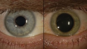
Gibb recruited children whose mothers had a history of major depressive disorder and measured their pupil dilation as they viewed angry, happy, and sad faces.

Follow-up assessments occurred over the next two years, during which structured interviews were used to assess for the children’s level of depressive symptoms, as well as the onset of depressive diagnoses.

Researchers found that a child’s reaction to faces can help predict the risk of developing short-term depression.

Specifically, children exhibiting relatively greater pupil dilation to sad faces experienced higher levels of depressive symptoms during the follow-up period. They also displayed a shorter time to the onset of a clinically significant depressive episode.

Interestingly, the type of emotions displayed by faces were a significant predictor of future depression. That is, the findings were specific to children’s pupil responses to sad faces and were not observed for children’s pupillary reactivity to angry or happy faces.

http://psychcentral.com/news/2015/07/09/new-predictive-test-for-childhood-depression/86632.html

American doctor declared free of Ebola finds the virus in his eye months later

American doctor Ian Crozier was treated for Ebola in Atlanta last year and declared free of the virus in his blood. But he had no way of knowing it still lurked in his eye.

About two months after being released from the hospital, he experienced a piercing pain in his left eye, he told The New York Times. The pressure in his eye elevated while his vision decreased.

After repeated tests, doctors discovered the virus was still living in his eye.

“It felt almost personal that the virus could be in my eye without me knowing it,” he told the paper.

His case has left doctors stunned and highlighted the need for eye checkups for Ebola survivors.

Crozier, 44, was hospitalized at Emory University Hospital for more than a month in September after contracting the disease in Sierra Leone, where he worked at a hospital.

At the time, the hospital said he was the sickest of all the four Ebola patients treated there.

Crozier was discharged in October, and about two months later, he developed eye problems and returned to Emory. Doctors stuck a needle in his eye and removed some fluid, which tested positive for the virus.

“Following recovery from Ebola virus disease, patients should be followed for the development of eye symptoms including pain, redness, light sensitivity and blurred vision, which may be signs of uveitis,” said Steven Yeh, associate professor of ophthalmology at Emory University School of Medicine.

Uveitis is an inflammation of the eye’s middle layer. Ebola is also known to live in semen months after it’s gone from the blood.

No risk of spreading the virus

Despite the presence of the virus in the eye, samples from tears and the outer eye membrane tested negative, which means the patient was not at risk of spreading the disease during casual contact, Emory said in a statement Thursday.

It did not name the patient, but The New York Times did. The New England Journal of Medicine also released a study on the case.

Though the patient was not at risk of spreading the virus, all health care providers treating survivors, including eye doctors, must follow Ebola safety protocols, said Jay Varkey, assistant professor at Emory University School of Medicine.

Ebola patient for a second time

When the virus was found in Crozier’s eye, the eye started losing its original blue hue, he told the paper.

Bewildered, doctors tried different forms of treatment as he relived his Ebola nightmare.

They gave him a steroid shot above his eyeball and had him take an experimental antiviral pill that required special approval from the Food and Drug Administration, the Times reported.

His eye gradually returned to normal, but it’s unclear whether it was as a result of the steroid shot, pill or his body’s immune system.

While Ebola survivors in West Africa have reported eye problems, it’s unclear how prevalent the condition is and how often it happens.

“These findings have implications for the thousands of Ebola virus disease survivors in West Africa and also for health care providers who have been evacuated to their home countries for ongoing care,” Varkey said. “Surveillance for the development of eye disease in the post-Ebola period is needed.”

http://www.cnn.com/2015/05/08/health/ebola-eye-american-doctor/index.html

The Purpose of Our Eyes’ Strange Wiring Is Unveiled


The reverse-wiring of the eyeball has long been a mystery, but new research shows a remarkable structural purpose: increasing and sharpening our color vision.

by Erez Ribak, at the Israel Institute of Technology

The human eye is optimised to have good colour vision at day and high sensitivity at night. But until recently it seemed as if the cells in the retina were wired the wrong way round, with light travelling through a mass of neurons before it reaches the light-detecting rod and cone cells. New research presented at a meeting of the American Physical Society has uncovered a remarkable vision-enhancing function for this puzzling structure.

About a century ago, the fine structure of the retina was discovered. The retina is the light-sensitive part of the eye, lining the inside of the eyeball. The back of the retina contains cones to sense the colours red, green and blue. Spread among the cones are rods, which are much more light-sensitive than cones, but which are colour-blind.

Before arriving at the cones and rods, light must traverse the full thickness of the retina, with its layers of neurons and cell nuclei. These neurons process the image information and transmit it to the brain, but until recently it has not been clear why these cells lie in front of the cones and rods, not behind them. This is a long-standing puzzle, even more so since the same structure, of neurons before light detectors, exists in all vertebrates, showing evolutionary stability.

Researchers in Leipzig found that glial cells, which also span the retinal depth and connect to the cones, have an interesting attribute. These cells are essential for metabolism, but they are also denser than other cells in the retina. In the transparent retina, this higher density (and corresponding refractive index) means that glial cells can guide light, just like fibre-optic cables.

n view of this, my colleague Amichai Labin and I built a model of the retina, and showed that the directional of glial cells helps increase the clarity of human vision. But we also noticed something rather curious: the colours that best passed through the glial cells were green to red, which the eye needs most for daytime vision. The eye usually receives too much blue—and thus has fewer blue-sensitive cones.

Further computer simulations showed that green and red are concentrated five to ten times more by the glial cells, and into their respective cones, than blue light. Instead, excess blue light gets scattered to the surrounding rods.

This surprising result of the simulation now needed an experimental proof. With colleagues at the Technion Medical School, we tested how light crosses guinea pig retinas. Like humans, these animals are active during the day and their retinal structure has been well-characterised, which allowed us to simulate their eyes just as we had done for humans. Then we passed light through their retinas and, at the same time, scanned them with a microscope in three dimensions. This we did for 27 colours in the visible spectrum.

The result was easy to notice: in each layer of the retina we saw that the light was not scattered evenly, but concentrated in a few spots. These spots were continued from layer to layer, thus creating elongated columns of light leading from the entrance of the retina down to the cones at the detection layer. Light was concentrated in these columns up to ten times, compared to the average intensity.

Even more interesting was the fact that the colours that were best guided by the glial cells matched nicely with the colours of the cones. The cones are not as sensitive as the rods, so this additional light allowed them to function better—even under lower light levels. Meanwhile, the bluer light, that was not well-captured in the glial cells, was scattered onto the rods in its vicinity.

These results mean that the retina of the eye has been optimised so that the sizes and densities of glial cells match the colours to which the eye is sensitive (which is in itself an optimisation process suited to our needs). This optimisation is such that colour vision during the day is enhanced, while night-time vision suffers very little. The effect also works best when the pupils are contracted at high illumination, further adding to the clarity of our colour vision.

http://www.scientificamerican.com/article/the-purpose-of-our-eyes-strange-wiring-is-unveiled/

Student goes blind after keeping her contact lenses in for six months and microscopic bug eat her eyeballs

A student in Taiwan who kept a pair of disposable contact lenses in her eyes for six months has been left blinded after a microscopic bug devoured her eyeballs.

The tiny single-cell amoeba ate away at undergraduate Lian Kao’s sight because she didn’t take out and clean the contacts once during that time.

According to a warning issued by doctors the case was a particularly severe example of a young person under pressure who did not take the time to carry out basic hygiene on their contact lenses

As well as being regularly cleaned, contact lenses should also be removed when swimming and washing.

The general advice is to avoid wearing contacts for more than eight hours a day.

Yet apparently 23-year-old Kao had even kept her contact lenses in at all times, even at the swimming pool.

Medics were horrified when they removed the contact lenses to find that the surface of the girl’s eyes had literally been eaten by the amoeba that had been able to breed in the perfect conditions that existed between the contact lens and the eye.

The director of ophthalmology at Taipei’s Wan Fang Hospital, Wu Jian-liang, said: ‘Contact lens wearers are a high-risk group that can easily be exposed to eye diseases.

‘A shortage of oxygen can destroy the surface of the epithelial tissue, creating tiny wounds into which the bacteria can easily infect, spreading to the rest of the eye and providing a perfect breeding ground.

‘The girl should have thrown the contact lenses away after a month but instead she overused them and has now permanently damaged her corneas.’

He said that she had been diagnosed with acanthamoeba keratitis, which although rare was always more common in the summer.

He confirmed and spoke about the girl’s case as a way of urging others to be more careful if they had to use contact lenses.

The problem is the condition can build up over several years – it’s only when it gets to an advanced stage that contacts wearers become aware of a problem, as that’s when it will cause red, irritated eyes, by which time it may be too late.

Acanthamoeba bugs stick to contact lenses and can then burrow their way through the cornea, causing acute pain.

It’s only at this stage that a sufferer would be aware they had a problem.

Prescription drugs may be able to treat the bug in the early stages, but specialists say it is very difficult to get rid of. In serious cases, the patient needs a corneal transplant but these have a high failure rate, resulting in sight loss.

Other steps to prevent the infection include never swimming or using a hot tub or shower when wearing contacts.

Each year, infections cause around 6,000 cases of a severe eye condition known as microbial keratitis – inflammation and ulceration of the cornea that can lead to vision loss.

Contact lens wearers are at a higher risk, since bacteria can get trapped in the lenses.

Read more: http://www.dailymail.co.uk/news/article-2687477/Student-goes-blind-keeping-contact-lenses-six-months-microscopic-bug-EATS-eyeballs.html#ixzz37dnGxTKv

Thanks to Pete Cuomo for bringing this to the attention of the It’s Interesting community.