Scientists explain the link between music and drugs

By Ian Hamilton

For centuries, musicians have used drugs to enhance creativity and listeners have used drugs to heighten the pleasure created by music. And the two riff off each other, endlessly. The relationship between drugs and music is also reflected in lyrics and in the way these lyrics were composed by musicians, some of whom were undoubtedly influenced by the copious amounts of heroin, cocaine and “reefer” they consumed, as their songs sometimes reveal.

Acid rock would never have happened without LSD, and house music, with its repetitive 4/4 beats, would have remained a niche musical taste if it wasn’t for the wide availability of MDMA (ecstasy, molly) in the 1980s and 1990s.

And don’t be fooled by country music’s wholesome name. Country songs make more references to drugs than any other genre of popular music, including hip hop.

Under the influence

As every toker knows, listening to music while high can make it sound better. Recent research, however, suggests that not all types of cannabis produce the desired effect. The balance between two key compounds in cannabis, tetrahydrocannabinol and cannabidiols, influence the desire for music and its pleasure. Cannabis users reported that they experienced greater pleasure from music when they used cannabis containing cannabidiols than when these compounds were absent.

Listening to music – without the influence of drugs – is rewarding, can reduce stress (depending upon the type of music listened to) and improve feelings of belonging to a social group. But research suggests that some drugs change the experience of listening to music.

Clinical studies that have administered LSD to human volunteers have found that the drug enhances music-evoked emotion, with volunteers more likely to report feelings of wonder, transcendence, power and tenderness. Brain imaging studies also suggest that taking LSD while listening to music, affects a part of the brain leading to an increase in musically inspired complex visual imagery.

Certain styles of music match the effects of certain drugs. Amphetamine, for example, is often matched with fast, repetitive music, as it provides stimulation, enabling people to dance quickly. MDMA’s (ecstasy) tendency to produce repetitive movement and feelings of pleasure through movement and dance is also well known.

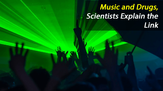
An ecstasy user describes the experience of being at a rave:

“I understood why the stage lights were bright and flashing, and why trance music is repetitive; the music and the drug perfectly complemented one another. It was as if a veil had been lifted from my eyes and I could finally see what everyone else was seeing. It was wonderful.”

There is a rich representation of drugs in popular music, and although studies have shown higher levels of drug use in listeners of some genres of music, the relationship is complex. Drug representations may serve to normalise use for some listeners, but drugs and music are powerful ways of strengthening social bonds. They both provide an identity and a sense of connection between people. Music and drugs can bring together people in a political way, too, as the response to attempts to close down illegal raves showed.

People tend to form peer groups with those who share their own cultural preferences, which may be symbolised through interlinked musical and substance choices. Although there are some obvious synergies between some music and specific drugs, such as electronic dance music and ecstasy, other links have developed in less obvious ways. Drugs are one, often minor, component of a broader identity and an important means of distinguishing the group from others.

Although it is important not to assume causality and overstate the links between some musical genres and different types of drug use, information about preferences is useful in targeting and tailoring interventions, such as harm reduction initiatives, at music festivals.

https://www.technologynetworks.com/neuroscience/articles/music-and-drugs-scientists-explain-the-link-296886?utm_campaign=NEWSLETTER_TN_Neuroscience_2017&utm_source=hs_email&utm_medium=email&utm_content=60340987&_hsenc=p2ANqtz–WbyIooqediqm4Mr6D09zjNCyCmjIe-6JF5OpygCiR3HaX93JSj3dyP1fGYyKLhvXSaI-EheJTPpuOIN_2UXpdsA4ewg&_hsmi=60340987

What happens in the brain before a bungee jump

Surjo R. Soekadar, psychiatrist and neuroscientist at the University of Tübingen, and his doctoral candidate Marius Nann have for the very first time succeeded in measuring the readiness potential, outside a laboratory and under extreme conditions, namely prior to a 192-meter bungee jump.

The readiness potential is a characteristic electrical voltage shift in the brain that indicates an upcoming willful act, and that appears even before a person becomes aware of his/her own conscious decision to act. The results of the study will be published in an international journal later this spring but are now available online: https://www.biorxiv.org/content/early/2018/01/27/255083 (DOI:
https://doi.org/10.1101/255083)

The readiness potential was first described in 1964 by Hans-Helmut Kornhuber and Lüder Deecke, who measured the brain waves of a test person over hundreds of finger movements and under strict laboratory conditions. Despite numerous studies, the readiness potential has never been measured in a real-life situation: Since the voltage shift is in the range of only a few millionths of a volt, only measurements under laboratory conditions were considered possible.

To advance the development of brain-machine interfaces, the researchers from Tübingen wanted to find out whether the readiness potential can be assessed in everyday environments. In addition, they were interested in whether the willpower necessary for initiating an act would influence the characteristics of the brain potential. For the study, two semi-professional cliff divers agreed to have their brain waves recorded before jumping from the second tallest bungee jumping platform in Europe, the 192-meter Europa Bridge near Innsbruck in Austria.

After only a few jumps, the researchers were able to measure the readiness potential beyond any doubt. “Once again, the current experiment shows that the boundaries of the possible are shifting and that neurotechnology might soon be part of our everyday life,” Soekadar says. “The small number of jumps necessary for the experiment shows that the readiness potential prior to a bungee jump is very well expressed”, Nann explains.

This article has been republished from materials provided by The University of Tuebingen. Note: material may have been edited for length and content. For further information, please contact the cited source.

Reference:
Nann, M., Cohen, L. G., Deecke, L., & Soekadar, S. R. (2018). To jump or not to jump: The Bereitschaftspotential required to jump into 192-meter abyss. arXiv preprint arXiv:1801.07244.

https://www.technologynetworks.com/neuroscience/news/taking-neuroscience-over-the-edge-exploring-the-brains-readiness-potential-prior-to-a-bungee-jump-296942?utm_campaign=Newsletter_TN_BreakingScienceNews&utm_source=hs_email&utm_medium=email&utm_content=60306590&_hsenc=p2ANqtz-8cO5QFZovVgMC9zlBl6UK9zld0IglpGEZRtDFFBjG_caWEqxM9VD_6ZqqTZPoGcZiMaQbIw-Kb5k_J2FTQkKGb0Odu3g&_hsmi=60306590

Some stroke patients can be successfully treated up to 16 hours after stroke

The time window for treating stroke patients may be considerably longer than doctors previously thought. Results from a multi-center clinical trial published Jan. 24 in the New England Journal of Medicine show that certain stroke patients can be successfully treated up to 16 hours after stroke.

Key to the success of the treatment was the use of advanced brain imaging that can identify stroke patients who still have salvageable brain tissue if blood flow can be restored. The study demonstrated that physically removing brain clots up to 16 hours after symptom onset in these selected patients led to improved outcomes compared to standard medical therapy.

University of Iowa Hospitals and Clinics was one of 38 stroke centers involved in the new study known as the Endovascular Therapy Following Imaging Evaluation for the Ischemic Stroke (DEFUSE 3) trial. The trial was led by researchers at Stanford University School of Medicine in California and funded by the National Institute of Neurological Disorders and Stroke (NINDS), part of the National Institutes of Health. The findings also were presented Jan. 24 at the International Stroke Conference in Los Angeles.

“This study will provide us with the opportunity to offer a life-saving and life-changing therapy to thousands of people,” says Santiago Ortega, MD, clinical assistant professor and neurointerventional surgery director in the UI Department of Neurology and principal investigator for the UI DEFUSE 3 study site. “As the top enrolling center in the study, the UI was an important contributor to this advance in the field of stroke and improving the treatment of this devastating disease.”


Saving brain tissue

Ischemic stroke occurs when a cerebral blood vessel becomes blocked, cutting off the delivery of oxygen and nutrients to brain tissue. The area immediately surrounding the blockage is known as the core. When a stroke occurs, brain tissue in the core typically cannot be saved, and the core can expand over time. However, physicians have long believed that tissue in the area surrounding the core (known as the ischemic penumbra) can potentially be saved based on how quickly blood flow can be restored.

The key to the new study was an advanced brain scan called perfusion imaging, which measures blood flow in brain tissue. Perfusion imaging can identify patients with brain tissue that can still be salvaged by removing the blockage.

Using an automated software to analyze perfusion MRI or CT scans, the DEFUSE 3 researchers identified patients thought to have salvageable tissue up to 16 hours after stroke onset. The participants were randomized to either receive endovascular thrombectomy plus standard medical therapy or medical therapy alone.

Endovascular thrombectomy, or the physical removal of the blockage, is currently approved for use up to six hours following onset of stroke symptoms. The DEFUSE 3 researchers discovered that this intervention can be effective up to 16 hours after symptoms begin in this select group of patients. The findings also showed that patients in the thrombectomy group had substantially better outcomes 90 days after treatment compared to those in the medical therapy control group. For example, 45 percent of the patients treated with the clot removal procedure achieved functional independence compared to 17 percent in the control group. Thrombectomy treatment was also associated with improved survival. According to the results, 14 percent of the treated group had died within 90 days of the study, compared to 26 percent in the control group.

The DEFUSE 3 results along with other recent trials are so compelling they have provided a basis for significant changes in the guidelines for managing acute stroke. The new guidelines, announced Jan. 24 by the American Heart Association/American Stroke Association, recommend thrombectomy in eligible patients six to 16 hours after a stroke.


DEFUSE 3 study at UI

The DEFUSE 3 study was supported by NINDS’ StrokeNet, a network of hospitals providing research infrastructure for multi-site clinical trials in stroke care. The UI is one of StrokeNet’s 25 Regional Coordinating Centers (RCC), and the UI site enrolled the highest number of participants (15 of 182) in the DEFUSE 3 study. Enrique Leira, MD, director of the UI Comprehensive Stroke Center is the principal investigator for the StrokeNet UIRCC. Leira, along with Harold Adams, MD, UI professor of neurology, have been instrumental in establishing the UI Comprehensive Stroke Center as a national leader in stroke care.

“UI Comprehensive Stroke Center team has worked hard on developing an acute endovascular protocol and infrastructure to expedite the assessment and treatment of patients suffering from this devastating disease in our state,” Ortega says. “Numerous members from different departments including, EMS personnel, emergency medicine physicians and nurses, radiology technologists, neurologists, anesthesiologist and OR nurses, and neurointerventional surgeons are constantly involved in the process and deserve recognition for their important contributions to the success of this study.”

Colin Derdeyn, MD, professor and DEO of the UI Department of Radiology, who also served on the DEFUSE 3 central steering committee, adds his praise for the study team’s work.

“Our success in this trial is related to several factors, one being Dr. Ortega’s great leadership and the work of his team, of which I am glad to be a part. Another major reason is the incredible network built by Dr. Adams and Dr. Leira to provide fast, state-of-the-art care to patients affected by acute stroke across Iowa,” Derdeyn says. “I am not aware of another institution in the country that supports so many small rural hospitals and accepts so many stroke patients in transfer for advanced care and access to really important trials like this.”

https://medcom.uiowa.edu/theloop/news/stroke-patients-can-be-successfully-treated-up-to-16-hours-after-stroke

Eyes and eardrums move in sync

Simply moving the eyes triggers the eardrums to move too, says a new study by Duke University neuroscientists.

The researchers found that keeping the head still but shifting the eyes to one side or the other sparks vibrations in the eardrums, even in the absence of any sounds.

Surprisingly, these eardrum vibrations start slightly before the eyes move, indicating that motion in the ears and the eyes are controlled by the same motor commands deep within the brain.

“It’s like the brain is saying, ‘I’m going to move the eyes, I better tell the eardrums, too,’” said Jennifer Groh, a professor in the departments of neurobiology and psychology and neuroscience at Duke.

The findings, which were replicated in both humans and rhesus monkeys, provide new insight into how the brain coordinates what we see and what we hear. It may also lead to new understanding of hearing disorders, such as difficulty following a conversation in a crowded room.

The paper appeared Jan. 23 in Proceedings of the National Academy of Sciences.

It’s no secret that the eyes and ears work together to make sense of the sights and sounds around us. Most people find it easier to understand somebody if they are looking at them and watching their lips move. And in a famous illusion called the McGurk Effect, videos of lip cues dubbed with mismatched audio cause people to hear the wrong sound.

But researchers are still puzzling over where and how the brain combines these two very different types of sensory information.

“Our brains would like to match up what we see and what we hear according to where these stimuli are coming from, but the visual system and the auditory system figure out where stimuli are located in two completely different ways,” Groh said. “The eyes are giving you a camera-like snapshot of the visual scene, whereas for sounds, you have to calculate where they are coming from based on differences in timing and loudness across the two ears.”

Because the eyes are usually darting about within the head, the visual and auditory worlds are constantly in flux with respect to one another, Groh added.

In an experiment designed by Kurtis Gruters, a formal doctoral student in Groh’s lab and co-first author on the paper, 16 participants were asked to sit in a dark room and follow shifting LED lights with their eyes. Each participant also wore small microphones in their ear canals that were sensitive enough to pick up the slight vibrations created when the eardrum sways back and forth.

Though eardrums vibrate primarily in response to outside sounds, the brain can also control their movements using small bones in the middle ear and hair cells in the cochlea. These mechanisms help modulate the volume of sounds that ultimately reach the inner ear and brain, and produce small sounds known as otoacoustic emissions.

Gruters found that when the eyes moved, both eardrums moved in sync with one another, one side bulging inward at the same time the other side bulged outward. They continued to vibrate back and forth together until shortly after the eyes stopped moving. Eye movements in opposite directions produced opposite patterns of vibrations.

Larger eye movements also triggered bigger vibrations than smaller eye movements, the team found.

“The fact that these eardrum movements are encoding spatial information about eye movements means that they may be useful for helping our brains merge visual and auditory space,” said David Murphy, a doctoral student in Groh’s lab and co-first author on the paper. “It could also signify a marker of a healthy interaction between the auditory and visual systems.”

The team, which included Christopher Shera at the University of Southern California and David W. Smith of the University of Florida, is still investigating how these eardrum vibrations impact what we hear, and what role they may play in hearing disorders. In future experiments, they will look at whether up and down eye movements also cause unique signatures in eardrum vibrations.

“The eardrum movements literally contain information about what the eyes are doing,” Groh said. “This demonstrates that these two sensory pathways are coupled, and they are coupled at the earliest points.”

Cole Jenson, an undergraduate neuroscience major at Duke, also coauthored the new study.

CITATION: “The Eardrums Move When the Eyes Move: A Multisensory Effect on the Mechanics of Hearing,” K. G. Gruters, D. L. K. Murphy, Cole D. Jensen, D. W. Smith, C. A. Shera and J. M. Groh. Proceedings of the National Academy of Sciences, Jan. 23, 2018. DOI: 10.1073/pnas.1717948115

Desire and Dislike Mapped in the Amygdala

The amygdala is a tiny hub of emotions where in 2016 a team led by MIT neuroscientist Kay Tye found specific populations of neurons that assign good or bad feelings, or “valence,” to experience. Learning to associate pleasure with a tasty food, or aversion to a foul-tasting one, is a primal function and key to survival.

In a new study in Cell Reports, Tye’s team at the Picower Institute for Learning and Memory returns to the amygdala for an unprecedentedly deep dive into its inner workings. Focusing on a particular section called the basolateral amygdala, the researchers show how valence-processing circuitry is organized and how key neurons in those circuits interact with others. What they reveal is a region with distinct but diverse and dynamic neighborhoods where valence is sorted out by both connecting with other brain regions and sparking cross-talk within the basolateral amygdala itself.

“Perturbations of emotional valence processing is at the core of many mental health disorders,” says Tye, associate professor of neuroscience at the Picower Institute of Learning and Memory and the Department of Brain and Cognitive Sciences. “Anxiety and addiction, for example, may be an imbalance or a misassignment of positive or negative valence with different stimuli.”

Despite the importance of valence assignment in both healthy behavior and psychiatric disorders, neuroscientists don’t know how the process really works. The new study therefore sought to expose how the neurons and circuits are laid out and how they interact.

Bitter, sweet

To conduct the study, lead author Anna Beyeler, a former postdoc in Tye’s lab and currently a faculty member at the University of Bordeaux in France, led the group in training mice to associate appealing sucrose drops with one tone and bitter quinine drops with another. They recorded the response of different neurons in the basolateral amygdala when the tones were played to see which ones were associated with the conditioned learned valence of the different tastes. They labeled those key neurons associated with valence encoding and engineered them to become responsive to pulses of light. When the researchers then activated them, they recorded the electrical activity not only of those neurons but also of many of their neighbors to see what influence their activity had in local circuits.

They also found, labeled, and made similar measurements among neurons that became active on the occasion that a mouse actually licked the bitter quinine. With this additional step, they could measure not only the neural activity associated with the learned valence of the bitter taste but also that associated with the innate reaction to the actual experience.

Later in the lab, they used tracing technologies to highlight three different kinds of neurons more fully, visualizing them in distinct colors depending on which other region they projected their tendrilous axons to connect with. Neurons that project to a region called the nucleus accumbens are predominantly associated with positive valence, and those that connect to the central amygdala are mainly associated with negative valence. They found that neurons uniquely activated by the unconditioned experience of actually tasting the quinine tended to project to the ventral hippocampus.

In all, the team mapped over 1,600 neurons.

To observe the three-dimensional configuration of these distinct neuron populations, the researchers turned the surrounding brain tissues clear using a technique called CLARITY, invented by Kwanghun Chung, assistant professor of chemical engineering and neuroscience and a colleague in the Picower Institute.

Neighborhoods without fences

Beyeler, Tye, and their co-authors were able to make several novel observations about the inner workings of the basolateral amygdala’s valence circuitry.

One finding was that the different functional populations of neurons tended to cluster together in neighborhoods, or “hotspots.” For example, picturing the almond-shaped amygdala as standing upright on its fat bottom, the neurons projecting to the central amygdala tended to cluster toward the point at the top and then on the right toward the bottom. Meanwhile the neurons that projected to the nucleus accumbens tended to run down the middle, and the ones that projected to the hippocampus were clustered toward the bottom on the opposite side from the central amygdala projectors.

Despite these trends, the researchers also noted that the neighborhoods were hardly monolithic. Instead, neurons of different types frequently intermingled creating a diversity where the predominant neuron type was never far from at least some representatives of the other types.

Meanwhile, their electrical activity data revealed that the different types exerted different degrees of influence over their neighbors. For example, neurons projecting to the central amygdala, in keeping with their association with negative valence, had a very strong inhibitory effect on neighbors, while nucleus accumbens projectors had a smaller influence that was more balanced between excitation and inhibition.

Tye speculates that the intermingling of neurons of different types, including their propensity to influence each other with their activity, may provide a way for competing circuits to engage in cross-talk.

“Perhaps the intermingling that there is might facilitate the ability of these neurons to influence each other,” says Tye.

Notably, Tye’s research has indicated the projections the different cell types may appear immutable, but the influence those cells have over each other is flexible. The basolateral amygdala may therefore be arranged to both assign valence and negotiate it, for instance in those situations when a mouse spies some desirable cheese, but that mean cat is also nearby.

“This helps us understand how form might give rise to function,” says Tye.

Reference:
Beyeler et al. “Organization of Valence-Encoding and Projection Defined Neurons in the Basolateral Amygdala” Cell Reports. https://doi.org/10.1016/j.celrep.2017.12.097

https://www.technologynetworks.com/neuroscience/news/valence-mapped-brain-study-reveals-roots-of-desire-and-dislike-in-the-amygdala-296668?utm_campaign=NEWSLETTER_TN_Neuroscience_2017&utm_source=hs_email&utm_medium=email&utm_content=60184122&_hsenc=p2ANqtz-_uyMIjTK1pmq-79zMcyJIvQNsa8i7gH9l8Tn-_75Taz2opCD4t1otYN6OBmeI-iAKoenGO8wKWNZ7VV6E_JcYum4fHlA&_hsmi=60184122

Pupil size changes with different stages of sleep, getting smaller as sleep gets deeper, in mice

When people are awake, their pupils regularly change in size. Those changes are meaningful, reflecting shifting attention or vigilance, for example. Now, researchers reporting in Current Biology on January 18 have found in studies of mice that pupil size also fluctuates during sleep. They also show that pupil size is a reliable indicator of sleep states.

“We found that pupil size rhythmically fluctuates during sleep,” says Daniel Huber of the University of Geneva in Switzerland. “Intriguingly, these pupil fluctuations follow the sleep-related brain activity so closely that they can indicate with high accuracy the exact stage of sleep—the smaller the pupil, the deeper the sleep.”

Studies of pupil size had always been a challenge for an obvious reason: people and animals generally sleep with their eyes closed. Huber says that he and his colleagues were inspired to study pupil size in sleep after discovering that their laboratory mice sometimes sleep with their eyes open. They knew that pupil size varies strongly during wakefulness. What, they wondered, happened during sleep?

To investigate this question, they developed a novel optical pupil-tracking system for mice. The device includes an infrared light positioned close to the head of the animal. That invisible light travels through the skull and brain to illuminate the back of the eye. When the eyes are imaged with an infrared camera, the pupils appear as bright circles. Thanks to this new method, it was suddenly possible to track changes in pupil size accurately, particularly when the animals snoozed naturally with their eyelids open.

Their images show that mouse pupils rhythmically fluctuate during sleep and that those fluctuations are not at all random; they correlate with changes in sleep states.

Further experiments showed that changes in pupil size are not just a passive phenomenon, either. They are actively controlled by the parasympathetic autonomic nervous system. The evidence suggests that in mice, at least, pupils narrow in deep sleep to protect the animals from waking up with a sudden flash of light.

“The common saying that ‘the eyes are the window to the soul’ might even hold true behind closed eyelids during sleep,” Özge Yüzgeç, the student conducting the study, says. “The pupil continues to play an important role during sleep by blocking sensory input and thereby protecting the brain in periods of deep sleep, when memories should be consolidated.”

Huber says they would like to find out whether the findings hold in humans and whether their new method can be adapted in the sleep clinic. “Inferring brain activity by non-invasive pupil tracking might be an interesting alternative or complement to electrode recordings,” he says.

Reference:

Yüzgeç, Ö., Prsa, M., Zimmermann, R., & Huber, D. (2018). Pupil Size Coupling to Cortical States Protects the Stability of Deep Sleep via Parasympathetic Modulation. Current Biology. doi:10.1016/j.cub.2017.12.049

https://www.technologynetworks.com/neuroscience/news/pupil-size-couples-to-cortical-states-to-protect-deep-sleep-stability-296519?utm_campaign=NEWSLETTER_TN_Neuroscience_2017&utm_source=hs_email&utm_medium=email&utm_content=60184122&_hsenc=p2ANqtz-_uyMIjTK1pmq-79zMcyJIvQNsa8i7gH9l8Tn-_75Taz2opCD4t1otYN6OBmeI-iAKoenGO8wKWNZ7VV6E_JcYum4fHlA&_hsmi=60184122

Brain Connections Set Creative Thinkers Apart

By Tereza Pultarova

Being creative is all about making connections — in your brain, that is.

In a new study, scientists found that the brains of highly creative people have more connections among three specific regions compared to the brains of less creative thinkers. Plus, the more-creative brains were better able to fire up these regions in coordinated way compared with other brains.

The three brain regions are ones that scientists understand well, said lead study author Roger Beaty, a postdoctoral fellow studying cognitive neuroscience at Harvard University. They include the default network, which is involved in spontaneous thinking and imagination; the salience network, which picks up on important information from the environment; and the executive control network, which is involved in cognitive control functions and evaluation.

And though the default network seems like it should be the key source of creativity, people need the salience and the executive control networks to act as a sort of inner critic that judges whether ideas are any good or useful for the given task, Beaty told Live Science.

“You have these three different systems that are all located in different parts of the brain, but they are all co-activated at once,” Beaty said. “People who are better able to co-activate them [came] up with more-creative responses.”

To measure creativity and brain connections, the researchers scanned the brains of about 160 participants using functional magnetic resonance imaging (fMRI), a tool that monitors brain activity by measuring changes in blood flow in various areas the brain. With their heads inside the scanner, the participants were asked to perform a creative-thinking task called divergent thinking. This involves coming up with creative ways to use common objects, such as a knife, a cup or a brick.

“Just thinking about new and unusual ways to use these [objects] has been shown to be a valid way of [measuring] creative thinking,” Beaty said.

The researchers found that performing the divergent-thinking task simultaneously activated the three different networks in the brain. And the greater the interconnectedness and synchronization of these three networks, the better the performance in the divergent-thinking task. In other words, the more connected and in-sync the brain is, the better it does on a creative task.

After establishing what distinguishes creative people’s brains from those of their less creative peers, the researchers wanted to see whether they could reverse the process and use brain activity as a predictor of creative performance.

“We had data sets of previously published studies were people were doing similar creative thinking tasks, and we wanted to see whether someone with weak connectivity in [these networks] has less-creative ideas than someone with stronger connectivity,” Beaty said. “And that’s what we found across three data sets.”

The researchers are now planning to look for similar patterns of brain activity in specific areas of creativity such as writing or music, Beaty said. In addition, the scientists want to find out if the brain activity can in fact change as people become more proficient at certain skills, he said.

The study was published today (Jan. 15) in the journal Proceedings of the National Academy of Sciences.

https://www.livescience.com/61428-brain-connections-creativity.html

Noninvasive brain intervention shown to reduce PTSD symptoms

A noninvasive intervention that uses brainwave mirroring technology improved symptoms of post-traumatic stress, insomnia, anxiety and depressive mood for up to 6 months in service members and veterans, according to pilot study findings published in Military Medical Research.

“Ongoing symptoms of post-traumatic stress, whether clinically diagnosed or not, are a pervasive problem in the military,” Charles H. Tegeler, MD, professor of neurology at Wake Forest School of Medicine, said in a press release. “Medications are often used to help control specific symptoms, but can produce side effects. Other treatments may not be well tolerated, and few show a benefit for the associated sleep disturbance. Additional noninvasive, nondrug therapies are needed.”

In this study, researchers used high-resolution, relational, resonance-based, electroencephalic mirroring (HIRREM), a noninvasive neurotechnology that translates dominant brain frequencies into audible tones to support the auto-calibration of neural oscillations in real time. They collected data on symptoms of post-traumatic stress (PTS), insomnia, depression and anxiety from 18 military personnel with military-related PTS who received an average of 19.5 HIRREM sessions over 12 days. Researchers conducted follow-up online interviews at 1-, 3- and 6-months.

Tegeler and colleagues observed clinically significant reductions in all symptom scores immediately after intervention completion, with durability through 6 months. Through the first two follow-up visits, 83% of participants reported PTS scores that were at least 10 points lower than baseline and 78% reported insomnia scores that were at least seven points lower than baseline. For 15 participants with a history of traumatic brain injury or concussion, there were durable reductions in concussion-related symptoms. Additionally, participants also experienced significant improvements in blood pressure measures such as heart rate variability and baroreflex sensitivity. No adverse events were reported.

“We observed reductions in post-traumatic symptoms, including insomnia, depressive mood and anxiety that were durable through 6 months after the use of HIRREM, but additional research is needed to confirm these initial findings,” Tegeler said in the release. “This study is also the first to report improvement in heart rate variability and baroreflex sensitivity — physiological responses to stress — after the use of an intervention for service members or veterans with ongoing symptoms of post-traumatic stress.” – by Savannah Demko

https://www.healio.com/psychiatry/ptsd/news/online/%7B8ae1a121-d40d-41a0-88df-d9ccd96c6523%7D/noninvasive-intervention-reduces-post-traumatic-stress-symptoms-in-military?utm_source=selligent&utm_medium=email&utm_campaign=psychiatry%20news&m_bt=1162769038120

Some video games are good for older adults’ brains

If you’re between 55 and 75 years old, you may want to try playing 3D platform games like Super Mario 64 to stave off mild cognitive impairment and perhaps even prevent Alzheimer’s disease.

That’s the finding of a new Canadian study by Université de Montréal psychology professors Gregory West, Sylvie Belleville and Isabelle Peretz. Published in PLOS One, it was done in cooperation with the Institut universitaire de gériatrie de Montréal (IUGM), Benjamin Rich Zendel of Memorial University in Newfoundland, and Véronique Bohbot of Montreal’s Douglas Hospital Research Centre.

In two separate studies, in 2014 and 2017, young adults in their twenties were asked to play 3D video games of logic and puzzles on platforms like Super Mario 64. Findings showed that the gray matter in their hippocampus increased after training.

The hippocampus is the region of the brain primarily associated with spatial and episodic memory, a key factor in long-term cognitive health. The gray matter it contains acts as a marker for neurological disorders that can occur over time, including mild cognitive impairment and Alzheimer’s.

West and his colleagues wanted to see if the results could be replicated among healthy seniors.

The research team recruited 33 people, ages 55 to 75, who were randomly assigned to three separate groups. Participants were instructed to play Super Mario 64 for 30 minutes a day, five days a week, take piano lessons (for the first time in their life) with the same frequency and in the same sequence, or not perform any particular task.

The experiment lasted six months and was conducted in the participants’ homes, where the consoles and pianos, provided by West’s team, were installed.

The researchers evaluated the effects of the experiment at the beginning and at the end of the exercise, six months later, using two different measurements: cognitive performance tests and magnetic resonance imaging (MRI) to measure variations in the volume of gray matter. This enabled them to observe brain activity and any changes in three areas:

the dorsolateral prefrontal cortex that controls planning, decision-making and inhibition;
the cerebellum that plays a major role in motor control and balance; and
the hippocampus, the centre of spatial and episodic memory.
According to the MRI test results, only the participants in the video-game cohort saw increases in gray matter volume in the hippocampus and cerebellum. Their short-term memory also improved.

The tests also revealed gray matter increases in the dorsolateral prefrontal cortex and cerebellum of the participants who took piano lessons, whereas some degree of atrophy was noted in all three areas of the brain among those in the passive control group.

What mechanism triggers increases in gray matter, especially in the hippocampus, after playing video games? “3-D video games engage the hippocampus into creating a cognitive map, or a mental representation, of the virtual environment that the brain is exploring.,” said West. “Several studies suggest stimulation of the hippocampus increases both functional activity and gray matter within this region.”

Conversely, when the brain is not learning new things, gray matter atrophies as people age. “The good news is that we can reverse those effects and increase volume by learning something new, and games like Super Mario 64, which activate the hippocampus, seem to hold some potential in that respect,” said West. Added Belleville: “These findings can also be used to drive future research on Alzheimer’s, since there is a link between the volume of the hippocampus and the risk of developing the disease.”

“It remains to be seen,” concluded West, “whether it is specifically brain activity associated with spatial memory that affects plasticity, or whether it’s simply a matter of learning something new.”

http://nouvelles.umontreal.ca/en/article/2017/12/06/some-video-games-are-good-for-older-adults-brains/

Huntington’s breakthrough may stop disease


Prof Sarah Tabrizi , from the UCL Institute of Neurology, led the trials

By James Gallagher

The defect that causes the neurodegenerative disease Huntington’s has been corrected in patients for the first time, the BBC has learned. An experimental drug, injected into spinal fluid, safely lowered levels of toxic proteins in the brain. The research team, at University College London, say there is now hope the deadly disease can be stopped.

Experts say it could be the biggest breakthrough in neurodegenerative diseases for 50 years.

Huntington’s is one of the most devastating diseases. Some patients described it as Parkinson’s, Alzheimer’s and motor neurone disease rolled into one.

Peter Allen, 51, is in the early stages of Huntington’s and took part in the trial: “You end up in almost a vegetative state, it’s a horrible end.”

Huntington’s blights families. Peter has seen his mum Stephanie, uncle Keith and grandmother Olive die from it. Tests show his sister Sandy and brother Frank will develop the disease. The three siblings have eight children – all young adults, each of whom has a 50-50 chance of developing the disease.

The unstoppable death of brain cells in Huntington’s leaves patients in permanent decline, affecting their movement, behaviour, memory and ability to think clearly.

Peter, from Essex, told me: “It’s so difficult to have that degenerative thing in you.

“You know the last day was better than the next one’s going to be.”
Huntington’s generally affects people in their prime – in their 30s and 40s
Patients die around 10 to 20 years after symptoms start
About 8,500 people in the UK have Huntington’s and a further 25,000 will develop it when they are older

Huntington’s is caused by an error in a section of DNA called the huntingtin gene. Normally this contains the instructions for making a protein, called huntingtin, which is vital for brain development. But a genetic error corrupts the protein and turns it into a killer of brain cells.

The treatment is designed to silence the gene.

On the trial, 46 patients had the drug injected into the fluid that bathes the brain and spinal cord. The procedure was carried out at the Leonard Wolfson Experimental Neurology Centre at the National Hospital for Neurology and Neurosurgery in London. Doctors did not know what would happen. One fear was the injections could have caused fatal meningitis. But the first in-human trial showed the drug was safe, well tolerated by patients and crucially reduced the levels of huntingtin in the brain.

Prof Sarah Tabrizi, the lead researcher and director of the Huntington’s Disease Centre at UCL, told the BBC: “I’ve been seeing patients in clinic for nearly 20 years, I’ve seen many of my patients over that time die. For the first time we have the potential, we have the hope, of a therapy that one day may slow or prevent Huntington’s disease . This is of groundbreaking importance for patients and families.”

Doctors are not calling this a cure. They still need vital long-term data to show whether lowering levels of huntingtin will change the course of the disease. The animal research suggests it would. Some motor function even recovered in those experiments.

Peter, Sandy and Frank – as well as their partners Annie, Dermot and Hayley – have always promised their children they will not need to worry about Huntington’s as there will be a treatment in time for them. Peter told the BBC: “I’m the luckiest person in the world to be sitting here on the verge of having that. “Hopefully that will be made available to everybody, to my brothers and sisters and fundamentally my children.”

He, along with the other trial participants, can continue taking the drug as part of the next wave of trials. They will set out to show whether the disease can be slowed, and ultimately prevented, by treating Huntington’s disease carriers before they develop any symptoms.

Prof John Hardy, who was awarded the Breakthrough Prize for his work on Alzheimer’s, told the BBC: “I really think this is, potentially, the biggest breakthrough in neurodegenerative disease in the past 50 years. That sounds like hyperbole – in a year I might be embarrassed by saying that – but that’s how I feel at the moment.”

The UCL scientist, who was not involved in the research, says the same approach might be possible in other neurodegenerative diseases that feature the build-up of toxic proteins in the brain. The protein synuclein is implicated in Parkinson’s while amyloid and tau seem to have a role in dementias.

Off the back of this research, trials are planned using gene-silencing to lower the levels of tau.

Prof Giovanna Mallucci, who discovered the first chemical to prevent the death of brain tissue in any neurodegenerative disease, said the trial was a “tremendous step forward” for patients and there was now “real room for optimism”.

But Prof Mallucci, who is the associate director of UK Dementia Research Institute at the University of Cambridge, cautioned it was still a big leap to expect gene-silencing to work in other neurodegenerative diseases.

She told the BBC: “The case for these is not as clear-cut as for Huntington’s disease, they are more complex and less well understood. But the principle that a gene, any gene affecting disease progression and susceptibility, can be safely modified in this way in humans is very exciting and builds momentum and confidence in pursuing these avenues for potential treatments.”

The full details of the trial will be presented to scientists and published next year.

The therapy was developed by Ionis Pharmaceuticals, which said the drug had “substantially exceeded” expectations, and the licence has now been sold to Roche.

http://www.bbc.com/news/health-42308341