For The First Time, Scientists Have Made Synthetic DNA With 4 Additional Letters

by Mike McRae

Earth might have a dizzying array of life forms, but our biology ultimately remains a solitary data point – we simply don’t have a reference for life based on DNA different from our own. Now, scientists have taken matters into their hands to push the boundaries on what life could be like.

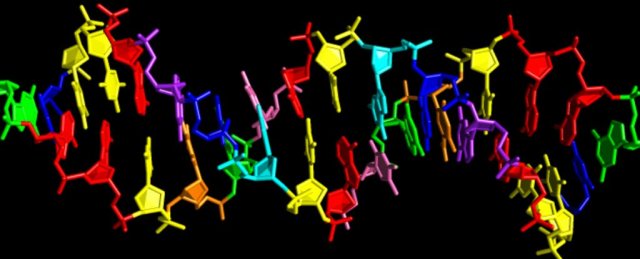
Research funded by NASA and led by the Foundation for Applied Molecular Evolution in the US has led to the creation of an entirely new flavour of the DNA double helix, one that has an additional four nucleotide bases.

It’s being called hachimoji DNA (from the Japanese words for ‘eight letters’) and it includes two new pairs to add to the existing partnerships of adenine (A) paired with thymine (T), and guanine (G) with cytosine (C).

This work to expand on nature’s own genetic recipe might sound a little familiar. The same scientists already successfully squeezed in two new letters in 2011. Only last year yet another version of an extended alphabet, also with six letters, was made to function inside a living organism.

Now, in what might seem like a case of overachievement, researchers have gone back to the drawing board to develop even more non-standard nucleotides.

They have a purpose for doubling the number of codes in the recipe book, though.

“By carefully analysing the roles of shape, size and structure in hachimoji DNA, this work expands our understanding of the types of molecules that might store information in extraterrestrial life on alien worlds,” says chemist Steven Benner.

We already know a lot about the stability and functionality of ‘natural’ DNA under a range of environmental conditions, and are slowly teasing apart possible scenarios describing its evolution from simpler organic materials to living chemistry.

But to really get a good sense of how a genetic system could evolve, we need to test the limits of its underlying chemistry.

Hachimoji DNA certainly allows for that. The new codes, labelled P, B, Z and S, are based on the same kind of nitrogenous molecules as existing ones, categorised as purines and pyrimidines.

Similarly, they link up with hydrogen bonds to form their own base pairs – S bonding with B, and P with Z.

That’s where the similarities fade out. These new ‘letters’ introduce dozens of new chemical parameters to the double helix structure that potentially affect how it zips and twists.

By devising models that predict the molecule’s stability and then observing actual structures made of this ‘alien’ DNA, researchers are better equipped what’s truly important when it comes to the fundamentals of a genetic template.

The researchers constructed hundreds of hachimoji helices made up of different configurations of natural and synthetic bases and then subjected them to a range of conditions to see how well they held up.

While there were a few minor differences in how the new letters behaved, there was no reason to believe hachimoji DNA wouldn’t work well as an information-carrying template that could mutate and evolve.

The team not only showed their synthetic letters could contribute to new codes without swiftly disintegrating, the sequences were also translated into synthetic RNA versions.

Their work falls well short of a second genesis. But a novel DNA format such as this is a step towards determining what living chemistry might – and might not – look like elsewhere in the Universe.

“Life detection is an increasingly important goal of NASA’s planetary science missions, and this new work will help us to develop effective instruments and experiments that will expand the scope of what we look for,” says NASA’s Planetary Science Division’s acting director, Lori Glaze.

Devising new bases that can operate alongside our own DNA also has applications closer to home, not only as a way to reprogram life with a different code base, but in our effort to build new kinds of nanostructures.

The sky really isn’t the limit with synthetic DNA. This is going to take us to the stars and back again.

This research was published in Science.

https://www.sciencealert.com/scientists-made-synthetic-dna-using-8-letters-and-it-could-help-us-find-aliens

The World Just Got Closer to a Controversial Mosquito ‘Wipe Out’ Experiment

by David Nield

Scientists are genetically modifying mosquitoes in a high-security lab – and they’re hoping the insects will help wipe out some of the mosquito-borne diseases that continue to plague communities worldwide.

It’s known as a gene drive: where mosquitoes modified to be incapable of passing on a particular virus are used to replace the existing population of insects over several generations, with the modified genes being passed on to all their offspring.

The idea has attracted controversy because it messes with the fundamentals of nature, but it’s now under consideration by the World Health Organisation (WHO). This particular testing has entered a new phase, NPR reports, with a large-scale release of genetically modified mozzies inside a facility in Terni, Italy.

“This will really be a breakthrough experiment,” entomologist Ruth Mueller, who runs the lab, told Rob Stein at NPR. “It’s a historic moment. It’s very exciting.”

Using the ‘molecular scissor’ editing technique CRISPR, a gene known as “doublesex” in the bugs has been altered. The gene transforms female mosquitoes, taking away their biting ability and making them infertile.

At the moment, the bugs are being released in cages designed to replicate their natural environments, with hot and humid air, and places to shelter. Artificial lights are used to simulate sunrise and sunset.

The idea is to see if the mosquitoes with CRISPR-edited genetic code can wipe out the unmodified insects inside the cages. It follows on from previous proof-of-concept studies that we’ve seen before.

Ultimately these mosquitoes could be released in areas hit by malaria, bringing the local mozzie population crashing down and saving human lives. The disease is responsible for more than 400,000 deaths every year – mostly young children.

Reducing those figures sounds like a great idea, so why the controversy? Well, many scientists are urging caution when it comes to altering genetic code at this fundamental level – we just don’t know what impact these genetically edited mosquitoes will have on the world around them.

For that reason the lab has been designed to minimise any chance that the specially engineered mosquitoes could escape. The testing has also been specifically located in Italy, where this mosquito species – Anopheles gambiae – wouldn’t be able to survive outside in the natural climate.

“This is a technology where we don’t know where it’s going to end,” Nnimmo Bassey, director of the Health of Mother Earth Foundation in Nigeria, told NPR. “We need to stop this right where it is. They’re trying to use Africa as a big laboratory to test risky technologies.”

Some experts think adding genetically modified mosquitoes to natural ecosystems could harm other plants and animals that depend on them. There are a lot of unknowns.

The team behind the new experiments counters the critique by saying they’re working slowly and methodically – and that the potential side effects are outweighed by the benefits of eradicating malaria.

At the moment scientists are targeting just one species of mosquito out of hundreds, and several more years of research and consultation are planned before genetically edited mozzies would ever be released.

“There’s going to be concerns with any technology,” one of the research team, Tony Nolan from Imperial College London in the UK, told NPR.

“But I don’t think you should throw out a technology without having done your best to understand what its potential is to be transformative for medicine. And, were it to work, this would be transformative.”

https://www.sciencealert.com/scientists-take-first-step-in-controversial-mosquito-gene-drive-experiment

A new form of neural communication in the brain has been discovered.

by PETER DOCKRILL

Scientists think they’ve identified a previously unknown form of neural communication that self-propagates across brain tissue, and can leap wirelessly from neurons in one section of brain tissue to another – even if they’ve been surgically severed.

The discovery offers some radical new insights about the way neurons might be talking to one another, via a mysterious process unrelated to conventionally understood mechanisms, such as synaptic transmission, axonal transport, and gap junction connections.

“We don’t know yet the ‘So what?’ part of this discovery entirely,” says neural and biomedical engineer Dominique Durand from Case Western Reserve University.

“But we do know that this seems to be an entirely new form of communication in the brain, so we are very excited about this.”

Before this, scientists already knew there was more to neural communication than the above-mentioned connections that have been studied in detail, such as synaptic transmission.

For example, researchers have been aware for decades that the brain exhibits slow waves of neural oscillations whose purpose we don’t understand, but which appear in the cortex and hippocampus when we sleep, and so are hypothesised to play a part in memory consolidation.

“The functional relevance of this input‐ and output‐decoupled slow network rhythm remains a mystery,” explains neuroscientist Clayton Dickinson from the University of Alberta, who wasn’t involved in the new research but has discussed it in a perspective article.

“But [it’s] one that will probably be solved by an elucidation of both the cellular and the inter‐cellular mechanisms giving rise to it in the first place.”

To that end, Durand and his team investigated slow periodic activity in vitro, studying the brain waves in hippocampal slices extracted from decapitated mice.

What they found was that slow periodic activity can generate electric fields which in turn activate neighbouring cells, constituting a form of neural communication without chemical synaptic transmission or gap junctions.

“We’ve known about these waves for a long time, but no one knows their exact function and no one believed they could spontaneously propagate,” Durand says.

“I’ve been studying the hippocampus, itself just one small part of the brain, for 40 years and it keeps surprising me.”

This neural activity can actually be modulated – strengthened or blocked – by applying weak electrical fields and could be an analogue form of another cell communication method, called ephaptic coupling.

The team’s most radical finding was that these electrical fields can activate neurons through a complete gap in severed brain tissue, when the two pieces remain in close physical proximity.

“To ensure that the slice was completely cut, the two pieces of tissue were separated and then rejoined while a clear gap was observed under the surgical microscope,” the authors explain in their paper.

“The slow hippocampal periodic activity could indeed generate an event on the other side of a complete cut through the whole slice.”

If you think that sounds freaky, you’re not the only one. The review committee at The Journal of Physiology – in which the research has been published – insisted the experiments be completed again before agreeing to print the study.

Durand et al. dutifully complied, but sound pretty understanding of the cautiousness, all things considered, given the unprecedented weirdness of the observation they’re reporting.

“It was a jaw-dropping moment,” Durand says, “for us and for every scientist we told about this so far.”

“But every experiment we’ve done since to test it has confirmed it so far.”

It’ll take a lot more research to figure out if this bizarre form of neural communication is taking place in human brains – let alone decoding what exact function it performs – but for now, we’ve got new science that’s shocking in all kinds of ways, as Dickson adroitly observes.

“While it remains to be seen if the [findings] are relevant to spontaneous slow rhythms that occur in both cortical and hippocampal tissue in situ during sleep and sleep‐like states,” Dickson writes, “they should probably (and quite literally) electrify the field.”

The findings are reported in The Journal of Physiology.

https://www.sciencealert.com/neuroscientists-say-they-ve-found-an-entirely-new-form-of-neural-communication

Scientists Produce Rigorous Study of Why Grapes Spark in the Microwave

by Ryan F. Mandelbaum

A paper published Monday in a well-known science journal begins with the following sentence: “It is a truth universally acknowledged that a pair of grape hemispheres exposed to intense microwave radiation will spark, igniting a plasma.” A universally acknowledged truth indeed… but what causes this microwave marvel?

If you’re not familiar, putting grapes into a microwave to make sparks has become a popular YouTube trick. This new research from Canadian scientists shows that worthwhile advances can come from anywhere, even by studying something sort of silly.

“This is a regime that hasn’t been significantly studied before,” one of the paper’s authors, Pablo Bianucci from Concordia University in Montreal, told Gizmodo.

The trick usually shows two grape halves connected by a thin sliver of skin. After a few seconds of being microwaved, they begin to spark. Though various explanations exist online, researchers wanted to study the phenomenon more rigorously.

The researcher imaged both sliced grapes and hydrogel beads—made from a material that absorbs lots of water—as they sparked in the microwave. They realized quickly that the grape skin wasn’t required in order to get the sparks, as evidenced by the sparking in the hydrogel beads, held together only by their weight and the shape of the dish they sat in, according to the research published in the Proceedings of the National Academy of Sciences.

The specific geometry of two touching water-filled circular objects in an electromagnetic field creates resonances concentrated at the point where the spheres or half-spheres intersect. This becomes a very small hotspot with a high energy density, enough to create plasma out of the ions in the region where the objects touch.

Is the research worth publishing in a journal as high-profile as PNAS? The paper’s editor, University of Illinois chemistry professor Catherine Murphy, certainly thought so. “The fact that they were rigorous enough to pass through the process of peer review is a testament that they’re doing a good job on the technical end,” she told Gizmodo.

But the paper is far more than a gimmick, Murphy said. This sort of research on directed energy could find important use in other directed-energy systems, such as explosives or high-intensity laser pulses. Additionally, the paper presents a way to image electric fields in these sorts of physical setups, and could lead to advances in photonics more generally.

https://gizmodo.com/scientists-produce-rigorous-study-of-why-grapes-spark-i-1832660386

Education Does not Improve Adaptability of Brain in Old Age, Study Suggests

By Alejandra Viviescas

A higher level of education is not related to better cognitive reserve — the ability of the adult brain to maintain normal cognitive function in the presence of neurodegeneration — in old age, a study suggests. However, the study, titled “Education and cognitive reserve in old age,” did find that it allowed people to store more information before reaching old age. It was published in the journal Neurology.

Higher education levels are widely associated with a higher cognitive reserve, lower risk of dementia, and delayed cognitive decline — the reduced storage capacity in the brain that usually occurs as a person ages. However, scientific evidence supporting these claims is controversial. Some studies suggest that this association is mostly due to the connection between education and a higher acquisition of knowledge rather than higher adaptability.

To assess the contribution of education to cognitive reserve in old age, researchers from Rush University in Chicago analyzed 2,899 participants (older than 50 years of age; average age of 77.8 years) who participated in two ongoing clinical studies: the Religious Orders Study, which began in 1994 and included older Catholic clergy members from across the U.S.; and the Memory and Aging Project, which began in 1997 and involved older laypeople from the Chicago metropolitan region.

At the time of enrollment, none of the participants had been diagnosed with dementia.

They were followed for an average of eight years; 2,143 (73.9%) were women, and 2,569 (88.6%) were white. All participants took cognitive tests once a year, and data were collected between 1994 and 2018.

Researchers evaluated two subgroups, the first one — the incident dementia subgroup — consisted of 696 participants who developed incident dementia during follow-up over a mean of 10.5 years. The second one — the incident dementia neuropathologically examined subgroup — included 405 individuals who died during follow-up and underwent an autopsy to assess if they had any neurodegenerative conditions.

Participants had a mean of 16.3 years of education, ranging from zero to 30. Higher education was associated with an initial higher rate of global cognition at a younger age but not with more significant cognitive change. This means that more educated people had a high storage capacity at the beginning of the study, but did not show greater cognitive adaptability.

There was a quicker decline in cognition in patients who developed dementia about 1.8 years before diagnosis. The level of education did not alter this decrease.

In the patients who had died, there was a faster cognition decline approximately 3.4 years before death. The level of education did not alter this decline, but researchers noted that in individuals with higher education, this decline started about 0.2 years earlier.

People with higher education were less likely to have areas of dead tissue in the brain. “There have been previous reports linking higher level of education with a lower risk of stroke consistent with the present findings,” according to the researchers. Higher education was not associated with any other neuropathology.

“The results suggest that the contribution of education to cognitive reserve is limited to its association with premorbid cognitive level and does not involve an association with cognitive aging trajectories,” the researchers wrote.

“That education apparently contributes little to cognitive reserve is surprising given its association with cognitive growth and changes in brain structure. However, formal education typically ends decades before old age begins … This implies that influences on cognitive reserve vary over time, with recent experiences more influential than remote experiences such as schooling,” they added.

The researchers noted that most individuals had some level of education, which might underestimate the effects on a non-educated group. Therefore, further studies that evaluate a higher sample of participants with less education would help them better understand the association between education and cognition.

Education Does not Improve Adaptability of Brain in Old Age, Study Suggests

Men Who Can Do More Than 40 Push-Ups Far Less Likely To Develop Heart Disease

Here’s one way to predict your heart health: get down and give me 41. A new study finds that men who can perform at least 40 push-ups in one attempt are much less likely to suffer from heart disease within the next 10 years.

Researchers from the Harvard T.H. Chan School of Public health say their report is the first to show how push-up capacity is linked to heart disease. They found that middle-aged men who can log more than 40 push-ups in a single try have a 96% reduced risk of developing the potentially deadly condition and other related ailments, such as heart failure, compared to those who can complete no more than 10 push-ups.

For their study, the authors reviewed health data from 1,104 active male firefighters taken annually from 2000 to 2010. At the start of the study, the average participant was about 40 years old with an average body mass index of 28.7. The firefighters were tasked with performing as many push-ups as they could, and their treadmill tolerance was also tested.

By the end of the study period, 37 participants suffered from a heart disease-related condition — and 36 of those men weren’t able to log more than 40 push-ups in the initial test. The results of the treadmill test were not as clearly linked to heart disease diagnoses.

“Our findings provide evidence that push-up capacity could be an easy, no-cost method to help assess cardiovascular disease risk in almost any setting,” says the study’s first author, Justin Yang, an occupational medicine resident at the school. Surprisingly, push-up capacity was more strongly associated with cardiovascular disease risk than the results of submaximal treadmill tests.”

The authors note that because the study was completed by middle-aged men with active occupations, the results shouldn’t be considered the same for women or men who are less active or of different ages.

This study was published in JAMA Network Open.

Imaging of the human brain reveals constellations of activity associated with conscious and unconscious states

by Ruth Williams

The brains of people in vegetative, partially conscious, or fully conscious states have differing profiles of activity as revealed by functional magnetic resonance imaging (fMRI), according to a report today (February 6) in Science Advances. The results of the study indicate that, compared with patients lacking consciousness, the brains of healthy individuals exhibit highly dynamic and complex connectivity.

“This new study provides a substantial advance in characterizing the ‘fingerprints’ of consciousness in the brain” Anil Seth, a neuroscientist at the University of Sussex, UK, who was not involved in the project, writes in an email to The Scientist. “It opens new doors to determining conscious states—or their absence—in a range of different conditions.”

A person can lose consciousness temporarily, such as during sleep or anesthesia, or more permanently as is the case with certain brain injuries. But while unconsciousness manifests behaviorally as a failure to respond to stimuli, such behavior is not necessarily the result of unconsciousness.

Some seemingly unresponsive patients, for example, can display brain activities similar to those of fully conscious individuals when asked to imagine performing a physical task such as playing tennis. Such a mental response in the absence of physical feedback is a condition known as cognitive-motor dissociation.

Researchers are therefore attempting to build a better picture of what is happening in the human brain during consciousness and unconsciousness. In some studies, electroencephalography (EEG) recordings of the brain’s electrical activities during sleep, under anesthesia, or after brain injury have revealed patterns of brain waves associated with consciousness. But, says Jacobo Sitt of the Institute of Brain and Spinal Cord in Paris, such measurements do not provide good spatial information about brain activity. With fMRI, on the other hand, “we know where the activity is coming from.”

Sitt and colleagues performed fMRI brain scans on a total of 47 healthy individuals and 78 patients who either had unresponsive wakefulness syndrome (UWS)—a vegetative state in which the patient’s eyes open, but they never exhibit voluntary movement—or were in a minimally conscious state (MCS)—having more complex behaviors, such as the ability to follow an object with their eyes, but remaining unable to communicate thoughts or feelings. The scans were performed by an international team of collaborators at three different facilities in Paris, New York, and Liège, Belgium.

Data from the fMRI scans, which generated roughly 400 images in approximately 20 minutes for each patient, was computationally analyzed for identifiable patterns of activity. Four patterns were reproducibly detected within the data from each facility. And, for two of these patterns, the likelihood of their occurrence in a given individual’s scan depended on diagnosis.

Healthy individuals, for example, were more likely than patients to display pattern 1—characterized by high spatial complexity and interregional connectivity indicating brain-wide coordination. Patients with UWS, on the other hand, rarely displayed pattern 1, most often displaying pattern 4—characterized by low complexity and reduced interregional connectivity. Generally speaking, MCS patients fell somewhere between. The occurrence of patterns 2 and 3 were equally likely across all groups.

The team went on to analyze a second set of 11 patients at a facility in Ontario, Canada. Again the four distinct patterns were detected within the fMRI images. Six of these patients had UWS and predominantly displayed pattern 4, while the remaining five, who had cognitive-motor dissociation, had higher rates of pattern 1, supporting previous evidence for consciousness in these patients.

With such a mix of patients, facilities, scanners, and researchers, the study “had every possibility of failing,” says neuroscientist Tristan Bekinschtein of the University of Cambridge, UK, who did not participate in the research. However, the results were “brutally consistent,” he says.

Having identifiable signatures of consciousness and unconsciousness might ultimately help doctors and families make difficult decisions about continuing life support for vegetative patients, says anesthesiology researcher Anthony Hudetz of the University of Michigan who was not involved with the work. It might also provide insight into whether particular rehabilitation methods or other treatments are working.

“All that hinges on a better understanding of what goes on in the brains of these patients versus healthy or aware [people],” Hudetz says. To that end, this paper “makes a major step forward.”

A. Demertzi et al., “Human consciousness is supported by dynamic complex patterns of brain signal coordination,” Sci Adv, 5: eaat7603, 2019.

https://www.the-scientist.com/news-opinion/neural-patterns-of-consciousness-identified-65433

‘Housing First’ – How Finland is working to end homelessness

by Noel Kirkpatrick

Europe is struggling with homelessness. According to the 2018 report from the European Federation of National Organizations Working with the Homeless (FEANTSA), the EU is facing a homelessness crisis.

“This past year has resolutely confirmed the existence of another Europe: a Europe not merely ignored but also misunderstood, not just despised but also forgotten — a Europe of the homeless. The homeless population has increased steadily in almost all EU countries.”

One of the exceptions is Finland. The country’s dedication to helping the homeless on a national scale has resulted in the homeless population dropping from a high of 18,000 in 30 years ago to 7,000 today, with 5,000 of those in some sort of temporary lodging situation with friends or relatives.

The Nordic country has managed to do this by putting housing first. In fact, that’s the name of Finland’s program on homelessness — a policy approach it borrowed from the United States.

Housing First in the U.S.

The Housing First approach “prioritizes providing permanent housing to people experiencing homelessness, thus ending their homelessness and serving as a platform from which they can pursue personal goals and improve their quality of life,” according to the National Alliance to End Homelessness fact sheet. This approach inverts the staircase model in which a homeless person complete certain steps, like obtaining a job or going through a drug rehabilitation program, before they’re provided with housing or housing assistance.

The staircase model positions housing as the goal while Housing First treats it as the starting line. From there, the sponsoring organization or government in charge of the housing will then supply various health and social services along with case management to the residents.

Housing First has been used in the U.S. in some form since the 1980s, but it has never been implemented on a national scale — and there lies the difference.

There has been some criticism of the Housing First approach in the U.S., most notably that Housing First tends to be used in a cookie cutter fashion instead of being customized to each area’s homeless. Another criticism is that Housing First policies don’t do enough to provide the necessary support after people have entered a residence, that there isn’t enough follow-through for a Housing Second or Housing Third step.

And that’s where Finland is taking the policy name and making it its own.

Ending homelessness, not managing it

Finland has worked to reduce homelessness in earnest since 1987, reports the Christian Science Monitor. The Finnish national government, along various cities, directed resources to help the homeless. While those efforts resulted in a decline in homelessness overall, long-term homelessness was not affected. In 2008, the Finnish government launched PAAVO I, a three-year plan developed using Housing First as its organizing principle with the goal of eliminating homelessness by 2015. Along with the national government, nine cities and various non-governmental organizations (NGOs) committed to the program.

“Basically, we decided that we wanted to end homelessness, rather than manage it,” Juha Kaakinen, CEO of the Y-Foundation, an NGO that helps to provide 16,500 low-cost apartments for the homeless, told the Christian Science Monitor. (You can listen to Kaakinen talk about the group’s philosophy and the impact of that approach in the great TEDx video below. One tidbit that sums it up: The name “Y” comes from the Finnish word “yksinainen,” which means “lonely” or “single.”)

Constructing that housing was the first step in implementing the Finnish plans. PAAVO I involved the construction of 1,250 new dwellings. By the end of the PAAVO I period in 2011, 1,519 units were up and running. In addition to the housing, the units needed to supply 24/7 on-site care for residents who required it.

One example, reported by the Christian Science Monitor, is Rukkila, a housing unit just outside of Helsinki that’s home to 20 people. Each resident has a modern apartment, and there’s a communal cooking and recreational area. Apart from rules governing overnight guests — residents need permissions first — residents are allowed to do whatever they want. Substance abuse rehabilitation is encouraged but not mandatory.

One resident is Fernando. He’s lived at Rukkila for three years now.

“I am dealing with my problems here,” he told the Christian Science Monitor. “In the meantime, it’s nice to know that whatever happens I have a roof over my head no matter what.”

This sort of mentality is what the Housing First approach in Finland encourages: wraparound services that aid the residents. Another housing unit, Väinölä, operated by the Salvation Army with support from the Y-Foundation, has over 10 people on staff to help residents with therapy or professional development. Residents are encouraged to help maintain the building through cleaning and gardening, and the building hosts open houses so members of the community can understand the goals of Väinölä and so the residents can be better integrated with the surrounding community.

“For a long time we dealt with homelessness in the traditional way,” Sanna Vesikansa, the deputy mayor of Helsinki told the Christian Science Monitor. “But it’s difficult for people to work on their problems if always in the morning they have to go out in the streets and then come back at night.”

The cost and the will

The 3,500 units constructed between 2008 and 2015 under PAAVO I and PAAVO II initiatives came in at just under $328 million, but supporters say the program has paid for itself.

Vesikansa cited a 2011 study that showed Finland saved $18,500 per homeless person who received a supported rental unit thanks to a reduction in the emergency and medical services no longer needed to assist them. She maintains the savings are probably higher now than they were in 2018.

“That doesn’t cover the contribution to the economy [from] residents who moved on from supported housing and got jobs,” she added.

For Kaakinen, whether or not the policies pay for themselves was of secondary concern.

“Of course the fact that the program pays for itself is important,” Kaakinen told the Christian Science Monitor, “but beyond that, from a moral point of view, as a society which cares for all of its citizens, we didn’t think we see an alternative. This, we felt, was the way to go forward. And we did.”

The Finnish government made a concentrated and dedicated effort to end homelessness in the country. As a FEANTSA study about Housing First policies reported, “In Finland, Housing First principles were implemented widely due to a strong political will to put an end to homelessness. All levels of government, regardless of political affiliation, have actively supported this process.”

Finland’s success is spurring on other countries, particularly the U.K., to adopt Housing First approaches modeled by the Nordic nation. The U.K. government will launch pilot projects in Greater Manchester, Merseyside and West Midlands beginning in 2019, reports the BBC. The plan is to provide 1,000 homes.

“If other countries are inspired by our example, that’s all for the better,” Kaakinen said. “There is no quick fix to all situations however, we found. A solid base can provide the foundation upon which to improve the lot of the homeless, and ultimately resolve this issue.

https://www.mnn.com/lifestyle/responsible-living/stories/finland-housing-first-solving-homeless-crisis?utm_source=Weekly+Newsletter&utm_campaign=872c994f83-RSS_EMAIL_CAMPAIGN_WED0206_2019&utm_medium=email&utm_term=0_fcbff2e256-872c994f83-40844241

Scientists Build Star Trek-like 3D Replicator

by Davide Castelvecchi

They nicknamed it ‘the replicator’ — in homage to the machines in the Star Trek saga that can materialize virtually any inanimate object.

Researchers in California have unveiled a 3D printer that creates an entire object at once, rather than building it layer by layer as typical additive-manufacturing devices do — bringing science-fiction a step closer to reality.

“This is an exciting advancement to rapidly prototype fairly small and transparent parts,” says Joseph DeSimone, a chemist at the University of North Carolina at Chapel Hill.

The device, described on 31 January in Science1, works like a computed tomography (CT) scan in reverse, explains Hayden Taylor, an electrical engineer at the University of California, Berkeley, who was part of the team that devised the replicator.

In CT machines, an X-ray tube rotates around the patient, taking multiple images of the body’s innards. Then, a computer uses the projections to reconstruct a 3D picture.

The team realized that the process could be reversed: given a computer model of a 3D object, the researchers calculated what it would look like from many different angles, and then fed the resulting 2D images into a ordinary slide projector. The projector cast the images into a cylindrical container filled with an acrylate, a type of synthetic resin.

As the projector cycled through the images, which covered all 360 degrees, the container rotated by a corresponding angle. “As the volume rotates, the amount of light received by any point can be independently controlled,” says Taylor. “Where the total amount exceeds a certain value, the liquid will become solid.”

This is because a chemical in the resin absorbs photons and, once it reaches a certain threshold, the acrylate undergoes polymerization — the resin molecules link together into chains to make a solid plastic.

The exposure process takes about two minutes for an object a few centimetres across; the team recreated a version of Auguste Rodin’s sculpture The Thinker a few centimetres tall.

The remaining liquid is then removed, leaving behind the solid 3D object.

The process is more flexible than conventional 3D printing, Taylor says; for example, it can create objects that enclose existing ones. The resulting structures also have smoother surfaces than can be achieved with typical 3D printers, which could be helpful for manufacturing optical components.

The scientists suggest the method could be used for printing medical components.

https://www.nature.com/articles/d41586-018-07798-9

Germs in Your Gut Are Talking to Your Brain. Scientists Want to Know What They’re Saying.

By Carl Zimmer

In 2014 John Cryan, a professor at University College Cork in Ireland, attended a meeting in California about Alzheimer’s disease. He wasn’t an expert on dementia. Instead, he studied the microbiome, the trillions of microbes inside the healthy human body.

Dr. Cryan and other scientists were beginning to find hints that these microbes could influence the brain and behavior. Perhaps, he told the scientific gathering, the microbiome has a role in the development of Alzheimer’s disease.

The idea was not well received. “I’ve never given a talk to so many people who didn’t believe what I was saying,” Dr. Cryan recalled.

A lot has changed since then: Research continues to turn up remarkable links between the microbiome and the brain. Scientists are finding evidence that microbiome may play a role not just in Alzheimer’s disease, but Parkinson’s disease, depression, schizophrenia, autism and other conditions.

For some neuroscientists, new studies have changed the way they think about the brain.

One of the skeptics at that Alzheimer’s meeting was Sangram Sisodia, a neurobiologist at the University of Chicago. He wasn’t swayed by Dr. Cryan’s talk, but later he decided to put the idea to a simple test.

“It was just on a lark,” said Dr. Sisodia. “We had no idea how it would turn out.”

He and his colleagues gave antibiotics to mice prone to develop a version of Alzheimer’s disease, in order to kill off much of the gut bacteria in the mice. Later, when the scientists inspected the animals’ brains, they found far fewer of the protein clumps linked to dementia.

Just a little disruption of the microbiome was enough to produce this effect. Young mice given antibiotics for a week had fewer clumps in their brains when they grew old, too.

“I never imagined it would be such a striking result,” Dr. Sisodia said. “For someone with a background in molecular biology and neuroscience, this is like going into outer space.”

Following a string of similar experiments, he now suspects that just a few species in the gut — perhaps even one — influence the course of Alzheimer’s disease, perhaps by releasing chemical that alters how immune cells work in the brain.

He hasn’t found those microbes, let alone that chemical. But “there’s something’s in there,” he said. “And we have to figure out what it is.”

‘It was considered crazy’

Scientists have long known that microbes live inside us. In 1683, the Dutch scientist Antonie van Leeuwenhoek put plaque from his teeth under a microscope and discovered tiny creatures swimming about.

But the microbiome has stubbornly resisted scientific discovery. For generations, microbiologists only studied the species that they could grow in the lab. Most of our interior occupants can’t survive in petri dishes.

In the early 2000s, however, the science of the microbiome took a sudden leap forward when researchers figured out how to sequence DNA from these microbes. Researchers initially used this new technology to examine how the microbiome influences parts of our bodies rife with bacteria, such as the gut and the skin.

Few of them gave much thought to the brain — there didn’t seem to be much point. The brain is shielded from microbial invasion by the so-called blood-brain barrier. Normally, only small molecules pass through.

“As recently as 2011, it was considered crazy to look for associations between the microbiome and behavior,” said Rob Knight, a microbiologist at the University of California, San Diego.

He and his colleagues discovered some of the earliest hints of these links. Investigators took stool from mice with a genetic mutation that caused them to eat a lot and put on weight. They transferred the stool to mice that had been raised germ-free — that is, entirely without gut microbiomes — since birth.

After receiving this so-called fecal transplant, the germ-free mice got hungry, too, and put on weight.

Altering appetite isn’t the only thing that the microbiome can do to the brain, it turns out. Dr. Cryan and his colleagues, for example, have found that mice without microbiomes become loners, preferring to stay away from fellow rodents.

The scientists eventually discovered changes in the brains of these antisocial mice. One region, called the amygdala, is important for processing social emotions. In germ-free mice, the neurons in the amygdala make unusual sets of proteins, changing the connections they make with other cells.

Studies of humans revealed some surprising patterns, too. Children with autism have unusual patterns of microbial species in their stool. Differences in the gut bacteria of people with a host of other brain-based conditions also have been reported.

But none of these associations proves cause and effect. Finding an unusual microbiome in people with Alzheimer’s doesn’t mean that the bacteria drive the disease. It could be the reverse: People with Alzheimer’s disease often change their eating habits, for example, and that switch might favor different species of gut microbes.

Fecal transplants can help pin down these links. In his research on Alzheimer’s, Dr. Sisodia and his colleagues transferred stool from ordinary mice into the mice they had treated with antibiotics. Once their microbiomes were restored, the antibiotic-treated mice started developing protein clumps again.

“We’re extremely confident that it’s the bacteria that’s driving this,” he said. Other researchers have taken these experiments a step further by using human fecal transplants.

If you hold a mouse by its tail, it normally wriggles in an effort to escape. If you give it a fecal transplant from humans with major depression, you get a completely different result: The mice give up sooner, simply hanging motionless.

As intriguing as this sort of research can be, it has a major limitation. Because researchers are transferring hundreds of bacterial species at once, the experiments can’t reveal which in particular are responsible for changing the brain.

Now researchers are pinpointing individual strains that seem to have an effect.

To study autism, Dr. Mauro Costa-Mattioli and his colleagues at the Baylor College of Medicine in Houston investigated different kinds of mice, each of which display some symptoms of autism. A mutation in a gene called SHANK3 can cause mice to groom themselves repetitively and avoid contact with other mice, for example.

In another mouse strain, Dr. Costa-Mattioli found that feeding mothers a high-fat diet makes it more likely their pups will behave this way.