Scientists are now able to take an MRI Scan of an atom

By Knvul Sheikh

As our devices get smaller and more sophisticated, so do the materials we use to make them. That means we have to get up close to engineer new materials. Really close.

Different microscopy techniques allow scientists to see the nucleotide-by-nucleotide genetic sequences in cells down to the resolution of a couple atoms as seen in an atomic force microscopy image. But scientists at the IBM Almaden Research Center in San Jose, Calif., and the Institute for Basic Sciences in Seoul, have taken imaging a step further, developing a new magnetic resonance imaging technique that provides unprecedented detail, right down to the individual atoms of a sample.

The technique relies on the same basic physics behind the M.R.I. scans that are done in hospitals.

When doctors want to detect tumors, measure brain function or visualize the structure of joints, they employ huge M.R.I. machines, which apply a magnetic field across the human body. This temporarily disrupts the protons spinning in the nucleus of every atom in every cell. A subsequent, brief pulse of radio-frequency energy causes the protons to spin perpendicular to the pulse. Afterward, the protons return to their normal state, releasing energy that can be measured by sensors and made into an image.

But to gather enough diagnostic data, traditional hospital M.R.I.s must scan billions and billions of protons in a person’s body, said Christopher Lutz, a physicist at IBM. So he and his colleagues decided to pack the power of an M.R.I. machine into the tip of another specialized instrument known as a scanning tunneling microscope to see if they could image individual atoms.


Four M.R.I. scans, combined, of a single titanium atom, showing the magnetic field of the atom at different strengths.CreditWillke et al.

The tip of a scanning tunneling microscope is just a few atoms wide. And it moves along the surface of a sample, it picks up details about the size and conformation of molecules.

The researchers attached magnetized iron atoms to the tip, effectively combining scanning-tunneling microscope and M.R.I. technologies.

When the magnetized tip swept over a metal wafer of iron and titanium, it applied a magnetic field to the sample, disrupting the electrons (rather than the protons, as a typical M.R.I. would) within each atom. Then the researchers quickly turned a radio-frequency pulse on and off, so that the electrons would emit energy that could be visualized. The results were described Monday in the journal Nature Physics.

“It’s a really magnificent combination of imaging technologies,” said A. Duke Shereen, director of the M.R.I. Core Facility at the Advanced Science Research Center in New York. “Medical M.R.I.s can do great characterization of samples, but not at this small scale.”

The atomic M.R.I. provides subångström-level resolution, meaning it can distinguish neighboring atoms from one another, as well as reveal which types of atoms are visible based on their magnetic interactions.

“It is the ultimate way to miniaturization,” Dr. Lutz said. He hopes the new technology could one day be used to design atomic-scale methods of storing information, for quantum computers.

Current transistors are thousands of atoms wide and need to switch on and off to store a single bit of information in a computer. The ability to corral individual atoms could drastically increase computing power and enable researchers to tackle complex calculations such as predicting weather patterns or diagnosing illnesses with artificial intelligence.

Moving an atom from one location to another in a composite could also change and lead to the development of new ones.

The technique might also help scientists study how proteins fold and develop new drugs that bind to specific curves in a biological structure.

“We can now see something that we couldn’t see before,” Dr. Lutz said. “So our imagination can go to a whole bunch of new ideas that we can test out with this technology.”

Arctic fox across polar ice. Makes 3506 km record run from Svalbard to Canada in 2.5 months

«We first did not believe it was true,» researchers Eva Fuglei says about the amazing run of the adventurous Arctic fox.

The animal that was carrying a satellite-tracked necklace set out from the Spitsbergen island on 26th March 2018. After 21 days, it arrived in Greenland. But it did not stop there. The fox subsequently continued its Arctic odyssey all the way to Ellesmere Island in Canada.

The distance of 3,506 km was completed in only 76 days, the Norwegian Polar Research Institute says. The average daily distance of the fox was 46 km. At most, the animal ran as much as 155 km per day.

«This is the quickest speed ever registered with an Arctic fox,» Fuglei says in a comment.

It used the Arctic ice as a trans-continental bridge, the researcher says.

Fuglei, a researcher at the Polar Research Institute, has conducted the study together with Arnaud Tarroux from the Norwegian Institute of Nature Research (NINA), and the results were recently published in an article in the Polar Research magazine.

It is the first ever study that in detail shows how an Arctic fox wanders between continents and different Arctic ecosystems, and the first ever documented migration from Svalbard to Canada.

The animal had an impressive speed, the researchers underline. It first crossed the polar ice between Svalbard and Greenland and then passed great glaciers before it again made it across the ice to Ellesmere Island.

The destiny of the small fox in Canada will be unknown to the researchers as the satellite transmitter stopped working in February this year. «But it will definitely have to change its food habits,» says Eva Fuglei. The Arctic fox population in Ellesmere Island eats mostly lemmings, while the Svalbard foxes find food in marine environments.

It is well known to researchers that Arctic foxes migrate across the Arctic, but Eva Fuglei and the Polar Reseach Institute are baffled by the long and quick run of the little super-fox.

Previously, Arctic fox populations migrated also between Iceland, Jan Mayen and other parts of the Arctic. But these populations are now isolated as the polar ice has vanished.

The ice has always provided animals with a platform for food and migration. However, with the warmer global climate and the melting Arctic ice life conditions for animals are under change.

https://thebarentsobserver.com/en/ecology/2019/06/arctic-fox-across-polar-ice-makes-record-run-svalbard-canada-25-month

Children wise to fear hand dryers, and 13-year-old proves it with published paper

Calgary student Nora Keegan has been studying decibel levels in hand dryers since she was 9 years old.

Children who say hand dryers “hurt my ears” are correct.

A new research paper by that very title has just been published in Paediatrics & Child Health, Canada’s premier peer-reviewed pediatric journal. And the researcher, 13-year-old Nora Keegan, has been studying the issue since she was nine years old.

“In Grade 4, I noticed that my ears kind of hurt after the hand dryer,” Keegan told the Calgary Eyeopener. “And then later, at the start of Grade 5, I also noticed that my ears were hurting after I used the hand dryer. So then I decided to test it to see if they were dangerous to hearing, and it turns out they are.”

Keegan used a decibel meter, and measured the noise at different heights and different distances from the wall.

“I thought it would be good to have a lot of children’s heights and also women’s height and men’s height, and then I measured 18 inches from the wall, which is the industry standard. And I also measured 12 inches from the wall since I thought the children might stand closer because their hands and arms are shorter.”

She discovered something even more alarming.

“And then one time I was testing on the decibel meter and my hand accidentally passed into the airstream flow, and the decibels shot up a lot,” she said. “So then I decided to make that another part of my testing method. So I also measured with hands in the air flow and without hands in the air.”

Keegan discovered that the sound was even louder with the hands in the airflow.

“And it was also really loud at children’s heights and manufacturers don’t measure for children’s height as much either.”

Eventually, Keegan determined that there are two models in particular that are harmful for children’s ears: the Dyson and XCelerator, which both operate at about 110 decibels. Health Canada has regulated that no toys operate at more than 100 decibels.

“So this is very loud, around the level of a rock concert,” Keegan said. “And this is also louder than Health Canada’s regulation for children’s toys, as they know that at this level it poses a danger to children’s hearing.”

Children have smaller ear canals and more sensitive follicles. And they tend to stand closer to the dryers because their bodies are smaller and their arms are shorter.

These are all things Keegan started documenting in a series of research projects.

“So it started out as a school science fair in Grade 5. And then I really enjoyed it, and I thought I could do more with it,” she said. “So then I continued working on it in Grade 6, and then Grade 7, I started writing the paper, and it just got published now in Paediatrics & Child Health.”

Keegan is a Grade 8 student at Branton Junior High School in Calgary. The full title of her paper is, “Children who say hand dryers ‘hurt my ears’ are correct: A real-world study examining the loudness of automated hand dryers in public places.”

But the young scientist, who says she hopes to have a career as a marine biologist, isn’t stopping with this personal success. She wants to do something about the problem.

By experimenting with different materials, she’s made a model that reduces the noise by 11 decibels.

Keegan’s synthetic air filter, which looks like a fuzzy handbag, absorbs the sound waves.

“The air comes down further so even though your hands still reach the airflow, then your ears are a greater distance from where the air comes out.”

Keegan conducted an informal test of the air filter at her school.

“I couldn’t really find a way to test it, but I installed it in my school’s washroom and I found that it didn’t (heat up). People seemed to enjoy it and it didn’t seem to have a problem.”

Keegan said she hasn’t tried to do anything official with the air filter — yet.

“I think I might go and talk to the manufacturers and also I might go and talk to Health Canada because even though this is a study, it’s still only one study. So it’d be better if they tested more hand dryers and found more about that loudness of hand dryers.”

Keegan assessed 44 different hand dryers, from places that kids would be using them all over Calgary — arenas, restaurants, schools, libraries and shopping malls.

https://www.cbc.ca/news/canada/calgary/calgary-student-nora-keegan-hand-dyer-research-decibel-1.5185853?utm_source=Nature+Briefing&utm_campaign=34225bcef1-briefing-dy-20190701&utm_medium=email&utm_term=0_c9dfd39373-34225bcef1-44039353

Soviet Union’s collapse led to massive drop in carbon emissions when people could not long afford meat-heavy diets.


A Soviet cow-fattening complex pictured in 1982.Credit: Nikolai Akimov/TASS

by Quirin Schiermeier

The collapse of the Soviet Union in 1991 led to a huge drop in greenhouse-gas emissions because the resulting economic crisis meant many people stopped eating meat.

Meat from domestic livestock farming was a main food staple during communist rule in the region. In 1990, Soviet citizens each consumed an average 32 kilograms of beef a year — 27% more than Western Europeans and four times more than the global average at the time.

But meat demand and livestock production in the region fell drastically when the prices of everyday consumer products soared and the purchasing power of the rouble dwindled in the post-communist economic crisis. An estimated one-third of late-Soviet cropland has been abandoned since.

These changes in the food and agriculture system in the former Soviet nations resulted in a net reduction of 7.6 billion tonnes of greenhouse gases in carbon dioxide equivalent from 1992 to 2011, researchers find from an analysis of data on livestock consumption and international trade1 (see ‘Soviet shocks’). The drop is equivalent to one-quarter of CO2 emissions from Amazon deforestation over the same period. Russia currently emits about 2.5 billion tonnes of greenhouse gases (CO2 equivalent) per year.

The figure considers emissions that result from domestic production of livestock and imported livestock, as well as carbon locked in soils and plants on abandoned Soviet cropland.

“There was a large drop in industrial production and emissions after the collapse of the Soviet Union, so it should be no surprise the same happened with food consumption and production,” says Glen Peters, a carbon-budget specialist at the Center for International Climate Research in Oslo, who was not involved in the analysis. “The study highlights the potential for carbon uptake in the former Soviet Union but also the risks to that carbon being released if agricultural production returns.”

Today, animal agriculture is responsible for 14.5% of human-caused greenhouse-gas emissions globally. Beef is the most emissions-intensive food because pastures are often created by clearing forests and savannahs.

Meat consumption — especially beef — and land-use changes in Russia and central Asia are a widely overlooked factor in calculations of greenhouse-gas emissions from land around the globe, says study author Florian Schierhorn at the Leibniz Institute of Agricultural Development in Transition Economies in Halle, Germany.

Trends in international trade suggest that emissions associated with meat consumption are on the rise again: Russia has over the past decade become a top destination for beef exported mainly from South America.

doi: 10.1038/d41586-019-02024-6

References
1. Schierhorn, F. et al. Environ. Res. Lett. 14, 065009 (2019).

https://www.nature.com/articles/d41586-019-02024-6?utm_source=Nature+Briefing&utm_campaign=34225bcef1-briefing-dy-20190701&utm_medium=email&utm_term=0_c9dfd39373-34225bcef1-44039353

Walking on bare feet preserves foot sensitivity while also offering protection, and may be superior to exclusively using cushioned footwear.


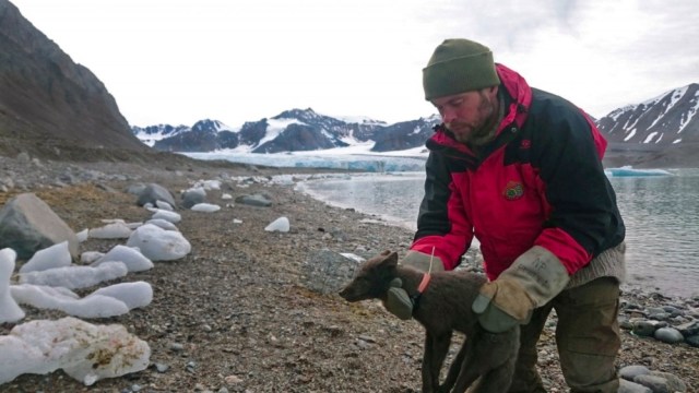
Villagers waiting to vote in Kenya. In this queue at a polling station, there are both barefoot and shod individuals. Holowka et al.5 studied people in Kenya and the United States who either are usually barefoot or usually wear shoes. The authors investigated whether the formation of thick patches of skin called calluses, which are usually thicker and harder in people who are normally barefoot than in shod individuals, affects foot sensitivity.Credit: Roberto Schmidt/AFP/Getty

By approximately 6 million years ago1,2, our hominin ancestors walked upright. Since then, ancient hominins, and eventually humans, have used their feet as their only point of contact with the ground. Evidence suggests that, long after our species evolved about 200,000 years ago to become anatomically modern humans (our current form)3, some people began to wear shoes for protection and for many other reasons — beginning about 40,000 years ago4. But wouldn’t it be great if foot protection existed that could preserve our sensation (termed tactile sensitivity) of the ground beneath our feet? Writing in Nature, Holowka et al.5 report that thick patches of foot skin, termed calluses, do just that. The authors reached this conclusion by studying callus thickness and hardness, plus foot sensitivity, in individuals in Kenya and the United States who usually either wear shoes or go barefoot.

Holowka and colleagues measured callus thickness using ultrasound. They report that people who were normally barefoot had calluses that were approximately 30% thicker than those of people who typically wore shoes. It could be assumed that thicker calluses provide more protection than thinner ones, all else being equal. But is all else indeed equal? To find out, the authors quantified the mechanical properties of foot soles using a device called a Shore durometer. This tool is commonly used in the footwear industry, and measures foot resistance to an indentation caused by the apparatus. The authors’ results show that, compared with skin on the feet of those who normally wore shoes, the skin of barefoot individuals was approximately 30% harder. This thicker, harder skin presumably protects their feet just like a shoe’s sole.

Our feet are remarkably sensitive, enabling pleasant sensations such as the feeling when walking barefoot on a beach, but also the experience of pain when stepping on a sharp rock. This sensitivity is useful because our body’s nerves use such information to fine-tune our posture and gait, in a similar way to how our sensitive fingertips enable us to precisely manipulate objects. As part of the system that aids this tactile sensitivity, a variety of mechanoreceptors in our skin sense mechanical stimuli such as pressure. If these receptors don’t work normally, as can occur in disease6 or during experimental manipulation7, people can have problems with their balance or gait8.

Using a device called a vibration exciter, Holowka and colleagues assessed the sensitivity of two types of mechanoreceptor, known as Meissner and Pacinian corpuscles, in their volunteers. These mechanoreceptors respond to high-frequency pressure stimulations (at 5–50 and 100–300 hertz, respectively) that occur when walking and running, especially when the foot strikes the ground. Holowka and colleagues’ key discovery is probably unexpected, given that one might predict that a thick layer of skin would be a barrier to the transmission of stimuli: mechanoreceptor sensitivity is not lower in habitually barefoot people than in people who usually wear shoes.

Barefoot walking with thick calluses is our biologically normal condition, and people who usually walk barefoot experience few problems doing so9,10, as I have also observed in my research in India11,12. Walkers who are habitually barefoot report no pain when walking on most terrains that shod walkers would find painful to walk on barefoot. However, habitually barefoot walkers might be at a higher risk of traumatic injury, given that shoes can offer better protection than can calluses13. Nevertheless, barefoot-walkers’ feet might be generally healthier than those of habitually shod people9, and foot problems such as bunions and fallen arches are rare in people who seldom wear shoes.

Should we now bin our shoes? Well, maybe not. Shoes can help people who have foot conditions14, and can also boost athletic performance15. In everyday life, shoes can keep our feet warm, and offer more protection than calluses can. Therefore, what kind of shoes we should wear becomes the more pressing question.

Holowka and colleagues argue that thick calluses preserve sensitivity because their hardness enables mechanical stimuli from the ground to be transmitted, with little dampening, to deep layers of the skin in which the key mechanoreceptors are located. If so, shoes with hard soles should be predicted to do the same job as calluses. Indeed, the hard-soled shoes used by drivers competing in Formula 1 races provide even greater than normal sensitivity at high frequencies of vibration16.

More research will be needed to fully understand the effect of shoe soles on gait. Humans are not like machines, in which just one variable at a time can be studied. Human movement is a complex, dynamic system, and changing even one variable, such as shoe-sole stiffness, will probably trigger other physiological and behavioural changes. For example, running when using cushioned soles, compared with running barefoot, triggers changes in how the foot makes contact with the ground (called the strike pattern)17, and also causes the arch of the foot to behave more stiffly18.

Holowka et al. conducted an experiment using a treadmill apparatus to quantify impact forces, which are the forces that the foot encounters immediately after it strikes the ground. They found that even if uncushioned shoes were used to mimic a callus-like sole, these shoes did not exactly mirror the effect of calluses during foot strike. Compared with their observations of unshod individuals, such footwear led to a slower rise in the impact force and a higher impulse (the product of the force and duration of the impact phase, which is when the foot hits the ground and slows abruptly).

It makes sense that preserving foot sensitivity is useful, especially if maintaining stability is challenging. This is true for gymnasts and also for older people, in whom faculties such as vision, balance and foot sensitivity decline naturally with age. Shoes with hard soles might therefore be a good idea for such individuals. Indeed, wearing hard-soled shoes can reduce the risk of older people falling19. Holowka and colleagues’ work helps to explain why this is so. Although this mystery has been solved, much remains to be discovered about what affects how humans walk.

doi: 10.1038/d41586-019-01953-6
References

1. Senut, B. et al. C. R. Acad. Sci. IIA 332, 137–144 (2001).

2. Brunet, M. et al. Nature 418, 145–151 (2002).

3. McDougall, I., Brown, F. H. & Fleagle, J. G. Nature 433, 733–736 (2005).

4. Trinkaus, E. & Shang, H. J. Archaeol. Sci. 35, 1928–1933 (2008).

5. Holowka, N. B. et al. Nature https://doi.org/10.1038/s41586-019-1345-6 (2019).

6. Alam, U. et al. Diabetes Ther. 8, 1253–1264 (2017).

7. Höhne, A., Ali, S., Stark, C. & Brüggemann, G.-P. Eur. J. Appl. Physiol. 112, 3829–3838 (2012).

8. Alfuth, M. & Rosenbaum, D. Footwear Sci. 4, 1–22 (2012).

9. Shulman, S. B. J. Natl Assoc. Chiropodists 49, 26–30 (1949).

10. Sim-Fook, L. & Hodgson, A. J. Bone Joint Surg. Am. 40, 1058–1062 (1958).

11. D’Août, K., Pataky, T. C., De Clercq, D. & Aerts, P. Footwear Sci. 1, 81–94 (2009).

12. Willems, C., Stassijns, G., Cornelis, W. & D’Août, K. Am. J. Phys. Anthropol. 162, 782–793 (2017).

13. Engle, E. T. & Morton, D. J. J. Bone Joint Surg. 13, 311–318 (1931).

14. Bus, S. A. et al. Diabetes/Metab. Res. Rev. 32 (suppl.), 99–118 (2016).

15. Hoogkamer, W. et al. Sports Med. 48, 1009–1019 (2018).

16. Schlee, G., Sterzing, T. & Milani, T. L. Eur. J. Appl. Physiol. 106, 305–309 (2009).

17. De Wit, B., De Clercq, D. & Aerts, P. J. Biomech.33, 269–278 (2000).

18. Kelly, L. A., Lichtwark, G. A., Farris, D. J. & Cresswell, A. J. R. Soc. Interface 13, 20160174 (2016).

19. Aboutorabi, A. et al. Prosthet. Orthot. Int. 40, 170–181 (2016).

https://www.nature.com/articles/d41586-019-01953-6

Hopes raised of cervical cancer eradication

Vaccination against the human papilloma virus, which causes most cervical cancers, began over a decade ago.

A Lancet review of 65 studies covering 60 million people showed a fall in HPV cases and in pre-cancerous growths.

Over decades, this should translate into a significant fall, and possible eradication, of the cancer they said.

Jo’s Cervical Cancer Trust said the data should boost faith in the jab.

What is the human papilloma virus (HPV)?

HPV is the name for a common group of viruses; there are more than 100 types of HPV
Many women will be infected with HPV over the course of their lifetime, with no ill effect
Most cervical cancers are caused by infection from a high-risk HPV
Others cause conditions including genital warts and cancers of the head and neck
The vaccine, given as two injections to girls aged 12 and 13, protects against four types of HPV – 16 and 18, which are linked to more than 70% of cervical cancers – and six and 11, which
cause about 90% of genital warts
Girls who miss the HPV jab at school can still get it for free on the NHS up to the age of 25
It is also available privately, costing around £150 per dose
Boys aged 12-13 will also be offered the jab from September this year
The vaccine does not protect against all the types of HPV that can cause cervical cancer, so women still need to go for regular screening

There are 3,200 cases of cervical cancer and 850 deaths from the disease each year.

‘Real-world’ evidence
The review covered studies in 14 high-income countries, including the UK. They looked at HPV rates, plus cases of genital warts and pre-cancerous cells in the cervix called CIN.

It found that when rates were compared before vaccination started and eight years after:

Cases of HPV 16 and 18 were down 83% in girls aged 15-19 – 66% in women 20-24
Genital warts cases fell 67% in girls 15-19 – 54% in women 20-24
Pre-cancerous growths were down by 51% in girls 15-19 – 31% in women 20-24
It also showed people who were not vaccinated benefited. Cases of genital warts in boys aged 15-19 fell by almost 50%, and also significantly in women over 30.

Rates fell more in countries where a wider age group was vaccinated and where coverage was higher.

Public Health England principal scientist Dr David Mesher said: “We are seeing reductions in HPV strains and in cervical disease as well, so there is every suggestion there will be reductions in cervical cancers too.”

Prof Marc Brisson, from Laval University, Canada, who led the review, said: “We will see reductions in women aged 20-30 within the next 10 years.”

He said cervical cancer elimination – defined as fewer than four cases per 100,000 – “might be possible if sufficiently high vaccination coverage can be achieved and maintained”.

Jo’s Cervical Cancer Trust said the findings “clearly showed” the impact of HPV vaccination.

“This study furthers the growing evidence to counteract those who don’t believe that this vaccine works, which is now extremely encouraging,” said chief executive Robert Music.

“We sincerely hope this will boost public faith in the HPV vaccine, so that more lives can be saved and we get closer to a world where cervical cancer is a thing of the past.”

https://www.bbc.com/news/health-48758730?utm_source=Nature+Briefing&utm_campaign=5a6f57394e-briefing-dy-20190627&utm_medium=email&utm_term=0_c9dfd39373-5a6f57394e-44039353

Trauma of Australia’s Indigenous ‘Stolen Generations’ is still affecting children today

Indigenous children in Australia who live in families that experienced forced separations in much of the twentieth century are more likely than other Indigenous children to have poor health and negative school experiences, according to a landmark government report released this month.

As many as one in three Aboriginal and Torres Strait Islander children were taken from their families and communities between 1910 and the 1970s, under racist government policies that tried to force Aboriginal people to assimilate with white Australians. The children were brought up in institutions or foster homes, or were adopted by white families. The Australian government formally apologized to members of these ‘Stolen Generations’ in 2008.

In the latest report, the Australian Institute of Health and Welfare, a government-funded statistics agency, used existing data from surveys of Aboriginal and Torres Strait Islander people to conduct the first national study of how the forced separations have affected children in subsequent generations. Previous reports looked at the impacts of these policies on the Stolen Generations themselves, and on their adult descendants.

“What all of this work around Stolen Generations is showing is that compared to other Aboriginal and Torres Strait Islander people, the Stolen Generations and their descendants are far worse off,” says Richard Weston, a descendant of the Meriam people from the Torres Strait, and chief executive of the Healing Foundation in Canberra, a government-funded organization that is working towards healing for the Stolen Generations and their descendants, and which commissioned the report. “Trauma stays with people, and its impacts are far-reaching and they’re profound,” says Weston.

Mostly worse off
The report examined health, cultural and socio-economic measures for about 7,900 Aboriginal and Torres Strait Islander children under the age of 15 who were living in households with at least one member of the Stolen Generations. These measures were compared with those of about 40,800 Indigenous children whose households included no adults who had been removed from their families as children. The relationships between the children and the members of the Stolen Generation in their households were not reported.

The analysis showed that 17.2% of Indigenous children living in Stolen Generations households reported having missed school without permission in the previous year, compared with 4.1% of the reference group. Children in Stolen Generation families were also nearly twice as likely to report having been “treated unfairly” at school because they were Indigenous, and 26% of Indigenous children living in Stolen Generations households rated their health as poor, compared with 19.2% of the comparison population.

Children living in Stolen Generations households were also 1.8 times as likely to have experienced stress in the previous 12 months, and 60% less likely to live in a home owned by a household member.

The analysis also considered the effects of other factors on the children’s health and socio-economic measures irrespective of whether they lived in a Stolen Generations household, such as age and gender, and whether the children lived in a remote area or in a household with someone who was employed, had completed school or had been incarcerated. The results show that removal has intergenerational effects even after controlling for these factors.

The report concludes that children living in a Stolen Generations household were more likely to experience adverse outcomes than were other Aboriginal and Torres Strait Islander children and that this “demonstrates a transfer of intergenerational poverty and trauma”.

“The results from the survey show how much suffering is still being endured from these policies,” says Maggie Walter, a Palawa woman from Tasmania and a sociologist at the University of Tasmania in Hobart. “It is ricocheting through generations.”

But the report did find that Indigenous children living in Stolen Generations households were twice as likely as Indigenous children not living in these households to identify with a clan, tribal or language group, and to recognize an area as homeland.

Weston says this could suggest that cultural identity has been a source of strength and resilience for members of the Stolen Generations.

Although Walters thinks the report shines a light on the difficulties these children face, she worries that focusing on households will inadvertently place the blame on them. “The risk with all of these things is that somehow it becomes Aboriginal families’ and communities’ fault that they are still living with the repercussions of those dreadful policies,” she says. “What we need to be looking at is wider social and cultural reality in which that family, both current and previous generations, have lived their lives.”

Addressing trauma
Weston thinks the trauma caused by racist policies such as the forced removal of children is the root cause of the fact that Indigenous Australians, on average, die about ten years earlier than non-Indigenous Australians. Studies of the effects of childhood trauma in the United States show that it can increase the risk of substance misuse and mental and physical ill-health, and can limit employment opportunities.

But Weston says government initiatives are not adequately addressing trauma, and this is why, despite numerous policies over the past ten years, the life expectancy of Indigenous Australians has yet to improve significantly. The government acknowledges that the country is not on track to meet its goal of closing the life-expectancy gap by 2031.

Some researchers are also worried that the trauma is being repeated today, in Indigenous children who are being removed from their families under state child-welfare laws. Aboriginal and Torres Strait Islander children represented 5.5% of Australian children under 18 in 2016–17, but 36.9% of all children placed in out-of-home care.

Walter says it is a contradiction for governments to say that they want to improve Indigenous life expectancy when Indigenous children are still being placed in out-of-home care. Removing children from their communities is contributing to these gaps, she says.

Although government policies state that Indigenous children should be placed with their extended family or families in their community before non-Indigenous carers, this isn’t always possible.

The high proportion of Aboriginal and Torres Strait Islander children in out-of-home care is a concern, says Ken Wyatt, a Noongar, Yamatji and Wongi man and the country’s minister for Indigenous Australians. Although child-protection systems are the responsibility of states and territories, Wyatt says, the national government is working to address the underlying factors that contribute to children being placed in out-of-home care, including intergenerational trauma.

Nature 570, 423-424 (2019)

doi: 10.1038/d41586-019-01948-3

https://www.nature.com/articles/d41586-019-01948-3?utm_source=Nature+Briefing&utm_campaign=611c8134ee-briefing-dy-20190626&utm_medium=email&utm_term=0_c9dfd39373-611c8134ee-44039353

Serotonin Linked to Somatic Awareness, a Condition Long Thought To Be Imaginary

An international team spearheaded by researchers at McGill University has discovered a biological mechanism that could explain heightened somatic awareness, a condition where patients experience physical discomforts for which there is no physiological explanation.

Patients with heightened somatic awareness often experience unexplained symptoms – headaches, sore joints, nausea, constipation or itchy skin – that cause emotional distress, and are twice as likely to develop chronic pain. The condition is associated with illnesses such as fibromyalgia, rheumatoid arthritis, and temporomandibular disorders, and is thought to be of psychological origin.

“Think of the fairy tale of the princess and the pea,” says Samar Khoury, a postdoctoral fellow at McGill’s Alan Edwards Centre for Research on Pain. “The princess in the story had extreme sensitivity where she could feel a small pea through a pile of 20 mattresses. This is a good analogy of how someone with heightened somatic awareness might feel; they have discomforts caused by a tiny pea that doctors can’t seem to find or see, but it’s very real.”

Thanks to an existing study on genetic association, Samar Khoury and her colleagues might have found the elusive pea capable of explaining somatic awareness.

Their work, recently published in the Annals of Neurology, used data available through the Orofacial Pain: Prospective Evaluation and Risk Assessment cohort and demonstrates that patients who suffer from somatic symptoms share a common genetic variant. The mutation leads to the malfunctioning of an enzyme critical for the production of serotonin, a neurotransmitter with numerous biological functions.

“I am very happy and proud that our work provides a molecular basis for heightened somatic symptoms,” says Luda Diatchenko, lead author of the new study and a professor in McGill’s Faculty of Dentistry. “We believe that this work is very important to patients because we can now provide a biological explanation of their symptoms. It was often believed that there were psychological or psychiatric problems, that the problem was in that patient’s head, but our work shows that these patients have lower levels of serotonin in their blood.”

The results of their study have laid the groundwork for the development of animal models that could be used to better characterize the molecular pathways in heightened somatic awareness. Above all, Diatchenko and Khoury hope their work will pave the way for treatment options.

“The next step for us would be to see if we are able to target serotonin levels in order to alleviate these symptoms,” says Diatchenko, who holds the Canada Excellence Research Chair in Human Pain Genetics.

This work was supported by the Canadian Institutes of Health Research, Natural Sciences and Engineering Research Council of Canada, the National Institutes of Health and the National Institute of Dental and Craniofacial Research.

https://reachmd.com/news/serotonin-linked-to-somatic-awareness-a-condition-long-thought-to-be-imaginary/1628160/?mkt_tok=eyJpIjoiTTJFM05EYzNORFZsTmpZMSIsInQiOiJ6dnNLckNwK0tZSTUwYnlBcmxBZ1dMSGg4ZTlFQ1FUd2xvOVV5bkpRV0hrOXB5aEs4cG95ckRFNDY2aTFCem41MXQxUTk0ZWtuNjdJMkJ5NUNqRTJzVFFKZkE3ZEpMS2xuMGFBZVBnQXM5WFBZVkpRZW1zZzNscmtUTlJIblJYOSJ9

Three Public Health Interventions Could Prevent 94 Million Premature Deaths

“Focusing our resources on the combination of these three interventions can have a huge potential impact on cardiovascular health through 2040,” said lead author Goodarz Danaei, associate professor of global health at Harvard Chan School.

Researchers used global data from multiple studies and estimates from the World Health Organization in making their calculations.

They estimated that scaling up treatment of high blood pressure to 70% of the world’s population could extend the lives of 39.4 million people. Cutting sodium intake by 30% could stave off another 40 million deaths and could also help decrease high blood pressure, a major risk factor for CVD. And eliminating trans fat could prevent 14.8 million early deaths.

More than half of all delayed deaths, and two-thirds of deaths delayed before age 70, are projected to be among men, who have the highest numbers of noncommunicable disease deaths globally, researchers found. Regions expected to benefit most from the interventions include East Asia, the Pacific, and South Asia, as well as countries in sub-Saharan Africa.

The authors said that a variety of programs and policies would be necessary to reduce premature CVD-related deaths. One important strategy would be to increase the use of blood pressure medications, many of which are safe and affordable.

The researchers acknowledged that scaling up the three interventions would be a “huge challenge,” requiring countries to commit additional resources to boost health care capacity and quality. But they added that previous analyses have shown that the interventions are achievable and affordable. For example, a Kaiser Permanente program in Northern California increased control of hypertension to 90% among thousands of the health system’s patients between 2001 and 2013, using strategies such as improved treatment protocols, patient-friendly services, and healthcare information systems that facilitate tracking people with hypertension. Similar approaches have been adapted and tested in some low- and middle-income countries, leading to notable improvements in hypertension treatment and control, the authors said.

“These are realistic goals that have been shown to be attainable on smaller scales,” said Danaei. “We need the commitment to scale up the programs to achieve them globally.”

Gut microbes can impact the efficacy of Parkinson’s disease medications

by DAVID NIELD

Dosing medicines can be a tricky process: How much of a medication actually ends up hitting its target can vary a lot between patients, sometimes for mysterious reasons. As it turns out, even the things living in our bodies could be gobbling up our drugs.

In a series of experiments with levodopa (L-dopa) drug treatments for Parkinson’s, a new study has found that the gut microbes Enterococcus faecalis and Eggerthella lenta can intercept L-dopa and chemically transform it before it reaches the brain.

While this research only focuses on a specific treatment for one condition, the team behind the work thinks we might be underappreciating the role that our gut microbiome plays in controlling the efficacy and potency of medicines.

“Maybe the drug is not going to reach its target in the body, maybe it’s going to be toxic all of a sudden, maybe it’s going to be less helpful,” says chemical biologist Maini Rekdal from Harvard University.

The job of L-dopa is to deliver dopamine to the brain, replacing the dopamine eaten up by Parkinson’s. However, since the introduction of L-dopa in the 1960s, scientists have known that enzymes in the gut can stop this delivery from happening, leading to some nasty side effects as dopamine “spills out” before reaching the brain.

A second drug, carbidopa, was introduced to keep L-dopa intact, but it doesn’t always seem to help. Even with this additional drug, the effectiveness of L-dopa can vary between patients. What this new research does is identify the specific bacteria to blame, out of trillions of potential species.

With reference to the Human Microbiome Project, the team found that not only our own gut enzymes can wreak havoc on the medication, but the bacterium E. faecalis can also convert L-dopa to dopamine before it reaches the brain. Sure enough, it ate up all the L-dopa in lab tests.

Using faecal samples and supplies of dopamine, the researchers identified that another strain of gut bacteria, E. lenta, then consumes the converted dopamine and produces the neuromodulator meta-thyramine as a byproduct.

Thus, E. faecalis and E. lenta are apparently working as a sort of microbe tag team, preventing the medication from reaching its target. Furthermore, while carbidopa is used to stop a human gut enzyme from converting L-dopa to dopamine in the digestive system, it doesn’t seem to work on the E. faecalis enzyme that’s doing the same.

The good news is that the researchers have already found a molecule, alpha-fluoromethyltyrosine (AFMT), that can stop E. faecalis from breaking down L-dopa without destroying the bacterium itself, by targeting a non-essential enzyme.

Ultimately, we might end up with a way of making L-dopa significantly more effective as a Parkinson’s treatment, without as many of the side effects – but that’s still a long way off.

“All of this suggests that gut microbes may contribute to the dramatic variability that is observed in side effects and efficacy between different patients taking L-dopa,” says chemical biologist Emily Balskus from Harvard University.

Even if we can’t fix the problem just yet, we now have a proof of concept that particular combinations of gut microbes can indeed cause havoc with our meds. Hopefully, this will give other researchers food for thought and we might see similar investigations of other medicines, too.

The research has been published in Science.

https://www.sciencealert.com/gut-microbes-could-be-eating-up-our-meds-before-they-get-chance-to-work