New discovery on brain chemistry of patients with schizophrenia and their relatives

katharine-thakkar

People with schizophrenia have different levels of the neurotransmitters glutamate and gamma-aminobutyric acidergic (GABA) than healthy people do, and their relatives also have lower glutamate levels, according to a study published online in Biological Psychiatry.

Using magnetic resonance spectroscopy, researchers discovered reduced levels of glutamate — which promotes the firing of brain cells — in both patients with schizophrenia and healthy relatives. Patients also showed reduced levels of GABA, which inhibits neural firing. Healthy relatives, however, did not.

Researchers are unsure why healthy relatives with altered glutamate do not show symptoms of schizophrenia or how they maintain normal GABA levels despite a predisposition to the illness.

“This finding is what’s most exciting about our study,” said lead investigator Katharine Thakkar, PhD, assistant professor of clinical psychology at Michigan State University, East Lansing. “It hints at what kinds of things have to go wrong for someone to express this vulnerability toward schizophrenia. The study gives us more specific clues into what kinds of systems we want to tackle when we’re developing new treatments for this very devastating illness.”

The study included 21 patients with chronic schizophrenia, 23 healthy relatives of other people with schizophrenia not involved in the study, and 24 healthy nonrelatives who served as controls.

Many experts believe there are multiple risk factors for schizophrenia, including dopamine and glutamate-GABA imbalance. Drugs that regulate dopamine do not work for all patients with schizophrenia. Dr. Thakkar believes magnetic resonance spectroscopy may help clinicians target effective treatments for specific patients.

“There are likely different causes of the different symptoms and possibly different mechanisms of the illness across individuals,” said Dr. Thakkar.

“In the future, as this imaging technique becomes more refined, it could conceivably be used to guide individual treatment recommendations. That is, this technique might indicate that one individual would benefit more from treatment A and another individual would benefit more from treatment B, when these different treatments have different mechanisms of action.”

—Jolynn Tumolo

References

Thakkar KN, Rösler L, Wijnen JP, et al. 7T proton magnetic resonance spectroscopy of GABA, glutamate, and glutamine reveals altered concentrations in schizophrenia patients and healthy siblings [publisehd online ahead of print April 19, 2016]. Biological Psychiatry.
Study uncovers clue to deciphering schizophrenia [press release]. Washington, DC: EurekAlert!; June 7, 2016.

New treatment protocol shows promise of improving cognition in patients with Alzheimer’s disease

Ten patients with early Alzheimer’s disease or its precursors showed improvement in memory after treatment with Metabolic Enhancement for NeuroDegeneration (MEND), a programmatic and personalized therapy protocol.

Researchers described results from the small trial, which used quantitative MRI and neuropsychological testing of participants before and after treatment, in the study published online in Aging.

“ The magnitude of the improvement is unprecedented,” researchers wrote, “providing additional objective evidence that this programmatic approach to cognitive decline is highly effective.”

Before starting the program, the 10 participants had well-defined mild cognitive impairment, subjective cognitive impairment, or had been diagnosed with Alzheimer’s disease. Their subsequent treatment consisted of a complex, 36-point therapeutic personalized program that included comprehensive changes in diet, brain stimulation, exercise, optimization of sleep, specific pharmaceuticals and vitamins, and multiple additional steps that affect brain chemistry.

Researcher Dale Bredesen, MD, a professor at the Buck Institute for Research on Aging and at the Easton Laboratories for Neurodegenerative Disease Research at UCLA, Los Angeles, believes the protocol’s broader-based approach is key to its apparent success in reversing cognitive decline.

“Imagine having a roof with 36 holes in it, and your drug patched one hole very well — the drug may have worked, a single ‘hole’ may have been fixed, but you still have 35 other leaks, and so the underlying process may not be affected much,” Dr. Bredesen said. “We think addressing multiple targets within the molecular network may be additive, or even synergistic, and that such a combinatorial approach may enhance drug candidate performance as well.”

Tests showed some participants “going from abnormal to normal,” Dr. Bredesen said.

In Aging , researchers describe the impact of MEND on all 10 patients, including:
•A 66-year-old man whose neuropsychological testing was compatible with a diagnosis of mild cognitive impairment. After 10 months on the MEND protocol, his hippocampal volume increased from the 17 th percentile for his age to the 75 th percentile, with an associated absolute increase in volume of nearly 12%.
•A 69-year-old entrepreneur with 11 years of progressive memory loss. After 22 months on the protocol, he showed marked improvements in all categories of neuropsychological testing, with long-term recall increasing from the 3 rd to 84 th percentile.
•A 49-year-old woman in the early stages of cognitive decline who, after 9 months on the protocol, no longer showed evidence on quantitative neuropsychological testing of cognitive decline.

Plans for larger studies are under way.

“Even though we see the far-reaching implications of this success,” Dr. Bredesen said, “we also realize that this is a very small study that needs to be replicated in larger numbers at various sites.”

http://www.psychcongress.com/article/mend-protocol-reverses-memory-loss-alzheimer%E2%80%99s-disease-27858

Urinary biomarker of Parkinson’s disease identified

New findings indicate that phosphorylated LRRK2 (leucine-rich repeat kinase 2) protein levels in urine are elevated in patients diagnosed with idiopathic Parkinson Disease (PD), and that urinary phosphorylated LRRK2 levels correlate with the presence and severity of symptoms such as cognitive impairment in individuals with PD. Researchers affiliated with the University of Alabama at Birmingham published their findings in Neurology and in Movement Disorders (1,2).

The etiology of PD is currently unknown and mechanisms of action are still not completely clarified. It is well established, however, that aging is the single most important risk factor. PD is the second most frequent age-related neurodegenerative disorder, and one of the key pathogenic features is slow and progressive neuronal death that is concomitant with cognitive dysfunction. Current therapeutic modalities are inadequate and clinical need is significant. More than 6 million individuals worldwide are diagnosed with PD.

To date, several common genetic variants, or single nucleotide polymorphisms (SNPs), have been identified that influence the risk for disease. For example, polymorphic variants in LRRK2 gene have previously been validated as genetic factors that confer susceptibility to PD.

Although the gene remains poorly characterized, five different mutations in the gene encoding LRRK2 are considered a common cause of inherited PD (3). One of the five mutations that are causal is the G2019S mutation in the LRRK2 kinase domain, a mutation that significantly increases phosphorylation activity (1,3).

“There are currently no known ways to predict which G2019S mutation carriers will develop PD,” the authors wrote in the Neurology publication. Investigators purified LRRK2 protein from urinary exosomes collected from a total of 76 men. (Exosomes are membrane vesicles of endosomal origin that are secreted by most cells in culture, and are present in most biological fluids such as urine, blood, and saliva.) Then, they compared the ratio of phosphorylated LRRK2 to total LRRK2 in urine exosomes. Results show that “elevated … phosphorylated LRRK2 predicted the risk” for onset of PD in LRRK2 G2019S mutation carriers (1).

In their follow-up study, which was published in Movement Disorders, investigators compared phosphorylated LRRK2 levels in urine samples of 79 individuals diagnosed with PD to those of 79 healthy control participants. Results show that phosphorylated LRRK2 levels were significantly elevated in patients with PD when compared to those of controls. Also, phosphorylated LRRK2 levels correlated with the severity of cognitive impairment in patients with PD (2).

“Because few viable biomarkers for PD exist … phosphorylated LRRK2 levels may be a promising candidate for further exploration,” the authors concluded in their publication.

References
1. Fraser KB, Moehle MS, Alcalay RN, et al. Urinary LRRK2 phosphorylation predicts parkinsonian phenotypes in G2019S LRRK2 carriers. Neurology. 2016;86:994-999.
2. Fraser KB, Rawlins AB, Clar RG, et al. Ser(P)-1292 LRRK2 in urinary exosomes is elevated in idiopathic Parkinson’s disease. Mov Disord. 2016. doi: 10.1002/mds.26686.
3. Greggio E, Cookson MR. Leucine-rich repeat kinase 2 mutations and Parkinson’s disease: three questions. ASN Neuro. 2009;1:e00002.

http://www.psychiatryadvisor.com/neurocognitive-disorders/urinary-biomarker-of-parkinson-disease-identified/article/508195/?DCMP=EMC-PA_Update_RD&cpn=psych_md,psych_all&hmSubId=&hmEmail=5JIkN8Id_eWz7RlW__D9F5p_RUD7HzdI0&NID=1710903786&dl=0&spMailingID=14919209&spUserID=MTQ4MTYyNjcyNzk2S0&spJobID=820575619&spReportId=ODIwNTc1NjE5S0

Toxoplasma infection might trigger neurodegenerative disease


Infection with the common parasite Toxoplasma gondii promotes accumulation of a neurotransmitter in the brain called glutamate, triggering neurodegenerative diseases in individuals predisposed to such conditions.

Written by Honor Whiteman

This is the finding of a new study conducted by researchers from the University of California-Riverside (UC-Riverside), recently published in PLOS Pathogens.

T. gondii is a single-celled parasite that can cause a disease known as toxoplasmosis.

Infection with the parasite most commonly occurs through eating undercooked, contaminated meat or drinking contaminated water.

It may also occur through accidentally swallowing the parasite after coming into contact with cat feces – by cleaning a litter tray, for example.

Though more than 60 million people in the United States are believed to be infected with T. gondii, few people become ill from it; a healthy immune system can normally stave it off.

As such, most people who become infected with the parasite are unaware of it.

Those who do become ill from T. gondii infection may experience flu-like symptoms – such as swollen lymph glands or muscle aches – that last for at least a month.

In severe cases, toxoplasmosis can cause damage to the eyes, brain, and other organs, though such complications usually only arise in people with weakened immune systems.

The new study, however, suggests there may be another dark side to T. gondii infection: it may lead to development of neurodegenerative disease in people who are predisposed to it.

To reach their findings, lead author Emma Wilson – an associate professor in the Division of Biomedical Sciences at the UC-Riverside School of Medicine – and colleagues focused on how T. gondii infection in mice affects glutamate production

How a build-up of glutamate can damage the brain

Glutamate is an amino acid released by nerve cells, or neurons. It is one of the brain’s most abundant excitatory neurotransmitters, aiding communication between neurons.

However, previous studies have shown that too much glutamate may cause harm; a build-up of glutamate is often found in individuals with traumatic brain injury (TBI) and people with certain neurodegenerative diseases, such as multiple sclerosis (MS) and amyotrophic lateral sclerosis (ALS).

The researchers explain that excess glutamate accumulates outside of neurons, and this build-up is regulated by astrocytes – cells in the central nervous system (CNS).

Astrocytes use a glutamate transporter called GLT-1 in an attempt to remove excess glutamate from outside of neurons and convert it into a less harmful substance called glutamine, which cells use for energy.

“When a neuron fires, it releases glutamate into the space between itself and a nearby neuron,” explains Wilson. “The nearby neuron detects this glutamate, which triggers a firing of the neuron. If the glutamate isn’t cleared by GLT-1 then the neurons can’t fire properly the next time and they start to die.”


T. gondii increases glutamate by inhibiting GLT-1

n mice infected with T. gondii, the researchers identified an increase in glutamate levels.

They found that the parasite causes astrocytes to swell, which impairs their ability to regulate glutamate accumulation outside of neurons.

Furthermore, the parasite prevents GLT-1 from being properly expressed, which causes an accumulation of glutamate and misfiring of neurons. This may lead to neuronal death, and ultimately, neurodegenerative disease.

“These results suggest that in contrast to assuming chronic Toxoplasma infection as quiescent and benign, we should be aware of the potential risk to normal neurological pathways and changes in brain chemistry.” – Emma Wilson

Next, the researchers gave the infected mice an antibiotic called ceftriaxone, which has shown benefits in mouse models of ALS and a variety of CNS injuries.

They found the antibiotic increased expression of GLT-1, which led to a reduction in glutamate build-up and restored neuronal function.

Wilson says their study represents the first time that T. gondii has been shown to directly disrupt a key neurotransmitter in the brain.

“More direct and mechanistic research needs to be performed to understand the realities of this very common pathogen,” she adds.

While their findings indicate a link between T. gondii infection and neurodegenerative disease, Wilson says they should not be cause for panic.

“We have been living with this parasite for a long time,” she says. “It does not want to kill its host and lose its home. The best way to prevent infection is to cook your meat and wash your hands and vegetables. And if you are pregnant, don’t change the cat litter.”

The team now plans to further investigate what causes the reduced expression of GLT-1 in T. gondii infection.

http://www.medicalnewstoday.com/articles/310865.php

Weighted blankets may help treat anxiety

tress, anxiety, and insomnia affect millions of people worldwide, and to alleviate the symptoms, there are a variety of routes one can take, including the ever-popular pharmaceutical pills. But as our world continues to break through the madness of synthetic options and expose each other to holistic options derived from both ancient teachings as well as present-day healers, it’s important we keep our eyes and ears open for our own good.

Anyone who suffers from the above disorders knows the word “simple” doesn’t quite fit with how they feel. In fact, it seems to be very much the opposite: a complex feeling that can barely be put into words. So, how can something as simple as sleeping with weighted blankets be a plausible solution to stress, anxiety, insomnia, and more?

Called deep pressure touch stimulation, (or DPTS), this type of therapy is similar to getting a massage. Pressure is exerted over the body and provides both physical and psychological benefits. Deep touch pressure, according to Temple Grandin, Ph.D., “is the type of surface pressure that is exerted in most types of firm touching, holding, stroking, petting of animals, or swaddling.” In comparison to very light touching, which has been found to alert the nervous system, deep pressure proves to be relaxing and calming.

Weighted blankets have been traditionally used by occupational therapists as a means to help children with sensory disorders, anxiety, stress, or issues related to autism, and research continues to support this practice. One study, using the Grandin’s Hug Machine device, which allows administration of lateral body pressure, investigated the effects of deep pressure as a tool for alleviating anxiety related to autism. The researchers found “a significant reduction in tension and a marginally significant reduction in anxiety for children who received the deep pressure compared with the children who did not.”

Of weighted blankets specifically, occupational therapist Karen Moore says in psychiatric care, “weighted blankets are one of our most powerful tools for helping people who are anxious, upset, and possibly on the verge of losing control.”

One study, published in Occupational Therapy in Mental Health in 2008, showed that weighted blankets helped with anxiety, and another study published in Australasian Psychiatry in 2012 confirmed this.

Weighted blankets are like warm hugs. They mold to your body to provide pressure that aids in relaxing the nervous system. Think of it like a baby being swaddled — the weight and pressure work to comfort and provide much-needed relief, encouraging the production of serotonin in order to uplift your mood. This same chemical naturally converts to melatonin, which signals your body to rest and relax. Weighted blankets are perfect for anyone looking to try out a non-drug therapy that is both safe and effective.

To weigh the blankets down, plastic poly pellets are typically used, being sewn into compartments throughout the blanket for even weight distribution. The weight of the blanket serves as a deep touch therapy, stimulating deep touch receptors all over your body that promote a more grounded and safe feeling to the individual.

Though the weight of the blanket depends on your size and personal preference, a standard weight for adults ranges from 15 to 30 pounds. It is recommended to speak with a doctor or occupational therapist regarding using one if you are suffering from a medical condition. It is also strongly advised not to use a weighted blanket should you be suffering from a respiratory, circulatory, or temperature regulation problem.

As for where you can buy them, there are many websites you can purchase them from, providing you with different weights, fabrics, colors, and sizes to personalize your experience. You can even make your own as well.

http://www.collective-evolution.com/2016/05/20/how-weighted-blankets-are-helping-people-with-anxiety/

Meditating in a tiny Iowa town to help recovery from war

By Supriya Venkatesan

At 19, I enlisted in the U.S. Army and was deployed to Iraq. I spent 15 months there — eight at the U.S. Embassy, where I supported the communications for top generals. I understand that decisions at that level are complex and layered, but for me, as an observer, some of those actions left my conscience uneasy.

To counteract my guilt, I volunteered as a medic on my sole day off at Ibn Sina Hospital, the largest combat hospital in Iraq. There I helped wounded Iraqi civilians heal or transition into the afterlife. But I still felt lost and disconnected. I was nostalgic for a young adulthood I never had. While other 20-somethings had traditional college trajectories, followed by the hallmarks of first job interviews and early career wins, I had spent six emotionally numbing years doing ruck marches, camping out on mountaintops near the demilitarized zone in South Korea and fighting someone else’s battle in Iraq.

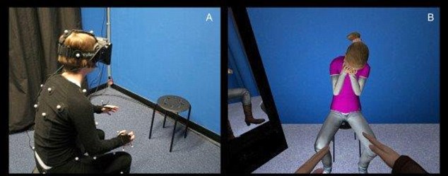
During my deployment, a few soldiers and I were awarded a short resort stay in Kuwait. There, I had a brief but powerful experience in a meditation healing session. I wanted more. So when I returned to the United States at the end of my service, I headed to Iowa.

Forty-eight hours after being discharged from the Army, I arrived on campus at Maharishi University of Management in Fairfield, Iowa. MUM is a small liberal arts college, smack dab in the middle of the cornfields, founded by Maharishi Mahesh Yogi, the guru of transcendental meditation. I joked that I was in a quarter-life crisis, but in truth my conscience was having a crisis. Iraq left me with questions about the world and grappling with my own mortality and morality.

Readjustment was a sucker punch of culture shock. While on a camping trip for incoming students, I watched girls curl their eyelashes upon waking up and burn incense and bundles of sage to ward off negative energy. I was used to being in a similar field environment but with hundreds of guys who spit tobacco, spoke openly of their sexual escapades and played video games incessantly. Is this what it looked like to be civilian woman? Is this what spirituality looked like?

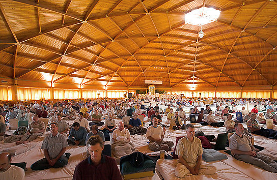
Mediation was mandatory for students on campus, and the rest of the town was composed mainly of former students or longtime followers of the maharishi. Shortly after arriving, I completed an advanced meditator course and began meditating three hours a day — a habit that is still with me five years later. Every morning, I went to a dome where students, teachers and the people of Fairfield gathered to practice meditation. In the evening, we met again for another round of meditation. During my time in Fairfield, even Oprah came to meditate in the dome.

I was incredibly lucky to have supportive mentors in the Army, but Fairfield embraced me in a maternal way. I cried for hours during post-meditation reflection. I released the trauma that is familiar to every soldier who has gone to war but is rarely discussed or even acknowledged. I let go, and I blossomed. I was emancipated of the unhealthy habits of binge-drinking and co-dependency in romantic interludes, as well as a fear that I didn’t know controlled me.

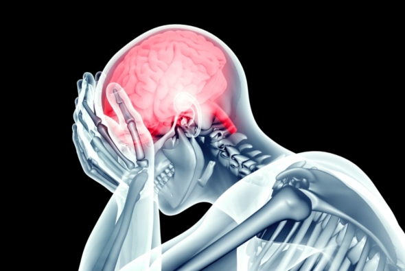
Suicide and other byproducts of post-traumatic stress disorder plague the military. In 2010, a veteran committed suicide every 65 minutes. In 2012, there were more deaths by suicide than by combat. In Iraq, one of my neighbors took his M16, put it in his mouth and shot himself. Overwhelmed with PTSD-related issues from back-to-back deployments and with no clear solution to the problem, in 2012, the Defense Department began researching meditation practices to see whether they would affect PTSD. The first study of meditation and the military population, done with Vietnam veterans in 1985, had shown 70 percent of veterans finding relief, but meditation never gained in popularity nor was it offered through veterans’ services. Even in 2010, when I learned TM, the military was alien to the concept.

But today, the results of the studies showcase immense benefits for veterans. According to the journal Military Medicine, meditation has shown a 40 percent to 55 percent reduction in symptoms of PTSD and depression among veterans. Furthermore, studies show that meditation correlates with a 42 percent reduction in insomnia and a 25 percent reduction in the stress hormone cortisol in the veteran population. To complement meditation, yoga has also been embraced as a tool for treatment by the military. With the growing acceptance of holistic approaches, psychological wounds are beginning to heal.

The four-day training course to learn TM is now available at every Veterans Affairs facility for those who have PTSD or traumatic brain injury. Even medical staff and counselors who help veterans at the VA are offered training in both TM and mindfulness meditation. Additionally, Norwich University, the oldest military college in the country, has done extensive research on TM and incoming cadets, and many military installations have integrated meditation programs into their mental health services. When I had first learned to meditate, many of my active-duty friends found it a bit too crunchy. But with the military’s recent efforts at researching meditation and funding it for all veterans, the stigma is gone, and my battle buddies see meditation as a tool for building resilience.

For me, meditation has created small but significant changes. One day, while going for a walk downtown, I stopped and patted a dog. A few minutes later, I came to a halt. I realized what I had done. While in Iraq, during a month when we were under heavy mortar attack, a bomb-sniffing K-9 had become traumatized and attacked me. This, coupled with a life-long fear of dogs, had left me guarded around the canines. I touched the scar on my elbow from where the K-9 had latched on and could no longer find the fear that had been there. Soon I was shedding all the things that held me back from living my life in an entirely unforeseen way.

For the first time in my life, I found forgiveness for those who had wronged me in the past. I literally stopped to smell the flowers on my way to work every day. And I smiled. All the freaking time. I even felt smarter. Research shows that meditation raises IQ. I’m not surprised. After graduation, I went on to complete my master’s at Columbia University.

Fairfield is also home to generations of Iowans who are born there, brought up there and die there. Many of these blue-collar Midwesterners have had animosity toward the meditators. Locals felt as if their town had been overtaken. They preferred steak to quinoa, beers at the bar to yoga and pickup trucks to carbon-reducing bicycles. And with MUM having a student body from more than 100 countries, the ethnic differences were a challenge. However, things are changing. Meditators and townspeople now fill less stereotypical roles. And with the economic boom that meditating entrepreneurs have provided the town, the differences are easier to ignore.

It was strange for me to live removed from the local Iowans. When I went shopping at the only Walmart the town had, I’d see the “Wall of Heroes” — a wall of photos of veterans from Fairfield. One day, I noticed a familiar face — a soldier from my last assignment. Fairfield and other socioeconomically depressed areas are where most military recruits come from. Here I was living among them, but not moving in step with them. Having that synchronous experience made me come back full circle. When I had first learned to meditate, my teacher had asked me what my goal was. I told her, “I want to be in the world, but not of it.” And that’s exactly what I got.

For me, this little Iowan town provided a place of respite and rejuvenation. It was easy for me to trade one lifestyle of order and discipline for another, and this provided me with nourishment and an understanding of self. Nowhere else in America can you find an entire town living and breathing the principles of Eastern mysticism. It goes way beyond taking a yoga class or going to the Burning Man festival. I continue my meditation practice and am grateful for the gifts it has provided me. But in the end, my time had come, and I had to leave. As residents would say, that was just my karma.

https://www.washingtonpost.com/posteverything/wp/2016/04/06/how-meditating-in-a-tiny-iowa-town-helped-me-recover-from-war/

Virtual Reality Therapy Shows Promise Against Depression

An immersive virtual reality therapy could help people with depression to be less critical and more compassionate towards themselves, reducing depressive symptoms, finds a new study from UCL (University College London) and ICREA-University of Barcelona.

The therapy, previously tested by healthy volunteers, was used by 15 depression patients aged 23-61. Nine reported reduced depressive symptoms a month after the therapy, of whom four experienced a clinically significant drop in depression severity. The study is published in the British Journal of Psychiatry Open and was funded by the Medical Research Council.

Patients in the study wore a virtual reality headset to see from the perspective of a life-size ‘avatar’ or virtual body. Seeing this virtual body in a mirror moving in the same way as their own body typically produces the illusion that this is their own body. This is called ’embodiment’.

While embodied in an adult avatar, participants were trained to express compassion towards a distressed virtual child. As they talked to the child it appeared to gradually stop crying and respond positively to the compassion. After a few minutes the patients were embodied in the virtual child and saw the adult avatar deliver their own compassionate words and gestures to them. This brief 8-minute scenario was repeated three times at weekly intervals, and patients were followed up a month later.

“People who struggle with anxiety and depression can be excessively self-critical when things go wrong in their lives,” explains study lead Professor Chris Brewin (UCL Clinical, Educational & Health Psychology). “In this study, by comforting the child and then hearing their own words back, patients are indirectly giving themselves compassion. The aim was to teach patients to be more compassionate towards themselves and less self-critical, and we saw promising results. A month after the study, several patients described how their experience had changed their response to real-life situations in which they would previously have been self-critical.”

The study offers a promising proof-of-concept, but as a small trial without a control group it cannot show whether the intervention is responsible for the clinical improvement in patients.

“We now hope to develop the technique further to conduct a larger controlled trial, so that we can confidently determine any clinical benefit,” says co-author Professor Mel Slater (ICREA-University of Barcelona and UCL Computer Science). “If a substantial benefit is seen, then this therapy could have huge potential. The recent marketing of low-cost home virtual reality systems means that methods such as this could potentially be part of every home and be used on a widespread basis.”

Publication: Embodying self-compassion within virtual reality and its effects on patients with depression. Falconer, CJ et al. British Journal of Psychiatry Open (February, 2016)

Risk of suicide increases 3X after a concussion

New research published in the Canadian Medical Association Journal shows that even mild concussions sustained in ordinary community settings might be more detrimental than anyone anticipated; the long-term risk of suicide increases threefold in adults if they have experienced even one concussion. That risk increases by a third if the concussion is sustained on a weekend instead of a weekday—suggesting recreational concussions are riskier long-term than those sustained on the job.

“The typical patient I see is a middle-aged adult, not an elite athlete,” says Donald Redelmeier, a senior scientist at the University of Toronto and one of the study’s lead authors. “And the usual circumstances for acquiring a concussion are not while playing football; it is when driving in traffic and getting into a crash, when missing a step and falling down a staircase, when getting overly ambitious about home repairs—the everyday activities of life.”

Redelmeier and his team wanted to examine the risks of the concussions acquired under those circumstances. They identified nearly a quarter of a million adults in Ontario who were diagnosed with a mild concussion over a timespan of 20 years—severe cases that resulted in hospital admission were excluded from the study—and tracked them for subsequent mortality due to suicide. It turned out that more than 660 suicides occurred among these patients, equivalent to 31 deaths per 100,000 patients annually—three times the population norm. On average, suicide occurred almost six years after the concussion. This risk was found to be independent of demographics or previous psychiatric conditions, and it increased with additional concussions.

For weekend concussions, the later suicide risk increased to four times the norm. Redelmeier and his fellow researchers had wondered whether the risk would differ between occupational and recreational concussions. They did not have information about how the concussions happened, so they used day of the week as a proxy. Although they do not know why weekend risk is indeed higher, they suspect it may be because on weekends medical staff may not be as available or accessible or people may not seek immediate care.

Although the underlying causes of the connection between concussion and suicide are not yet known, Redelmeier says that there were at least three potential explanations. A concussion may be a marker but not necessarily a mechanism of subsequent troubles—or, in other words, people who sustain concussions may already have baseline life imbalances that increase their risks for depression and suicide. “But we also looked at the subgroup of patients who had no past psychiatric history, no past problems, and we still found a significant increase in risk. So I don’t think that’s the entire story,” he notes. One of the more likely explanations, he says, is that concussion causes brain injury such as inflammation (as has been found in some studies) from which the patient may never fully recover. Indeed, a study conducted in 2014 found that sustaining a head injury leads to a greater risk of mental illness later in life. The other possibility is that some patients may not give themselves enough time to get better before returning to an ordinary schedule, leading to strain, frustration and disappointment—which, in turn, may result in depression and ultimately even suicide.

Lea Alhilali, a physician and researcher at the Barrow Neurological Institute who did not participate in this study, uses diffusion tensor imaging (an MRI technique) to measure the integrity of white matter in the brain. Her team has found similarities between white matter degeneration patterns in patients with concussion-related depression and noninjured patients with major depressive disorder—particularly in the nucleus accumbens, or the “reward center” of the brain. “It can be difficult to tease out what’s related to an injury and what’s related to the circumstances surrounding the trauma,” Alhilali says. “There could be PTSD, loss of job, orthopedic injuries that can all influence depression. But I do believe there’s probably an organic brain injury.”

Alhilali points to recent studies on chronic traumatic encephalopathy (CTE), a progressive degenerative brain disease associated with repeated head traumas. Often linked to dementia, depression, loss of impulse control and suicide, CTE was recently diagnosed in 87 of 91 deceased NFL players. Why, then, she says, should we not suspect that concussion causes other brain damage as well?

This new study may only represent the tip of the iceberg. “We’re only looking at the most extreme outcomes, at taking your own life,” Redelmeier says. “But for every person who dies from suicide, there are many others who attempt suicide, and hundreds more who think about it and thousands more who suffer from depression.”

More research needs to be done; this study was unable to take into account the exact circumstances under which the concussions were sustained. Redelmeier’s research examined only the records of adults who sought medical attention, it did not include more severe head injuries that required hospitalization or extensive emergency care. To that extent, his findings may have underestimated the magnitude of the absolute risks at hand.

Yet many people are not aware of these risks.

Redelmeier is adamant that people should take concussions seriously. “We need to do more research about prevention and recovery,” he says. “But let me at least articulate three things to do: One, give yourself permission to get some rest. Two, when you start to feel better, don’t try to come back with a vengeance. And three, even after you’re feeling better, after you’ve rested properly, don’t forget about it entirely. If you had an allergic reaction to penicillin 15 years ago, you’d want to mention that to your doctor and have it as a permanent part of your medical record. So, too, if you’ve had a concussion 15 years ago.”

http://www.scientificamerican.com/article/a-single-concussion-may-triple-the-long-term-risk-of-suicide1/

New research links subgroups of schizophrenia to specific visualized brain anomalies

An international team of researchers has linked specific symptoms of schizophrenia with various anatomical characteristics in the brain, according to research published in NeuroImage.

By analyzing the brain’s anatomy with magnetic resonance imaging (MRI), researchers from the University of Granada, Washington University in St. Louis, and the University of South Florida have demonstrated the existence of distinctive subgroups among patients with schizophrenia who suffer from different symptoms.

These findings could herald a significant step forward in diagnosing and treating schizophrenia.

To perform the study, the researchers conducted the MRI technique “diffusion tensor imaging” on 36 healthy participants and 47 schizophrenic participants.

The researchers found that tests on schizophrenic participants revealed various abnormalities in parts of the corpus callosum, a bundle of neural fibers that connects the left and right cerebral hemispheres and is essential for effective interhemispheric communication.

Different anomalies in the corpus callosum were associated with different symptoms in the schizophrenic participants. An anomaly in one part of the brain structure was associated with strange and disorganized behavior; another anomaly was associated with disorganized thought and speech, as well as negative symptoms such as a lack of emotion; and other anomalies were associated with hallucinations.

In 2014, this same research group proved that schizophrenia is not a single illness. The team demonstrated the existence of 8 genetically distinct disorders, each with its own symptoms. Igor Zwir, PhD, and Javier Arnedo from the University of Granada’s Department of Computer Technology and Artificial Intelligence found that different sets of genes were strongly linked with different clinical symptoms.

“The current study provides further evidence that schizophrenia is a heterogeneous group of disorders, as opposed to a single illness, as was previously thought to be case,” Dr Zwir said in a statement.

While current treatments for schizophrenia tend to be generic regardless of the symptoms exhibited by each patient, the researchers believe that in the future, analyzing how specific gene networks are linked to various brain features and specific symptoms will help develop treatments that are adapted to each patient’s individual disorder.

To conduct the analysis of the gene groups and brain scans, the researchers developed a new, complex analysis of the relationships between different types of data and recommendations regarding new data. The system is similar to that used by companies such as Netflix to determine what movies they want to broadcast.

“To conduct the research, we did not begin by studying individuals who had certain schizophrenic symptoms in order to determine whether they had the corresponding brain anomalies,” said Dr Zwir in a statement. “Instead, we first analyzed the data, and that’s how we discovered these patterns. This type of information, combined with data on the genetics of schizophrenia, will someday be of vital importance in helping doctors treat the disorders in a more precise and effective way.”

Reference
Arnedo J, Mamah D, Baranger DA, et al. Decomposition of brain diffusion imaging data uncovers latent schizophrenias with distinct patterns of white matter anisotropy. NeuroImage. 2015; doi:10.1016/j.neuroimage.2015.06.083.

http://www.psychiatryadvisor.com/schizophrenia-and-psychoses/types-subgroups-schizophrenia-linked-various-different-brain-anomalies-corpus-callosum/article/470226/?DCMP=EMC-PA_Update_rd&cpn=psych_md&hmSubId=&hmEmail=5JIkN8Id_eWz7RlW__D9F5p_RUD7HzdI0&NID=&dl=0&spMailingID=13630678&spUserID=MTQ4MTYyNjcyNzk2S0&spJobID=720090900&spReportId=NzIwMDkwOTAwS0

Research uncovers genetic cause underlying schizophrenia

Excessive activity in complement component 4 (C4) genes linked to the development of schizophrenia may explain the excessive pruning and reduced number of synapses in the brains of patients with schizophrenia, according to a study published in Nature.

The study, co-funded by the Office of Genomics Research Coordination at the National Institute of Mental Health and the Stanley Center for Psychiatric Research at the Broad Institute in Cambridge, Massachusetts, analyzed various structurally diverse versions of the C4 gene.

Led by Steve McCarroll, PhD, of the Broad Institute of Harvard and MIT, researchers analyzed the genomes of 65 000 study participants and 700 postmortem brains, detecting a link between specific gene versions and the biological process that causes some cases of schizophrenia.

The team—including Beth Stevens, PhD; Michael Carroll, PhD; and Aswin Sekar, BBS— determined that C4 genes generate varying levels of C4A and C4B proteins; the more C4A found in a person, the higher his or her risk of developing schizophrenia. The researchers found that during critical periods of brain maturation, C4 identifies synapses for pruning. Overexpression of C4 results in higher amounts of C4A, which could cause excessive pruning during the late teens and early adulthood, “conspicuously corresponding to the age-of-onset of schizophrenia symptoms,” the researchers noted.

“It has been virtually impossible to model [schizophrenic] disorder in cells or animals,” said Dr McCarroll. “The human genome is providing a powerful new way into this disease. Understanding these genetic effects on risk is a way of prying open that black box, peering inside, and starting to see actual biological mechanisms.”

Research suggests that future schizophrenia treatments may be developed to target and suppress excessive levels of pruning, halting a process that has the potential to develop into psychotic illness.

Reference

Sekar A, Bialas AR, de Rivera H, et al. Schizophrenia risk from complex variation of complement component 4. Nature. 2016; doi: 10.1038/nature16549.