The Reanima Project – Scientists Are Attempting to Reanimate the Brain Dead


Model of the human brain. The Reanima Project aims to regrow parts of the brain stem.

by Philip Perry

Imagine this, your loved one gets into a serious accident. You and your family gather at the hospital. In the I.C.U. the doctor makes a grim announcement, they‘re brain dead. It is highly unlikely they will ever come out of a vegetative state. Today, there is no way past such horror, save for a miracle. But if one biotech firm has its way, soon doctors would be able to regrow the person’s brain, using a new procedure and a host of technologies, which could theoretically restore them to who they were before. Even so, there are lots of questions and ethical dilemmas surrounding this procedure, and the advancements it may someday thrust upon the world.

The idea originates from nature, as certain fish and amphibians can actually heal whole sections of the brain, brain stem, and other portions of the central nervous system, even after significant injury. Scientists believe they can someday mimic this process in human patients.

This study surrounds Bioquark, Inc., a Philadelphia-based company, who has received ethical approval by a U.S. and Indian Institutional Review Board. Bioquark will collaborate with Revita Life Sciences, led by famed specialist Dr. Himanshu Bansaa. The team will run a pilot study of 20 clinically brain dead patients, each having suffered a traumatic brain injury (TBI). Taking place at Anupam Hospital in India, Bioquark is currently recruiting patients for the study, expected to take place over six weeks.

Known as the “Reanima Project,” several different therapies will be employed in combination, including stem cells injected into the brain to try and regrow damaged portions, lasers, nerve stimulation techniques—which have been successful in waking patients out of a coma, and a combination of different peptides. The peptides will be introduced daily through a spinal cord pump, and the stem cells injected every other week. The patients will be evaluated and monitored for months with brain imaging technology and an EEG to see if the brain, particularly the upper spinal cord or lower brain stem region, is regenerated. This is the oldest part of the brain which controls breathing and heartbeat.

The CEO of Bioquark Inc. Dr. Ira Pastor, said in a statement that this was the first step toward the “eventual reversal of death in our lifetime.” He believes they will achieve results within the first couple of months or so. This is the seminal stage, a “proof of concept” study. If you are afraid of the zombie apocalypse, Dr. Pastor says a common sense protocol, adopted industry-wide, should avoid any nasty scenarios from taking place. But every technology or advanced method is always thought ironclad at the onset. He believes this study will show that brain death is recoverable. Dr. Bansal has attempted a similar procedure on two brain dead patients, one in Europe and another in the Persian Gulf. They are currently in a “minimal conscious state,” but may still come out of it.

According to Dr. Bansal, “We are now trying to create a definitive study in 20 subjects and prove that the brain death is reversible. This will open the door for future research and especially for people who lose their dear ones suddenly.” Brain stem death is defined as the loss of such functions as breathing and consciousness. When a person’s brain stem has stopped functioning, there is no chance for recovery, as it stands.

Those on life support deemed brain dead still have active bodies which grow, mature, heal, digest, circulate blood, and excrete waste. A woman can even gestate and deliver a baby in this state. Some new studies suggest that even after brain death, blood flow and limited electrical activity take place inside the brain. But it isn’t enough to repair the damage, nor live without life support.

Dr. Sergei Paylian is the founder, president, and chief science officer of Bioquark Inc. He said that this experiment is not only important in developing our understanding of brain death, but also the vegetative and minimally conscious states, coma, and even neurodegenerative conditions, like Parkinson’s and Alzheimer’s. Critics urge that though these areas may not be irreparable, one pilot study is far from a complete neurological transformation. Truly it will take years or even decades for such a technique to be refined, should it even work.

Beyond that, advancements in science are always a mixed blessing. The splitting of the atom brought the microwave, the horrors of Hiroshima and Nagasaki, and generations afterward living under constant fear of nuclear annihilation. The internal combustion engine has wrought the transportation industry and climate change. What could reanimating a human brain after such trauma ultimately produce?

One wonders if neurons will grow back exactly as they were, or will the person be a blank slate? The attempt will try and engage a functional epimorphic event. Epimorphic cells are those that can wipe their memory banks clean and start anew. So is this what will happen with the brain dead, should their brains be neuro-regenerated? Think of the emotional trauma to families who aren’t recognized by a healed loved one, not to mention the trauma to the person themselves? Will adults be like walking babies and need to relearn everything over again? Will it be like with amnesia? There’s no way to tell at this point.

http://bigthink.com/philip-perry/scientists-attempt-to-reanimate-the-brain-dead-what-are-the-implications?utm_source=feedburner&utm_medium=feed&utm_campaign=Feed%3A+bigthink%2Fmain+%28Big+Think+Main%29

Copper discovered to be key component of burning fat


A fluorescent probe creates a heat map of copper in white fat cells. Higher levels of copper are shown in yellow and red. The left panel shows normal levels of copper from fat cells of control mice, and the right panel shows cells deficient in copper.
Credit: Lakshmi Krishnamoorthy and Joseph Cotruvo Jr./UC Berkeley

A new study is further burnishing copper’s reputation as an essential nutrient for human physiology. A research team led by a scientist at the Department of Energy’s Lawrence Berkeley National Laboratory (Berkeley Lab) and at the University of California, Berkeley, has found that copper plays a key role in metabolizing fat.

Long prized as a malleable, conductive metal used in cookware, electronics, jewelry and plumbing, copper has been gaining increasing attention over the past decade for its role in certain biological functions. It has been known that copper is needed to form red blood cells, absorb iron, develop connective tissue and support the immune system.

The new findings, to appear in the July print issue of Nature Chemical Biology but published online today, establishes for the first time copper’s role in fat metabolism.

The team of researchers was led by Chris Chang, a faculty scientist at Berkeley Lab’s Chemical Sciences Division, a UC Berkeley professor of chemistry and a Howard Hughes Medical Institute investigator. Co-lead authors of the study are Lakshmi Krishnamoorthy and Joseph Cotruvo Jr, both UC Berkeley postdoctoral researchers in chemistry with affiliations at Berkeley Lab.

“We find that copper is essential for breaking down fat cells so that they can be used for energy,” said Chang. “It acts as a regulator. The more copper there is, the more the fat is broken down. We think it would be worthwhile to study whether a deficiency in this nutrient could be linked to obesity and obesity-related diseases.”

Dietary copper

Chang said that copper could potentially play a role in restoring a natural way to burn fat. The nutrient is plentiful in foods such as oysters and other shellfish, leafy greens, mushrooms, seeds, nuts and beans.

According to the Food and Nutrition Board of the Institute of Medicine, an adult’s estimated average dietary requirement for copper is about 700 micrograms per day. The Food and Nutrition Board also found that only 25 percent of the U.S. population gets enough copper daily.

“Copper is not something the body can make, so we need to get it through our diet,” said Chang. “The typical American diet, however, doesn’t include many green leafy vegetables. Asian diets, for example, have more foods rich in copper.”

But Chang cautions against ingesting copper supplements as a result of these study results. Too much copper can lead to imbalances with other essential minerals, including zinc.

Copper as a ‘brake on a brake’

The researchers made the copper-fat link using mice with a genetic mutation that causes the accumulation of copper in the liver. Notably, these mice have larger than average deposits of fat compared with normal mice.

The inherited condition, known as Wilson’s disease, also occurs in humans and is potentially fatal if left untreated.

Analysis of the mice with Wilson’s disease revealed that the abnormal buildup of copper was accompanied by lower than normal lipid levels in the liver compared with control groups of mice. The researchers also found that the white adipose tissue, or white fat, of the mice with Wilson’s disease had lower levels of copper compared with the control mice and correspondingly higher levels of fat deposits.

They then treated the Wilson’s disease mice with isoproterenol, a beta agonist known to induce lipolysis, the breakdown of fat into fatty acids, through the cyclic adenosine monophosphate (cAMP) signaling pathway. They noted that the mice with Wilson’s disease exhibited less fat-breakdown activity compared with control mice.

The results prompted the researchers to conduct cell culture analyses to clarify the mechanism by which copper influences lipolysis. The researchers used inductively coupled plasma mass spectroscopy (ICP-MS) equipment at Berkeley Lab to measure levels of copper in fat tissue.

“It had been noted in cattle that levels of copper in the feed would affect how fatty the meat was,” said Chang. “This effect on fat deposits in animals was in the agricultural literature, but it hadn’t been clear what the biochemical mechanisms were linking copper and fat.”

The new work builds upon prior research from Chang’s lab on the roles of copper and other metals in neuroscience. In support of President Barack Obama’s BRAIN Initiative, Berkeley Lab provided Chang seed funding in 2013 through the Laboratory Directed Research and Development program. Chang’s work continued through the BRAIN Tri-Institutional Partnership, an alliance with Berkeley Lab, UC Berkeley and UC San Francisco.

Of the copper in human bodies, there are particularly high concentrations found in the brain. Recent studies, including those led by Chang, have found that copper helps brain cells communicate with each other by acting as a brake when it is time for neural signals to stop.

While Chang’s initial focus was on the role of copper in neural communications, he branched out to investigations of metals in fat metabolism and other biological pathways. This latest work was primarily funded by the National Institutes of Health.

https://www.sciencedaily.com/releases/2016/06/160606200439.htm

They found that copper binds to phosphodiesterase 3, or PDE3, an enzyme that binds to cAMP, halting cAMP’s ability to facilitate the breakdown of fat.

“When copper binds phosphodiesterase, it’s like a brake on a brake,” said Chang. “That’s why copper has a positive correlation with lipolysis.”

Hints from cows

The connection between copper and fat metabolism is not altogether surprising. The researchers actually found hints of the link in the field of animal husbandry.

First-Ever In-Home Toilets Spotted for Ants

ants

Other than dung beetles, most animals try their best to avoid poop. Humans typically build entire rooms designed to flush the stuff away. The ick factor evolved for good reason: fecal matter is a great place for microorganisms to live and grow, some of which can lead to serious infection and illness.
Like us, many insects that live in colonies have evolved ways of keeping their nests and hives sanitary. Honeybees perform so-called defecation flights, in which they leave the nest to do their business. Some ants, like leaf-cutters, use their feces as manure for gardens that grow fungal food, but only certain “sanitation workers” are permitted to handle it. Ants in general are well known for their cleanliness—disposing of the dead outside the nest and leaving food scraps and other waste in special refuse chambers.

Thus, University of Regensburg biologist Tomer J. Czaczkes was surprised when he noticed dark patches accumulating in the corners of the white plaster nests in which his black garden ants, Lasius niger, lived. Over seven years of observations, he became convinced the dark patches were made of feces.
To confirm his suspicion, Czaczkes added artificial coloring to the ants’ food for 21 colonies. Sure enough, the dark patches started showing up in brilliant shades of red and blue. Because the piles of ant poo never contained food scraps, corpses or other debris, Czaczkes and his colleagues conclude that referring to these spots as “toilets” is apt. The results were detailed in the February issue of PLOS ONE.

No one is sure why black garden ants keep their feces inside the nest, especially given that Formicidae are otherwise fastidious housekeepers. Perhaps it is used for defense, for territory demarcation or as a building material. Or it could serve as a source of salt or other nutrients. Another possibility, according to Czaczkes, is that the waste is stored precisely because it is stinky. “Ants tell friend from foe apart by their smell,” he explains. “Perhaps newly emerged ants go to the toilet and sort of ‘bathe’ in it, to pick up the colony smell quickly.” Each explanation is plausible, so more research will be necessary to determine the best one.

“The next obvious step is a lot of boring observation, where I hope to catch the ants using the toilets,” he says. To covertly watch them do their business, Czaczkes will have to make nests with see-through lids and work under red light, which the ants cannot see. Onward, entomology.

http://www.scientificamerican.com/article/first-ever-in-home-toilets-spotted-for-ants/

Do you think you’ll live to be 100? The answer may be in your genes.

By Marlene Cimons

Mary Harada’s father lived to 102, healthy and sharp to the end. She wouldn’t mind living that long, if she could stay as mentally and physically fit as he was. “He died sitting in his chair,’’ says Harada, 80, a retired history professor who lives in West Newbury, Mass. “He was in excellent shape until his heart stopped.’’

She may, in fact, have a good chance of getting there. Longevity experts believe that extreme old age — 100 or older — runs in families, and often is strikingly apparent in families where there are several siblings or other close relatives who have reached that milestone. (Harada’s great-aunt — her father’s aunt — also lived an extremely long life, to 104.)

Moreover, researchers are finding that many of those who live to extreme old age remain in remarkably good condition, delaying the onset of such chronic and debilitating age-related illnesses as cancer, heart disease and diabetes until close to the end of their lives, and a certain percentage don’t get them at all.

“It’s one thing to live to be 100 and quite another to live to be 100 and be in good shape,’’ says Winifred K. Rossi, deputy director of the Division of Geriatrics and Clinical Gerontology at the National Institute on Aging. The institute is sponsoring an ongoing study of more than 500 families with long-lived members that involves nearly 5,000 individuals. “Something is going on that has protected them from the bad stuff that causes problems for other people earlier in life.’’

Experts attribute healthy longevity to a combination of good genes and good behaviors. Good behaviors play a greater role than genes in getting you to your mid-to-late 80s — don’t smoke or drink alcohol, exercise regularly and eat healthfully — while getting beyond 90, and to 100 or even older, probably depends more heavily on genes, they say. Families with a cluster of members with exceptional longevity don’t occur by chance, they say, but probably from familial factors they all share.

Growing numbers

Centenarians have become a fast-growing group in this country. In 1980, there were 32,194 Americans age 100 or older. By 2010, the number had grown to 53,364, or 1.73 centenarians per 10,000 people, according to the Census Bureau. This represents a 65.8 percent increase during that period, compared with a 36.3 percent rise in the general population.

Moreover, the number of Americans 90 and older nearly tripled during the past three decades, reaching 1.9 million in 2010, and is expected to more than quadruple between 2010 and 2050, according to the bureau. Globally, the number of centenarians is expected to increase tenfold during that time, according to the aging institute.

This is probably due to numerous factors, among them improved health care, dietary changes and reduced rates of smoking.

“When I started practicing, it was rare to see someone of 100, but now it’s not that strange at all,’’ says Anne B. Newman, director of the Center for Healthy Aging at the University of Pittsburgh. “More people have had the opportunity to get there,’’ largely because of advances in public health and medicine.

But as the numbers of very old have increased and the examination of human genetics has become more sophisticated, researchers have been trying to discover the genetic and biological factors that contribute to a life span of 100 or older and why some centenarians stay healthy for so long. Not surprisingly, what they are finding is complicated and far from a one-size-fits-all answer.

“Aging is not simple,’’ says Thomas Perls, a professor of medicine at Boston University and director of the New England Centenarian Study at Boston Medical Center. “There are many different biological mechanisms involved in aging, so it makes sense that there are different genes involved. We are still in the infancy of figuring this out.’’

Nir Barzilai, director of the Institute for Aging Research at the Albert Einstein College of Medicine in New York, has been conducting several studies that focus on inherited genetic and biological influences that promote longevity.

In 2003, for example, his team discovered that centenarians, especially women, and their offspring have significantly higher HDL, or good cholesterol, which protects against heart disease, hypertension and metabolic syndrome, a series of risk factors that raise the chances of heart disease, diabetes and stroke.

Health & Science
Do you think you’ll live to be 100? The answer may be in your genes.
By Marlene Cimons December 14, 2015
Mary Harada’s father lived to 102, healthy and sharp to the end. She wouldn’t mind living that long, if she could stay as mentally and physically fit as he was. “He died sitting in his chair,’’ says Harada, 80, a retired history professor who lives in West Newbury, Mass. “He was in excellent shape until his heart stopped.’’

She may, in fact, have a good chance of getting there. Longevity experts believe that extreme old age — 100 or older — runs in families, and often is strikingly apparent in families where there are several siblings or other close relatives who have reached that milestone. (Harada’s great-aunt — her father’s aunt — also lived an extremely long life, to 104.)

Moreover, researchers are finding that many of those who live to extreme old age remain in remarkably good condition, delaying the onset of such chronic and debilitating age-related illnesses as cancer, heart disease and diabetes until close to the end of their lives, and a certain percentage don’t get them at all.

[Tech Titan’s Latest Project: Defying Death]

“It’s one thing to live to be 100 and quite another to live to be 100 and be in good shape,’’ says Winifred K. Rossi, deputy director of the Division of Geriatrics and Clinical Gerontology at the National Institute on Aging. The institute is sponsoring an ongoing study of more than 500 families with long-lived members that involves nearly 5,000 individuals. “Something is going on that has protected them from the bad stuff that causes problems for other people earlier in life.’’

( Martin Tognola for The Washington Post)
Experts attribute healthy longevity to a combination of good genes and good behaviors. Good behaviors play a greater role than genes in getting you to your mid-to-late 80s — don’t smoke or drink alcohol, exercise regularly and eat healthfully — while getting beyond 90, and to 100 or even older, probably depends more heavily on genes, they say. Families with a cluster of members with exceptional longevity don’t occur by chance, they say, but probably from familial factors they all share.

Growing numbers
Centenarians have become a fast-growing group in this country. In 1980, there were 32,194 Americans age 100 or older. By 2010, the number had grown to 53,364, or 1.73 centenarians per 10,000 people, according to the Census Bureau. This represents a 65.8 percent increase during that period, compared with a 36.3 percent rise in the general population.

Moreover, the number of Americans 90 and older nearly tripled during the past three decades, reaching 1.9 million in 2010, and is expected to more than quadruple between 2010 and 2050, according to the bureau. Globally, the number of centenarians is expected to increase tenfold during that time, according to the aging institute.

This is probably due to numerous factors, among them improved health care, dietary changes and reduced rates of smoking.

“When I started practicing, it was rare to see someone of 100, but now it’s not that strange at all,’’ says Anne B. Newman, director of the Center for Healthy Aging at the University of Pittsburgh. “More people have had the opportunity to get there,’’ largely because of advances in public health and medicine.

But as the numbers of very old have increased and the examination of human genetics has become more sophisticated, researchers have been trying to discover the genetic and biological factors that contribute to a life span of 100 or older and why some centenarians stay healthy for so long. Not surprisingly, what they are finding is complicated and far from a one-size-fits-all answer.

“Aging is not simple,’’ says Thomas Perls, a professor of medicine at Boston University and director of the New England Centenarian Study at Boston Medical Center. “There are many different biological mechanisms involved in aging, so it makes sense that there are different genes involved. We are still in the infancy of figuring this out.’’

The average American can expect to live for about 80 years. But that may change as scientists develop new ways to prolong human life. In this game, you will have access to seven promising tools. Play to learn more. Can you make it to 100 years or beyond? VIEW GRAPHIC
Nir Barzilai, director of the Institute for Aging Research at the Albert Einstein College of Medicine in New York, has been conducting several studies that focus on inherited genetic and biological influences that promote longevity.

In 2003, for example, his team discovered that centenarians, especially women, and their offspring have significantly higher HDL, or good cholesterol, which protects against heart disease, hypertension and metabolic syndrome, a series of risk factors that raise the chances of heart disease, diabetes and stroke.

The results, which found HDL levels of 60 and higher within this group — anything lower than 50 raises the risk of heart disease — suggest a heritable trait “that promotes healthy aging,’’ he says. This isn’t surprising, considering that women outlive men overall and — in 2010 — nearly 83 percent of centenarians were female, according to the Census Bureau.

Unusual chemistry

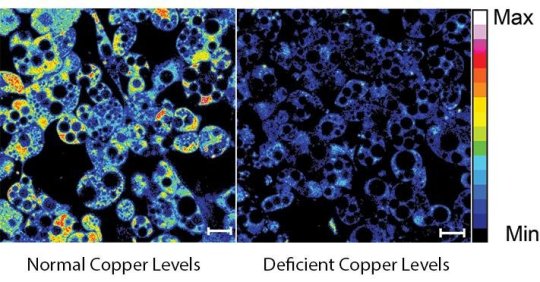
The Einstein researchers also have found that centenarians and their offspring often make unusually large amounts of a peptide (a short chain of amino acids) called humanin, which declines with age in most people and whose loss contributes to the development of Type 2 diabetes and Alzheimer’s disease. This may help explain why those who produce higher levels of humanin enjoy greater protection against those diseases and experience exceptionally long lives. For these individuals, humanin diminishes as they age, too, but the levels are much higher to start with than those of average people.

Barzilai believes the propensity for high levels of both HDL and humanin is heritable: “Offspring of centenarians have higher levels of humanin than their parents. Same with HDL. It declines with age, so it’s more apparent in the offspring.’’

Perls and his colleagues, in a study released almost four years ago, concluded there is no single common gene variant responsible for exceptional longevity. Rather, after examining about 280 gene variations, they discovered a series of gene combinations — nearly two dozen, in fact — that they believe contribute to long lives, “meaning there are different ways to get to these old ages,’’ Perls says. “It’s like playing the lottery. If you get all seven numbers, you’ll hit the jackpot.’’

These genetic groupings also seem to be involved in protecting against developing age-related diseases, since the scientists did not find an absence of disease-causing genes in their study group. “They have just as many as everybody else, which was a big surprise to us,’’ Perls says.

Also, the researchers found that the children of these healthy centenarians stay healthy longer than their same-age counterparts. The offspring of centenarians show 60 percent less heart disease, stroke, diabetes and hypertension, and 80 percent fewer overall deaths when they are in their early 70s, than those who were born at the same time but who do not have longevity in their families.

“They remain incredibly healthy into their 70s and 80s, and their mortality rate is very low, compared to others born at the same time,’’ Perls says.

Perls has studied 2,300 centenarians since 1995, including “super-centenarians’’ of 110 or older, and their offspring. He says about 45 percent of those who reach 100 manage to delay chronic age-related diseases until after they turn 80, and about 15 percent never get them at all.

Furthermore, he found that “semi-super-centenarians’’ — that is, those who are 105 to 109 — and super-centenarians don’t develop those diseases until roughly the final 5 percent of their very long lives. “They are dealing with diseases much better than the average person,’’ he says, who is more likely to develop these diseases in the 60s and 70s.

Many eventually die from the same diseases as non-centenarians, “but they do it 30 years later,’’ Barzilai says.

‘An additional 10 years’

Perls says that if you want to know whether you will live to 100, “you don’t have to do all this complicated genetic testing. Just look at your family and your health-related behaviors.’’ If you engage in healthful practices, you could reach your late 80s. “If you have the genes for longevity and you fight them [with risky behaviors], you will chop time off,’’ he says. “But if there is longevity in your family and you don’t do those things, you might get an additional 10 years past 90.’’

Newman agrees. “Don’t underestimate how powerful lifestyle is in longevity,’’ she says. “Even if longevity runs in your family, your life expectancy still will be more influenced by how you take care of yourself. If you have a centenarian parent, don’t count on living to 100 if you smoke, drink, eat a high-fat diet, and are sedentary and sleep-deprived.’’

Mary Harada thinks less about her genes and more about the unexpected event — breaking a bone, for example — that could make her a burden to her adult children.

“I don’t spend much time thinking about how long I’m going to live,’’ she says. “Whatever happens, happens. I spend more time thinking about how long I’m going to stay in my current house.’’

She has no age-related diseases and always has taken good care of herself. She has been a runner for 47 years, and she lifts weights. She shuns smoking and avoids most processed foods. She lives alone — her husband died last year — and she does most of the maintenance in and around her four-bedroom house, including leaf removal, routine yard work and spending two hours every 10 days in spring and summer mowing a very hilly lawn.

“I’ve lived here for 40 years, and I like living in this house and in this town,’’ she says. “If I could be like my father, and not break anything, I would stay here another five to 10 years. That would be wonderful.’’

https://www.washingtonpost.com/national/health-science/do-you-have-genes-that-will-let-you-live-to-age-100/2015/12/09/1460f234-953d-11e5-a2d6-f57908580b1f_story.html

Dead or Alive, Schrödinger’s Cat Can Be in 2 Boxes at Once, New Research Using Light Particles Reveals

schrodinger-cat-two-boxes

By Tia Ghose

Bizarrely behaving light particles show that the famous Schrödinger’s cat thought experiment, meant to reveal the strange nature of subatomic particles, can get even weirder than physicists thought.

Not only can the quantum cat be alive and dead at the same time — but it can also be in two places at once, new research shows.

“We are showing an analogy to Schrödinger’s cat that is made out of an electromagnetic field that is confined in two cavities,” said study lead author Chen Wang, a physicist at Yale University. “The interesting thing here is the cat is in two boxes at once.”

The findings could have implications for cracking unsolvable mathematicalproblems using quantum computing, which relies on the ability of subatomic particles to be in multiple states at once, Wang said.

Cat experiment

The famous paradox was laid out by physicist Erwin Schrödinger in 1935 to elucidate the notion of quantum superposition, the phenomenon in which tiny subatomic particles can be in multiple states at once.

In the paradox, a cat is trapped in a box with a deadly radioactive atom. If the radioactive atom decayed, the cat was a goner, but if it had not yet decayed, the cat was still alive. Because, according to the dominant interpretation of quantum mechanics, particles can exist in multiple states until they are measured, logic dictated that the cat would be both alive and dead at the same time until the radioactive atom was measured.

Cat in two boxes

The setup for the new study was deceptively simple: The team created two aluminum cavities about 1 inch (2.5 centimeters) across, and then used a sapphire chip to produce a standing wave of light in those cavities. They used a special electronic element, called a Josephson Junction, to superimpose a standing wave of two separate wavelengths of light in each cavity. The end result was that the cat, or the group of about 80 photons in the cavities, was oscillating at two different wavelengths at once — in two different places. Figuring out whether the cat is dead or alive, so to speak, requires opening both boxes.

Though conceptually simple, the physical setup required ultrapure aluminum and highly precise chips and electromagnetic devices to ensure that the photons were as isolated from the environment as possible, Wang said.

That’s because at large scales, quantum superposition tends to disappear almost instantaneously, as soon as these superimposed subatomic particles whose fates are linked interact with the environment. Most of the time, this so-called decoherence would happen so quickly that researchers would have no time to observe the superposition, Wang said. So devices that keep coherence (or keep the particles in superposition) for long periods of time, known as the quality factor, is extremely important, Wang added.

“The quality of these things determines once you put a single excitation into the system, how long does it live, or does it die away,” Wang told Live Science.

If the excitation of the system — the production of the electromagnetic standing wave — is similar to the swing of a pendulum, then “our pendulum swings essentially tens of billions of times before it stops.”

The new findings could make for easier error correction in quantum computing, Wang said. In quantum computing, bits of information are encoded in the fragile superposition states of particles, and once that superposition is lost or corrupted, the data is also corrupted. So most quantum computing concepts involve a lot of redundancy.

“It’s well understood that 99 percent of computation or more will be done to correct for errors, rather than computation itself,” Wang said.

Their system could conceivably get around this problem by encoding the redundancy in the size of the cavity itself rather than in separate, calculated bits, Wang said.

“Demonstrating this cat in a ‘two boxes state’ is basically the first step in our architecture,” Wang said.

See more at: http://www.livescience.com/54890-schrodinger-cat-can-be-in-two-places.html#sthash.X4gB2Mc1.dpuf

That New Superbug Was Found in a UTI and That’s Key

BR3GWM bacteria streaked and grows on an agar plate in the lab

by SARAH ZHANG

THE WOMAN HARBORING E. coli resistant to colistin did not know it, and it’s only luck that we do. Her doctor would have never prescribed that last-resort antibiotic for a routine urinary tract infection—it can cause serious kidney damage. But her doctor did take a urine sample, which ended up at the Walter Reed National Military Medical Center, where researchers had recently started testing for colistin resistance. The test came back positive. Then the came scary headlines about a new superbug in the US.

Superbugs are bacteria with genetic mutations that let them survive humanity’s harshest weapons in germ warfare: antibiotics. The gene behind this E. coli’s colistin resistance is called mcr-1. It first emerged last year when Chinese researchers found it in samples from hospital patients and raw pork. Why pork? Colistin’s serious side effects mean it’s no longer used as a human antibiotic in many countries. But in China, farmers have been adding it by the pound into feed to fatten animals up.

Once epidemiologists knew to look for mcr-1, they found it in Malaysia, England and then the rest of Europe. It was only a matter of time before colistin resistance turned up in the US. On the same day news came out about this woman’s colistin-resistant UTI, the Department of Health and Human Services also announced it found mcr-1 in a sample from a pig intestine.

Colistin is not used in animal feed in the US, so it’s unclear how colistin-resistant bacteria ended up infecting that woman—or that pig. But food and people move freely across borders. And more even seriously, US animal farmers do use other antibiotics—even human ones—on chicken, pigs, and cows. A growing body of research has linked antibiotic use in food animals to drug-resistant bouts of food poisoning from salmonella, campylobacter, and MRSA. Even more interesting is a possible link between antibiotics on meat and urinary tract infections, which science journalist Maryn McKenna has covered extensively. The Food and Drug Administration issued a guidance last year for farms to phase out medically important antibiotics, though only voluntarily.


The Rise of the Drug-Resistant UTI

Urinary tract infections are damn common—annoyingly common if you ask many women. And antibiotic resistant UTIs are on the rise, too: From 2000 to 2010, the number of UTIs resistant to the antibiotic Cipro went from 3 percent to 17.1 percent. Because UTIs afflict so many people, they’re fairly representative antibiotic resistance out there in people community—especially compared to the resistant infections that epidemiologists tend to study most intensely, like ones that kill already sick hospital patients. “UTIs are a good picture of what people are being exposed to on a daily basis” says Amee Manges, an epidemiologist at the University of British Columbia. Case in point: That colistin-resistant bacteria in the woman from Philadelphia.

Manges has spent the past fifteen years studying the link between antibiotic use in meat production, especially poultry, and UTIs. Back when she was working on her doctoral thesis at the University of California, Berkeley, she kept seeing young, otherwise healthy students with UTIs. Originally, she thought she was going to track sexual transmission of the E. coli that caused such infections. With that kind of sporadic sexual transmission, she should have seen many different strains. But when she DNA fingerprinted the bacteria, she found they were all the same strain—the same pattern you’d see from a single source, like if the campus cafeteria gave everyone food poisoning. She was never able to trace those UTI cases back to the original source, but she’s been working on the question ever since.

UTIs are so hard to trace because the infection might not set in until long after a patient first acquired to bacteria. Say a woman eats some undercooked chicken. “The bacteria just hangs out in your intestine for months or possibly years,” says Manges. Then you get risk factor for UTI—sex or a catheter insertion—and that bacteria makes its way from, ahem, the end of your gut to the urethra. But getting people to remember what they ate a week ago is hard. Getting people to remember what they ate a year ago? Hahaha.

The Surveillance Net
Nevertheless, Manges and others have found that strains on meat match strains found in UTIs. Because of the difficulty in tracing UTIs, that evidence is not as ironclad as the evidence for antibiotics use and antibiotic-resistant food poisoning. With routine surveillance of UTIs though, epidemiologists could get a better handle of not only resistant bacteria that come from meat—but also other sources like drinking water or travel or family members being in the hospital. But that surveillance doesn’t happen. “There’s no organized infrastructure to get a good handle about resistance rates across communities,” says Kalpana Gupta, an infectious disease specialist at Boston University.

When patients walk in with UTIs, doctors will often hand out antibiotics without doing a urine culture. Growing the bacteria takes two days—testing for antibiotic-resistance a third—and by that time the patient is usually on the mend already. The fact that the women in Philadelphia got tested was unusual. The fact that her sample was tested against colistin even more so. As Gupta says, “Colistin is not something we would even use to treat UTIs.” (Resistance to another class of antibiotics triggered that extra test in this case.)

The Centers for Disease Control and Prevention is now following up with the woman in Philadelphia to find out she ended up with that colistin-strain of E. coli, which has never been found in the US before. Her infection was fortunately not resistant to all antibiotics. But what makes the colistin-resistance gene mcr-1 so worrisome is that it’s on a small loop of DNA that different bacteria easily swap back and forth. Someday, another bacteria already immune to all other antibiotics will pick up mcr-1, too. It’s only a matter of time.

The wider the surveillance net though, the more quickly we’ll find it.

In Search For Cures, Scientists Create Embryos That Are Both Animal And Human

A handful of scientists around the United States are trying to do something that some people find disturbing: make embryos that are part human, part animal.

The researchers hope these embryos, known as chimeras, could eventually help save the lives of people with a wide range of diseases.

One way would be to use chimera embryos to create better animal models to study how human diseases happen and how they progress.

Perhaps the boldest hope is to create farm animals that have human organs that could be transplanted into terminally ill patients.

But some scientists and bioethicists worry the creation of these interspecies embryos crosses the line. “You’re getting into unsettling ground that I think is damaging to our sense of humanity,” says Stuart Newman, a professor of cell biology and anatomy at the New York Medical College.

The experiments are so sensitive that the National Institutes of Health has imposed a moratorium on funding them while officials explore the ethical issues they raise.

Nevertheless, a small number of researchers are pursuing the work with alternative funding. They hope the results will persuade the NIH to lift the moratorium.

“We’re not trying to make a chimera just because we want to see some kind of monstrous creature,” says Pablo Ross, a reproductive biologist at the University of California, Davis. “We’re doing this for a biomedical purpose.”

The NIH is expected to announce soon how it plans to handle requests for funding.

Recently, Ross agreed to let me visit his lab for an unusual look at his research. During the visit, Ross demonstrated how he is trying to create a pancreas that theoretically could be transplanted into a patient with diabetes.

The first step involves using new gene-editing techniques to remove the gene that pig embryos need to make a pancreas.

Working under an elaborate microscope, Ross makes a small hole in the embryo’s outer membrane with a laser. Next, he injects a molecule synthesized in the laboratory to home in on and delete the pancreas gene inside. (In separate experiments, he has done this to sheep embryos, too.)

After the embryos have had their DNA edited this way, Ross creates another hole in the membrane so he can inject human induced pluripotent stem cells, or iPS for short, into the pig embryos.

Like human embryonic stem cells, iPS cells can turn into any kind of cell or tissue in the body. The researchers’ hope is that the human stem cells will take advantage of the void in the embryo to start forming a human pancreas.

Because iPS cells can be made from any adult’s skin cells, any organs they form would match the patient who needs the transplant, vastly reducing the risk that the body would reject the new organ.

But for the embryo to develop and produce an organ, Ross has to put the chimera embryos into the wombs of adult pigs. That involves a surgical procedure, which is performed in a large operating room across the street from Ross’s lab.

The day Ross opened his lab to me, a surgical team was anesthetizing an adult female pig so surgeons could make an incision to get access to its uterus.

Ross then rushed over with a special syringe filled with chimera embryos. He injected 25 embryos into each side of the animal’s uterus. The procedure took about an hour. He repeated the process on a second pig.

Every time Ross does this, he then waits a few weeks to allow the embryos to develop to their 28th day — a time when primitive structures such as organs start to form.

Ross then retrieves the chimeric embryos to dissect them so he can see what the human stem cells are doing inside. He examines whether the human stem cells have started to form a pancreas, and whether they have begun making any other types of tissues.

The uncertainty is part of what makes the work so controversial. Ross and other scientists conducting these experiments can’t know exactly where the human stem cells will go. Ross hopes they’ll only grow a human pancreas. But they could go elsewhere, such as to the brain.

“If you have pigs with partly human brains you would have animals that might actually have consciousness like a human,” Newman says. “It might have human-type needs. We don’t really know.”

That possibility raises new questions about the morality of using the animals for experimentation. Another concern is that the stem cells could form human sperm and human eggs in the chimeras.

“If a male chimeric pig mated with a female chimeric pig, the result could be a human fetus developing in the uterus of that female chimera,” Newman says. Another possibility is the animals could give birth to some kind of part-human, part-pig creature.

“One of the concerns that a lot of people have is that there’s something sacrosanct about what it means to be human expressed in our DNA,” says Jason Robert, a bioethicist at Arizona State University. “And that by inserting that into other animals and giving those other animals potentially some of the capacities of humans that this could be a kind of violation — a kind of, maybe, even a playing God.”

Ross defends what his work. “I don’t consider that we’re playing God or even close to that,” Ross says. “We’re just trying to use the technologies that we have developed to improve peoples’ life.”

Still, Ross acknowledges the concerns. So he’s moving very carefully, he says. For example, he’s only letting the chimera embryos develop for 28 days. At that point, he removes the embryos and dissects them.

If he discovers the stem cells are going to the wrong places in the embryos, he says he can take steps to stop that from happening. In addition, he’d make sure adult chimeras are never allowed to mate, he says.

“We’re very aware and sensitive to the ethical concerns,” he says. “One of the reasons we’re doing this research the way we’re doing it is because we want to provide scientific information to inform those concerns.”

Ross is working with Juan Carlos Izpisua Belmonte from the Salk Intitute for Biological Studies in La Jolla, Calif., and Hiromitsu Nakauchi at Stanford University. Daniel Garry of the University of Minnesota and colleagues are conducting similar work. The research is funded in part by the Defense Department and the California Institute for Regenerative Medicine (CIRM).

http://www.npr.org/sections/health-shots/2016/05/18/478212837/in-search-for-cures-scientists-create-embryos-that-are-both-animal-and-human

Thanks to Kebmodee for bringing this to the attention of the It’s Interesting community.

New science research shows that most people have only about 50% of the friends that they think they have.

As it turns out, we can be pretty terrible at knowing who our friends are: In what may be among the saddest pieces of social-psychology research published in quite some time, a study in the journal PLoS One recently made the case that as many as half the people we consider our friends don’t feel the same way.

The study authors gave a survey to 84 college students in the same class, asking each one to rate every other person in the study on a scale of zero (“I do not know this person”) to five (“One of my best friends”), with three as the minimum score needed to qualify for friendship. The participants also wrote down their guesses for how each person would rate them.

Overall, the researchers documented 1,353 cases of friendship, meaning instances where one person rated another as a three or higher. And in 94 percent of them, the person doing the ranking guessed that the other person would feel the same way.

Which makes sense — you probably wouldn’t call someone a friend, after all, unless you thought that definition was mutual. That’s why we have terms to capture more one-sided relationships, like friend crush or hey, I don’t really know her but I think she’s neat. Both of which, come to think of it, might have been better descriptors of a lot of the relationships in the study. In reality, only 53 percent of the friendships — a small, sad, oh honey number of them — were actually reciprocal.

Some caveats: The study was small, and all the subjects were undergraduates; friendships change over the course of a lifetime, and it’s certainly possible that, over time, many tenuous lopsided friendships can dwindle to a more solid few. But the study authors also looked at a handful of previous surveys on friendship, ranging in size from 82 people to 3,160, and found similar results: Among those, the highest proportion of reciprocal friendships was 53 percent, and the lowest was a bummer, at 34 percent.

“These findings suggest a profound inability of people to perceive friendship reciprocity, perhaps because the possibility of non-reciprocal friendship challenges one’s self-image,” the study authors wrote.

http://nymag.com/scienceofus/2016/05/half-of-your-friends-probably-dont-think-of-you-as-a-friend.html

Lightning storms make it rain diamonds on Saturn and Jupiter

saturn

t sounds like a wacky fantasy, but scientists believe that it rains diamonds in the clouds of Saturn and Jupiter.

Diamonds are made from highly compressed and heated carbon. Theoretically, if you took a charcoal bricket out of your grill and heated it and pressed it hard enough for long enough, you could make a diamond.

On Earth, diamonds form about 100 miles underground. Volcanic magma highways then bring them closer to the surface, providing us with shiny gemstones that we stick in rings and ear studs.

But in the dense atmospheres of planets like Jupiter and Saturn, whose massive size generates enormous amounts of gravity, crazy amounts of pressure and heat can squeeze carbon in mid-air — and make it rain diamonds.

Scientists have speculated for years that diamonds are abundant in the cores of the smaller, cooler gas giants, Neptune and Uranus. They believed that the larger gaseous planets, Jupiter and Saturn, didn’t have suitable atmospheres to forge diamonds.

But when researchers recently analyzed the pressures and temperatures for Jupiter’s and Saturn’s atmospheres, then modeled how carbon would behave, they determined that diamond rain is very likely.

Diamonds seem especially likely to form in huge, storm-ravaged regions of Saturn, and in enormous quantities — Kevin Baines, a researcher at University of Madison-Wisconsin and NASA JPL, told BBC News it may rain as much as 2.2 million pounds of diamonds there every year.

The diamonds start out as methane gas. Powerful lightning storms on the two huge gas giants then zap it into carbon soot.

“As the soot falls, the pressure on it increases,” Baines told the BBC. “And after about 1,000 miles it turns to graphite – the sheet-like form of carbon you find in pencils.”

And the graphite keeps falling. When it reaches the deep atmosphere of Saturn, for example — around 3,700 miles down — the immense pressure squeezes the carbon into diamonds, which float in seas of liquid methane and hydrogen.

Eventually the gems sink toward the interior of the planet (a depth of 18,600 miles), where nightmarish pressure and heat melts the diamonds into molten carbon.

“Once you get down to those extreme depths,” Baines told the BBC, “the pressure and temperature is so hellish, there’s no way the diamonds could remain solid.”

http://www.techinsider.io/diamond-rain-saturn-jupiter-2016-4

New study may explain gene’s role in major psychiatric disorders

A new study shows the death of newborn brain cells may be linked to a genetic risk factor for five major psychiatric diseases, and at the same time shows a compound currently being developed for use in humans may have therapeutic value for these diseases by preventing the cells from dying.

In 2013, the largest genetic study of psychiatric illness to date implicated mutations in the gene called CACNA1C as a risk factor in five major forms of neuropsychiatric disease — schizophrenia, major depression, bipolar disorder, autism, and attention deficit hyperactivity disorder (ADHD). All the conditions also share the common clinical feature of high anxiety. By recognizing an overlap between several lines of research, scientists at the University of Iowa and Weill Cornell Medicine of Cornell University have now discovered a new and unexpected role for CACNA1C that may explain its association with these neuropsychiatric diseases and provide a new therapeutic target.

The new study, recently published in eNeuro, shows that loss of the CACNA1C gene from the forebrain of mice results in decreased survival of newborn neurons in the hippocampus, one of only two regions in the adult brain where new neurons are continually produced – a process known as neurogenesis. Death of these hippocampal neurons has been linked to a number of psychiatric conditions, including schizophrenia, depression, and anxiety.

“We have identified a new function for one of the most important genes in psychiatric illness,” says Andrew Pieper, MD, PhD, co-senior author of the study, professor of psychiatry at the UI Carver College of Medicine and a member of the Pappajohn Biomedical Institute at the UI. “It mediates survival of newborn neurons in the hippocampus, part of the brain that is important in learning and memory, mood and anxiety.”

Moreover, the scientists were able to restore normal neurogenesis in mice lacking the CACNA1C gene using a neuroprotective compound called P7C3-A20, which Pieper’s group discovered and which is currently under development as a potential therapy for neurodegenerative diseases. The finding suggests that the P7C3 compounds may also be of interest as potential therapies for these neuropsychiatric conditions, which affect millions of people worldwide and which often are difficult to treat.

Pieper’s co-lead author, Anjali Rajadhyaksha, associate professor of neuroscience in Pediatrics and the Feil Family Brain and Mind Research Institute at Weill Cornell Medicine and director of the Weill Cornell Autism Research Program, studies the role of the Cav1.2 calcium channel encoded by the CACNA1C gene in reward pathways affected in various neuropsychiatric disorders.

“Genetic risk factors that can disrupt the development and function of brain circuits are believed to contribute to multiple neuropsychiatric disorders. Adult newborn neurons may serve a role in fine-tuning rewarding and environmental experiences, including social cognition, which are disrupted in disorders such as schizophrenia and autism spectrum disorders,” Rajadhyaksha says. “The findings of this study provide a direct link between the CACNA1C risk gene and a key cellular deficit, providing a clue into the potential neurobiological basis of CACNA1C-linked disease symptoms.”

Several years ago, Rajadhyaksha and Pieper created genetically altered mice that are missing the CACNA1C gene in the forebrain. The team discovered that the animals have very high anxiety.

“That was an exciting finding, because all of the neuropsychiatric diseases in which this gene is implicated are associated with symptoms of anxiety,” says Pieper who also holds appointments in the UI Departments of Neurology, Radiation Oncology, Molecular Physiology and Biophysics, the Holden Comprehensive Cancer Center, and the Iowa City VA Health Care System.

By studying neurogenesis in the mice, the research team has now shown that loss of the CACNA1C gene from the forebrain decreases the survival of newborn neurons in the hippocampus – only about half as many hippocampal neurons survive in mice without the gene compared to normal mice. Loss of CACNA1C also reduces production of BDNF, an important brain growth factor that supports neurogenesis.

The findings suggest that loss of the CACNA1C gene disrupts neurogenesis in the hippocampus by lowering the production of BDNF.

Pieper had previously shown that the “P7C3-class” of neuroprotective compounds bolsters neurogenesis in the hippocampus by protecting newborn neurons from cell death. When the team gave the P7C3-A20 compound to mice lacking the CACNA1C gene, neurogenesis was restored back to normal levels. Notably, the cells were protected despite the fact that BDNF levels remained abnormally low, demonstrating that P7C3-A20 bypasses the BDNF deficit and independently rescues hippocampal neurogenesis.

Pieper indicated the next step would be to determine if the P7C3-A20 compound could also ameliorate the anxiety symptoms in the mice. If that proves to be true, it would strengthen the idea that drugs based on this compound might be helpful in treating patients with major forms of psychiatric disease.

“CACNA1C is probably the most important genetic finding in psychiatry. It probably influences a number of psychiatric disorders, most convincingly, bipolar disorder and schizophrenia,” says Jimmy Potash, MD, professor and DEO of psychiatry at the UI who was not involved in the study. “Understanding how these genetic effects are manifested in the brain is among the most exciting challenges in psychiatric neuroscience right now.”

http://www.news-medical.net/news/20160427/Study-reveals-new-function-for-CACNA1C-gene-in-psychiatric-diseases.aspx