Pluto May Have Formed from 1 Billion Comets


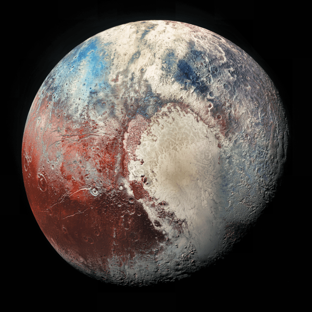
This view of Pluto’s Sputnik Planitia nitrogen-ice plain was captured by NASA’s New Horizons spacecraft during its flyby of the dwarf planet in July 2015.

At its heart, Pluto may be a gigantic comet.

Researchers have come up with a new theory about the dwarf planet’s origins after taking a close look at Sputnik Planitia, the vast nitrogen-ice glacier that constitutes the left lobe of Pluto’s famous “heart” feature.

“We found an intriguing consistency between the estimated amount of nitrogen inside the glacier and the amount that would be expected if Pluto was formed by the agglomeration of roughly a billion comets or other Kuiper Belt objects similar in chemical composition to 67P, the comet explored by Rosetta,” Chris Glein, a scientist at the Southwest Research Institute (SwRI) in San Antonio, said in a statement.

The European Space Agency’s Rosetta mission orbited Comet 67P/Churyumov-Gerasimenko from 2014 through 2016. The orbiting mothership also dropped a lander named Philae onto the icy body, pulling off the first-ever soft touchdown on a comet’s surface. (The Kuiper Belt is the ring of frigid objects beyond Neptune’s orbit; Pluto is the belt’s largest resident.)

Glein and his SwRI colleague Hunter Waite devised the new Pluto-formation scenario after analyzing data from Rosetta and NASA’s New Horizons mission, which flew by Pluto in July 2015.

The scientists also made some inferences about the dwarf planet’s evolution in their new study, which was published online Wednesday (May 23) in the journal Icarus.

“Our research suggests that Pluto’s initial chemical makeup, inherited from cometary building blocks, was chemically modified by liquid water, perhaps even in a subsurface ocean,” Glein said.

Glein and Waite aren’t claiming to have nailed down Pluto’s origin definitively; a “solar model,” in which the dwarf planet coalesced from cold ices with a chemical composition closer to that of the sun, also remains in play, the duo said.

“This research builds upon the fantastic successes of the New Horizons and Rosetta missions to expand our understanding of the origin and evolution of Pluto,” Glein said.

“Using chemistry as a detective’s tool, we are able to trace certain features we see on Pluto today to formation processes from long ago,” he added. “This leads to a new appreciation of the richness of Pluto’s ‘life story,’ which we are only starting to grasp.”

Rosetta’s mission ended in September 2016, when the probe’s handlers steered it to an intentional crash-landing on 67P’s surface. New Horizons’ work, however, is far from done. The NASA spacecraft is speeding toward a flyby of a small Kuiper Belt object known officially as 2014 MU69 (and unofficially as Ultima Thule). This close encounter, which will occur on Jan. 1, 2019, about 1 billion miles (1.6 billion kilometers) beyond Pluto’s orbit, is the centerpiece of New Horizons’ extended mission.

https://www.space.com/40687-pluto-formation-1-billion-comets.html

A Hangover Pill? Tests on drunk mice show promise

“Civilization begins with distillation,” said William Faulkner, a writer and drinker. Although our thirst for alcohol dates back to the Stone Age, nobody has figured out a good way to deal with the ensuing hangover after getting drunk.

As a chemical engineering professor and wine enthusiast, I felt I needed to find a solution. As frivolous as this project may sound, it has serious implications. Between 8 and 10 percent of emergency room visits in America are due to acute alcohol poisoning. Alcohol is the leading risk factor for premature deaths and disability among people aged 15-49 and its abuse leads to serious health problems, including cardiovascular and liver cancer. Despite these sobering facts, current treatments for alcohol overdose largely rely on the body’s own enzymes to break down this drug.

I decided to design an antidote that could help people enjoy wine or cocktails or beer without a hangover, and at the same time create a lifesaving therapy to treat intoxication and overdose victims in the ER. I chose to create capsules filled with natural enzymes usually found in liver cells to help the body process the alcohol faster.

Together with professor Cheng Ji, an expert in liver diseases from Keck School of Medicine at the University of Southern California, and my graduate student Duo Xu, we developed an antidote and tested it in mice.

Inspired by the body’s approach for breaking down alcohol, we chose three natural enzymes that convert alcohol into harmless molecules that are then excreted. That might sound simple, because these enzymes were not new, but the tricky part was to figure out a safe, effective way to deliver them to the liver.

To protect the enzymes, we wrapped each of them in a shell, using a material the U.S. Food and Drug Administration had already approved for pills. We then injected these nanocapsules into the veins of drunk mice where they hurtled through the circulatory system, eventually arriving in the liver where they entered the cells and served as mini–reactors to digest alcohol.

We showed that in inebriated mice (which fall asleep much faster than drunk humans), the treatment decreased the blood alcohol level by 45 percent in just four hours compared to mice that didn’t receive any. Meanwhile, the blood concentration of acetaldehyde – a highly toxic compound that is carcinogenic, causes headaches and vomiting, makes people blush after drinking, and is produced during the normal alcohol metabolism – remained extremely low. The animals given the drug woke from their alcohol-induced slumber faster than their untreated counterparts – something all college students would appreciate.

The ability to efficiently break down alcohol quickly should help patients wake up earlier and prevent alcohol poisoning. It should also protect their liver from alcohol–associated stress and damage.

We are currently completing tests to ensure that our nanocapsules are safe and don’t trigger unexpected or dangerous side effects. If our treatments prove effective in animals, we could begin human clinical trials in as early as one year.

This sort of antidote won’t stop people from going too far when consuming alcohol, but it could help them recover quicker.

https://www.technologynetworks.com/neuroscience/articles/a-hangover-pill-tests-on-drunk-mice-show-promise-302970?utm_campaign=NEWSLETTER_TN_Neuroscience_2017&utm_source=hs_email&utm_medium=email&utm_content=63148685&_hsenc=p2ANqtz-_9-CBGC564lH1Jr5Fxrauf8vQZ42sDx9gSSQj_dPJTj3gm3QDvY74R4WiynR1vM5L7tdtTLBIV40iEWBKcEB7JzwFUnQ&_hsmi=63148685

Scientists plan DNA testing of Loch Ness lake

Prof Neil Gemmell, a New Zealand scientist leading the project, said he did not believe in Nessie, but was confident of finding genetic codes for other creatures.

He said a “biological explanation” might be found to explain some of the stories about the Loch Ness Monster.

The team will collect tiny fragments of skin and scales for two weeks in June.

Prof Gemmell, from the University of Otago in Dunedin, said: “I don’t believe in the idea of a monster, but I’m open to the idea that there are things yet to be discovered and not fully understood.

“Maybe there’s a biological explanation for some of the stories.”

The University of the Highlands and Islands’ UHI Rivers and Lochs Institute in Inverness is assisting in the project.

Other organisms

After the research team’s trip to Loch Ness, the samples will be sent to laboratories in New Zealand, Australia, Denmark and France to be analysed against a genetic database.

Prof Gemmell said: “There’s absolutely no doubt that we will find new stuff. And that’s very exciting.

“While the prospect of looking for evidence of the Loch Ness monster is the hook to this project, there is an extraordinary amount of new knowledge that we will gain from the work about organisms that inhabit Loch Ness – the UK’s largest freshwater body.”

The scientist said the team expected to find sequences of DNA from plants, fish and other organisms.

He said it would be possible to identify these plants and animals by comparing the sequences of their DNA against sequences held on a large, international database.

Prof Gemmell added: “There is this idea that an ancient Jurassic Age reptile might be in Loch Ness.

“If we find any reptilian DNA sequences in Loch Ness, that would be surprising and would be very, very interesting.”

The Loch Ness Monster is one of Scotland’s oldest and most enduring myths. It inspires books, TV shows and films, and sustains a major tourism industry around its home.

The story of the monster can be traced back 1,500 years when Irish missionary St Columba is said to have encountered a beast in the River Ness in 565AD.

Later, in the 1930s, The Inverness Courier reported the first modern sighting of Nessie.

Whale-like creature

In 1933, the newspaper’s Fort Augustus correspondent, Alec Campbell, reported a sighting by Aldie Mackay of what she believed to be Nessie.

Mr Campbell’s report described a whale-like creature and the loch’s water “cascading and churning”.

The editor at the time, Evan Barron, suggested the beast be described as a “monster”, kick starting the modern myth of the Loch Ness Monster.

Over the years various efforts have tried and failed to find the beast.

In tourism terms, there are two exhibitions dedicated to the monster and there is not a tourist shop in the Highlands, and even more widely across Scotland, where a cuddly toy of Nessie cannot be found.

In 2016, the inaugural Inverness Loch Ness International Knitting Festival exhibited knitted Nessie’s made from all parts of the world.

‘Record high’

In popular culture, the Loch Ness Monster has reared its head many times, including in 1975’s four-part Doctor Who – Terror of the Zygons, the 1980s cartoon The Family-Ness as well as The Simpsons and 1996’s Loch Ness starring Ted Danson.

In 2014, it was reported that for the first time in almost 90 years no “confirmed sightings” had been made of the Loch Ness Monster.

Gary Campbell, who keeps a register of sightings, said no-one had come forward in 18 months to say they had seen the monster.

But last year, sightings hit a record high.

http://www.bbc.com/news/uk-scotland-highlands-islands-44223259

Biomaterial developed at UCLA helps regrow brain tissue after stroke in mice

by Leigh Hopper

Tnew stroke-healing gel created by UCLA researchers helped regrow neurons and blood vessels in mice whose brains had been damaged by strokes. The finding is reported May 21 in Nature Materials.

“We tested this in laboratory mice to determine if it would repair the brain and lead to recovery in a model of stroke,” said Dr. S. Thomas Carmichael, professor of neurology at the David Geffen School of Medicine at UCLA. “The study indicated that new brain tissue can be regenerated in what was previously just an inactive brain scar after stroke.”

The results suggest that such an approach could some day be used to treat people who have had a stroke, said Tatiana Segura, a former professor of chemical and biomolecular engineering at UCLA who collaborated on the research. Segura is now a professor at Duke University.

The brain has a limited capacity for recovery after stroke. Unlike the liver, skin and some other organs, the brain does not regenerate new connections, blood vessels or tissue structures after it is damaged. Instead, dead brain tissue is absorbed, which leaves a cavity devoid of blood vessels, neurons or axons — the thin nerve fibers that project from neurons.

To see if healthy tissue surrounding the cavity could be coaxed into healing the stroke injury, Segura engineered a hydrogel that, when injected into the cavity, thickens to create a scaffolding into which blood vessels and neurons can grow. The gel is infused with medications that stimulate blood vessel growth and suppress inflammation, since inflammation results in scars and impedes functional tissue from regrowing.

After 16 weeks, the stroke cavities contained regenerated brain tissue, including new neuronal connections — a result that had not been seen before. The mice’s ability to reach for food improved, a sign of improved motor behavior, although the exact mechanism for the improvement wasn’t clear.

“The new axons could actually be working,” Segura said. “Or the new tissue could be improving the performance of the surrounding, unharmed brain tissue.”

The gel was eventually absorbed by the body, leaving behind only new tissue.

The research was designed to explore recovery in acute stroke, the period immediately following a stroke — in mice, that period lasts five days; in humans, it’s two months. Next, Carmichael and Segura plan to investigate whether brain tissue can be regenerated in mice long after the stroke injury. More than 6 million Americans are living with long-term effects of stroke, which is known as chronic stroke.

The other authors of the paper are Lina Nih and Shiva Gojgini, both of UCLA.

The study was supported by the National Institutes of Health.

http://newsroom.ucla.edu/releases/biomaterial-ucla-regrow-brain-tissue-after-stroke-mice

Molecular link between long-term memory and neurodegenerative disease discovered

Scientists have just discovered that a small region of a cellular protein that helps long-term memories form also drives the neurodegeneration seen in motor neuron disease (MND). This small part of the Ataxin-2 protein thus works for good and for bad. When a version of the protein lacking this region was substituted for the normal form in fruit flies (model organisms), the animals could not form long-term memories – but, surprisingly, the same flies showed a remarkable resistance to neurodegeneration.

The popular “ice bucket challenge” highlighted the social significance of MND, as well as the need to better understand and treat neurodegenerative conditions. This new research identifies a very specific basic mechanism that facilitates progression of neuronal loss in an animal model of MND, and, by shedding light on a potential way to protect against cell death in MND, it should inform strategies for the development of therapeutics to treat or manage these devastating conditions, which are currently incurable.

The Science Foundation Ireland-funded research, involving scientists from the Trinity College Institute of Neuroscience, NCBS Bangalore and HMMI, University of Colorado, Boulder, has just been published in the leading international journal Neuron.

Professor of Neurogenetics at Trinity College Dublin, Mani Ramaswami, said: “This work, by collaborating young researchers based in Irish, Indian and American laboratories, provides a great example of the ability of fundamental research in model organisms to produce biologically and clinically interesting information.”

A common feature of neurodegenerative diseases is the presence of specific protein aggregates in nerve cells, which accumulate and clump together — usually as protein fibres called amyloid filaments. Such aggregates are believed to trigger processes that cause the neuronal death associated with these debilitating diseases. For example, amyloid-beta (Aβ) aggregates are associated with Alzheimer’s disease, while TDP-43, FUS and Ataxin-2 proteins are commonly found in MND patients.

The scientists behind the current study set out to test this “amyloid hypothesis” to see whether it may explain how MND develops. The scientists genetically engineered fruit flies with mutations designed to reduce Ataxin-2 protein assembly into aggregates without affecting other functions of the protein.

Arnas Petrauskas, Trinity, said: “The flies with this altered, non-aggregating version of the protein showed a striking resistance to neurodegeneration. This suggests the normal Ataxin-2 protein and its ability to form aggregates is required for the progression of at least some forms of MND, which means these results provide support for the amyloid hypothesis.”

“What really surprised us though was that this same protein region seems to be required for the flies to develop long-term memory, as those with the altered version of Ataxin-2 showed normal short-term but defective long-term memories.”

Fruit flies normally respond strongly to new odorants, but weakly to familiar odorants through a process called habituation. This memory of the familiar can be of the short-term kind – to an odorant encountered for half-an-hour, or of the long-term kind, to odorants encountered for days (think of it as remembering a phone number of a new acquaintance versus remembering your own phone number). Flies lacking this small domain of Ataxin-2 showed greatly reduced long-term memory.

So how is long-term memory formation and disease progression connected? It turns out that proteins like the TDP-43, FUS and Ataxin-2 found in MND are also involved in the natural control and management of protein expression in the cell. The very same region of Ataxin-2 is needed to form RNP granules that store RNAs (essentially blueprints, or recipes for specific proteins) in a silent form until they are unpackaged by a signal and used to produce molecules when they are required. This local control of RNAs is required for long-term changes at neuronal synapses that underlie long-term memory.

The new discovery shows that Ataxin-2 concentrates several RNA-binding proteins used in the process of memory storing, but in doing so, it creates a biological environment that can help these proteins aggregate into disease-causing amyloids. A “trade-off” therefore exists in nature where the Ataxin-2 gene increases the danger of neurodegeneration, but helps our cells control RNA and form long-term memories.

In a commentary on the research published in the same issue of the journal Neuron, Aaron Gitler, Professor of Genetics in the Stanford Neuroscience Institute, an independent expert in MND research said: “This data suggest that manipulating RNP granule formation by genetically manipulating ataxin-2’s IDRs, or by other means could be therapeutic in ALS. Beyond ataxin-2, the race is now on to discover additional proteins that help build RNP granules.”

https://www.tcd.ie/news_events/articles/link-between-long-term-memory-and-neurodegenerative-disease/8941

Jellyfish, which don’t have brains, found to have what may be an early form of sleep

The purpose and evolutionary origins of sleep are among the biggest mysteries in neuroscience. Every complex animal, from the humblest fruit fly to the largest blue whale, sleeps — yet scientists can’t explain why any organism would leave itself vulnerable to predators, and unable to eat or mate, for a large portion of the day. Now, researchers have demonstrated for the first time that even an organism without a brain — a kind of jellyfish — shows sleep-like behaviour, suggesting that the origins of sleep are more primitive than thought.

Researchers observed that the rate at which Cassiopea jellyfish pulsed their bell decreased by one-third at night, and the animals were much slower to respond to external stimuli such as food or movement during that time. When deprived of their night-time rest, the jellies were less active the next day.

“Everyone we talk to has an opinion about whether or not jellyfish sleep. It really forces them to grapple with the question of what sleep is,” says Ravi Nath, the paper’s first author and a molecular geneticist at the California Institute of Technology (Caltech) in Pasadena. The study was published in Current Biology.

“This work provides compelling evidence for how early in evolution a sleep-like state evolved,” says Dion Dickman, a neuroscientist at the University of Southern California in Los Angeles.

Mindless sleep
Nath is studying sleep in the worm Caenorhabditis elegans, but whenever he presented his work at research conferences, other scientists scoffed at the idea that such a simple animal could sleep. The question got Nath thinking: how minimal can an animal’s nervous system get before the creature lacks the ability to sleep? Nath’s obsession soon infected his friends and fellow Caltech PhD students Michael Abrams and Claire Bedbrook. Abrams works on jellyfish, and he suggested that one of these creatures would be a suitable model organism, because jellies have neurons but no central nervous system. Instead, their neurons connect in a decentralized neural net.

Cassiopea jellyfish, in particular, caught the trio’s attention. Nicknamed the upside-down jellyfish because of its habit of sitting on the sea floor on its bell, with its tentacles waving upwards, Cassiopea rarely moves on its own. This made it easier for the researchers to design an automated system that used video to track the activity of the pulsing bell. To provide evidence of sleep-like behaviour in Cassiopea (or any other organism), the researchers needed to show a rapidly reversible period of decreased activity, or quiescence, with decreased responsiveness to stimuli. The behaviour also had to be driven by a need to sleep that increased the longer the jellyfish was awake, so that a day of reduced sleep would be followed by increased rest.

Other researchers had already documented a nightly drop in activity in other species of jellyfish, but no jellyfish had been known to display the other aspects of sleep behaviour. In a 35-litre tank, Nath, Abrams and Bedbrook tracked the bell pulses of Cassiopea over six days and nights and found that the rate, which was an average of one pulse per second by day, dropped by almost one-third at night. They also documented night-time pulse-free periods of 10–15 seconds, which didn’t occur during the day.

Restless night
Without an established jellyfish alarm clock, the scientists used a snack of brine shrimp and oyster roe to try to rouse the snoozing Cassiopea. When they dropped food in the tank at night, Cassiopea responded to its treat by returning to a daytime pattern of activity. The team used the jellyfish’s preference for sitting on solid surfaces to test whether quiescent Cassiopea had a delayed response to external stimuli. They slowly lifted the jellyfish off the bottom of the tank using a screen, then pulled it out from under the animal, leaving the jelly floating in the water. It took longer for the creature to begin pulsing and to reorient itself when this happened at night than it did during the day. If the experiment was immediately repeated at night, the jellyfish responded as if it were daytime. Lastly, when the team forced Cassiopea to pull an all-nighter by keeping it awake with repeated pulses of water, they found a 17% drop in activity the following day.

“This work shows that sleep is much older than we thought. The simplicity of these organisms is a door opener to understand why sleep evolved and what it does,” says Thomas Bosch, an evolutionary biologist at Kiel University in Germany. “Sleep can be traced back to these little metazoans — how much further does it go?” he asks.

That’s what Nath, Abrams and Bedbrook want to find out. Amid the chaos of finishing their PhD theses, they have begun searching for ancient genes that might control sleep, in the hope that this might provide hints as to why sleep originally evolved.

https://www.nature.com/news/jellyfish-caught-snoozing-give-clues-to-origin-of-sleep-1.22654

New Inherited Neurodevelopmental Disease Discovered

Writing in the journal eLife, the team reveals that this disease is caused by a recessive mutation in CAMK2A – a gene that is well known for its role in regulating learning and memory in animals. The findings suggest that dysfunctional CAMK2 genes may contribute to other neurological disorders, such as epilepsy and autism, opening up potential new avenues for treating these conditions.

“A significant number of children are born with growth delays, neurological defects and intellectual disabilities every year across the world,” explains senior author Bruno Reversade, Research Director at the Institute of Medical Biology and Institute of Molecular and Cell Biology, A*STAR, Singapore, who supervised the study. “While specific genetic mutations have been identified for some patients, the cause remains unknown in many cases. Identifying novel mutations would not only advance our understanding of neurological diseases in general, but would also help clinicians diagnose children with similar symptoms and/or carry out genetic testing for expecting parents.”

The team’s research began when they identified a pair of siblings who demonstrated neurodevelopmental delay with frequent, unexplained seizures and convulsions. While the structure of their bodies developed normally, they did not gain the ability to walk or speak. “We believed that the children had novel mutations in CAMK2A, and we wanted to see if this were true,” says Reversade.

The fully functional CAMK2A protein consists of multiple subunits. Using a genomic technique called exome sequencing, the team discovered a single coding error affecting a key residue in the CAMK2A gene that prevents its subunits from assembling correctly.

Moving their studies into the roundworm Caenorhabditis elegans, the scientists saw that this mutation disrupts the ability of CAMK2A to ensure proper neuronal communication and normal motor function. This suggests that the mutation is indeed the cause of the neurodevelopmental defects seen in the siblings.

To the best of the team’s knowledge, this new disorder represents the first human disease caused by inherited mutations on both copies of the CAMK2A gene. In addition, another report* published recently identified single-copy mutations on both CAMK2A and CAMK2B that caused intellectual disabilities as soon as the mutations occurred. “We would like to bring these findings to the attention of those working in the area of paediatric genetics, such as clinicians and genetic counsellors, as there are likely more undiagnosed children with similar symptoms who have mutations in their CAMK2A gene,” explains co-first author Franklin Zhong, Research Scientist in Reversade’s lab at A*STAR.

“Neuroscientists working to understand childhood brain development, neuronal function and memory formation also need to consider this new disease, since CAMK2A is associated with these processes. In future, it would be interesting to test whether restoring CAMK2A activity can bring therapeutic benefit to patients with this condition, as well as those with related neurological disorders.”

The paper ‘A homozygous loss-of-function CAMK2A mutation causes growth delay, frequent seizures and severe intellectual disability‘ can be freely accessed online at https://doi.org/10.7554/eLife.32451. Contents, including text, figures and data, are free to reuse under a CC BY 4.0 license.

*Küry, S., van Woerden, G.M., Besnard, T., Proietti Onori, M., Latypova, X., Towne, M.C., Cho, M.T., Prescott, T.E., Ploeg, M.A., Sanders, S., et al. (2017). De Novo Mutations in Protein Kinase Genes CAMK2A and CAMK2B Cause Intellectual Disability. The American Journal of Human Genetics 101, 768-788.

https://www.technologynetworks.com/neuroscience/news/new-inherited-neurodevelopmental-disease-discovered-303233?utm_campaign=Newsletter_TN_BreakingScienceNews&utm_source=hs_email&utm_medium=email&utm_content=63149617&_hsenc=p2ANqtz-_AJri5fciUzcysqtDye56dm2VpMIbIwRqkV2di9BmqZhzk9xuPEv5CWgKF24BpT8_OB1uWAjitxNXhmduWHyW2XKGlhw&_hsmi=63149617

Could a Dose of Sunshine Make You Smarter?

By Ruth Williams

The sun’s ultraviolet (UV) radiation is a major cause of skin cancer, but it offers some health benefits too, such as boosting production of essential vitamin D and improving mood. A recent report in Cell adds enhanced learning and memory to UV’s unexpected benefits.

Researchers have discovered that, in mice, exposure to UV light activates a molecular pathway that increases production of the brain chemical glutamate, heightening the animals’ ability to learn and remember.

“The subject is of strong interest, because it provides additional support for the recently proposed theory of ultraviolet light’s regulation of the brain and central neuroendocrine system,” dermatologist Andrzej Slominski of the University of Alabama who was not involved in the research writes in an email to The Scientist.

“It’s an interesting and timely paper investigating the skin-brain connection,” notes skin scientist Martin Steinhoff of University College Dublin’s Center for Biomedical Engineering who also did not participate in the research. “The authors make an interesting observation linking moderate UV exposure to . . . [production of] the molecule urocanic acid. They hypothesize that this molecule enters the brain, activates glutaminergic neurons through glutamate release, and that memory and learning are increased.”

While the work is “fascinating, very meticulous, and extremely detailed,” says dermatologist David Fisher of Massachusetts General Hospital and Harvard Medical School, “it does not imply that UV is actually good for you. . . . Across the board, for humanity, UV really is dangerous.”

Wei Xiong of the University of Science and Technology of China who led the research did not set out to investigate the effects of UV light on the brain or the skin-brain connection. He stumbled upon his initial finding “almost accidentally,” he explains in an email to The Scientist. Xiong and his colleagues were using a mass spectrometry technique they had recently developed for analyzing the molecular contents of single neurons, when their results revealed the unexpected presence of urocanic acid—a little-known molecule produced in the skin in response to UV light.

“It was a surprise because we checked through all the literature and found no reports of the existence of this small molecule in the central nervous system,” writes Xiong.

With little information to go on, Xiong and his colleagues decided to see whether UV light could also boost levels of urocanic acid in the brain. They exposed shaved mice to a low-dose of UVB—responsible for sunburn in humans—for 2 hours, then performed mass spectrometry on the animals’ individual brain cells. Sure enough, levels of urocanic acid increased in neurons of the animals exposed to the light, but not in those of control animals.

Urocanic acid can absorb UV rays and, as a result, may be able to protect skin against the sun’s harmful effects. But in the liver and other peripheral tissues, the acid is also known to be an intermediate molecule generated in the metabolic pathway that converts histidine to glutamate. Given glutamate’s role in the brain as an excitatory neurotransmitter, Xiong and his colleagues were interested to test whether the observed UV-dependent increase in urocanic acid in neurons might be coupled with increased glutamate production. It was.

Next, the team showed that UV light enhanced electrical transmission between glutaminergic neurons in brain slices taken from animals exposed to UV, but not in those from control animals. This UV-induced effect was prevented when the researchers inhibited activity of the enzyme urocanase, which converts urocanic acid to glutamate, indicating that the acid was indeed the mediator of the UV-induced boost in glutaminergic activity.

Lastly, the team showed that mice exposed to UV performed better in motor learning and recognition memory tasks than their unexposed counterparts. And, as before, treating the animals with a urocanase inhibitor prevented the UV-induced improvements in learning and memory. Administering urocanic acid directly to animals not exposed to ultraviolet light also spurred similar learning and memory improvements to those achieved with UV exposure.

Whether the results obtained in mice, which are nocturnal and rarely see the sun, will hold true in humans is yet to be determined. But, Fisher says, if the results do hold, the finding that urocanic acid alone can enhance learning and memory might suggest “a way to utilize this information to benefit people without exposing them to the damaging effects of UV.”

H. Zhu et al., “Moderate UV exposure enhances learning and memory by promoting a novel glutamate biosynthetic pathway in the brain,” Cell, doi: 10.1016/j.cell.2018.04.014, 2018.

https://www.the-scientist.com/?articles.view/articleNo/54603/title/Could-a-Dose-of-Sunshine-Make-You-Smarter-/

Nanoparticles normally used to fight cancer could also help rescue malnourished crops

Synthetic nanoparticles used to fight cancer could also heal sickly plants.

The particles, called liposomes, are nanosized, spherical pouches that can deliver drugs to specific parts of the body (SN: 12/16/06, p. 398). Now, researchers have filled these tiny care packages with fertilizing nutrients. The new liposomes, described online May 17 in Scientific Reports, soak into plant leaves more easily than naked nutrients. That allows the nanoparticles to give malnourished crops a more potent pick-me-up than the free-floating molecules in ordinary nutrient spray.

Each liposome is a hollow sphere about 100 nanometers across, and is made of fatty molecules extracted from soybean plants. Once a plant leaf absorbs these nanoparticles, the liposomes spread to cells in the plant’s other leaves and its roots, where the fatty envelopes break down and release their molecular cargo.

Researchers first exposed tomato plants to either liposomes packed with a rare earth metal called europium, or free-floating europium molecules. Europium doesn’t naturally exist in plants or soil, so it’s easy to trace how much of this element plants soaked up after treatment. Three days after exposure, plants treated with liposomes had absorbed up to 33 percent of the nanoparticles. Plants exposed to free-floating europium took in less than 0.1 percent of the molecules

The researchers then spritzed iron- and magnesium-deficient tomato plants with either a standard spray containing iron and magnesium, or a solution containing liposomes packed with those nutrients. Two weeks later, the leaves on plants treated with free-floating nutrients were still tinged yellow and curled. Plants that received liposome treatment sported healthy, green leaves.

Avi Schroeder, a chemical engineer at the Israel Institute of Technology in Haifa, and colleagues don’t know exactly why liposomes are more palatable to plants than plain nutrients. But sprays that contain nutrient-loaded liposomes could help farmers rejuvenate frail plants more efficiently than existing mixtures, Schroeder says.

Liposome-based spray would need to be tested on a variety of vegetation before it could enter widespread use, says Ramesh Raliya, a nanobiotechnology researcher at Washington University in St. Louis not involved in the work. That’s because the pores on leaves where liposomes are assumed to enter plants can range from 50 to 150 nanometers across. If a plant’s pores are smaller than 100 nanometers, the liposomes can’t squeeze inside.

Mariya Khodakovskaya, a biologist at the University of Arkansas at Little Rock, is wary of the potential cost of this new technique. Fashioning liposomes is expensive. That’s not a problem for making liposome-based medication, which requires only a small amount of nanoparticles. But for any new agricultural practice to take root, she says, “it has to be massive, and it has to be cheap.”

A. Karny et al. Therapeutic nanoparticles penetrate leaves and deliver nutrients to agricultural crops. Scientific Reports. Published online May 17, 2018. doi: 10.1038/s41598-018-25197-y.

Nanoparticles could help rescue malnourished crops

Earth-Like Alien Planets Could Experience Snowball States


A NASA artist visualized what Earth would look like if it entered the “snowball state” predicted by new research from the University of Washington

By Chelsea Gohd

Earth-like planets with severe tilts and orbits could enter abrupt “snowball states,” in which entire oceans freeze and surface life cannot survive, according to new research.

Researchers at the University of Washington (UW) have found a new reason why, just because a planet is located in a “habitable zone” — meaning it’s close enough to its host star to sustain liquid water — it isn’t necessarily habitable. The team found that the axial tilt and orbital dynamics of planets in the habitable zone around “G dwarf” stars like our own sun can lead to “snowball states,” which are essentially extreme ice ages.

This new research looked at how a planet’s obliquity, or the angle at which a planet’s rotation axis tilts, and its orbital eccentricity, a parameter that determines the amount that an orbit deviates from a perfect circle, could affect that planet’s potential to be habitable.

Previous research suggested that planets in a habitable zone with a sun-like star that had a severe axial tilt or tilting orbit would be warmer, according to the statement. The team’s research found that the opposite holds true, which was quite a shock, they said.”We found that planets in the habitable zone could abruptly enter ‘snowball’ states if the eccentricity or the semi-major axis variations — changes in the distance between a planet and star over an orbit — were large or if the planet’s obliquity increased beyond 35 degrees,” Russell Deitrick, lead author of the new work and a postdoctoral researcher at the University of Bern who completed this research at UW, said in a statement.

Luckily, Earth’s axial tilt varies ever so slightly, leaving Earth “a relatively calm planet, climate-wise,” co-author Rory Barnes, an astronomer at UW, said in the statement. But, as it pertains to exoplanets, Deitrick “has essentially shown that ice ages on exoplanets can be much more severe than on Earth, that orbital dynamics can be a major driver of habitability and that the habitable zone is insufficient to characterize a planet’s habitability,” Barnes said.

A planet’s position in the habitable zone is typically a major factor in considering whether it may be habitable. However, this new research shows that even if a planet seems Earth-like and is orbiting at the right distance from its star, if “its orbit and obliquity oscillate like crazy, another planet might be better for follow-up with telescopes of the future,” Deitrick said.

With this research in mind, orbital dynamics should be considered an important part of determining a planet’s habitability, Deitrick added.

The work will be published in The Astronomical Journal, according to the statement.

https://www.space.com/40606-exoplanets-sudden-ice-age-snowball-states.html