Scientists Have Witnessed a Single-Celled Algae Evolve Into a Multicellular Organism

by Fiona MacDonald

Most of us know that at some point in our evolutionary history around 600 million years ago, single-celled organisms evolved into more complex multicellular life.

But knowing that happened and actually seeing it happen in real-time in front of you is an entirely different matter altogether.

And that’s exactly what researchers from the George Institute of Technology and University of Montana have witnessed – and captured in the breathtaking, time-lapse footage below.

The evolution took just 50 weeks, and was triggered by the introduction of a simple predator.

In this incredible experiment, the team was trying to figure out exactly what drove single-celled organisms to become multicellular all those years ago.

One hypothesis is that it was predation that put selective pressure on single-celled organisms, causing them to become more complex.

So to test the validity of this in the lab, the team led by evolutionary biologist William Ratcliff, took populations of single-celled green alga Chlamydomonas reinhardtii.

They then put a single-celled filter-feeding predator in the mix, Paramecium tetraurelia and watched what happened.

Incredibly, the researchers watched as in just 50 weeks – less than the span of a year – two out of five experimental populations of the single-celled creatures evolved into multicellular life.

“Here we show that de novo origins of simple multicellularity can evolve in response to predation,” the team write in their paper.

Fifty weeks is a relative blink of an eye on the evolutionary scale. For the algae it was a little longer – 750 generations. But that’s still quite impressive when you think that they evolved entirely new life cycles.

Being able to witness something like this is not only absolutely mind-blowing, but it also suggests that predation could have played some kind of role in at least part of the evolution of multicellularity.

Not only that, but the resulting multicellular organisms were all incredibly varied. Just like you’d expect in natural evolution.

“Considerable variation exists in the evolved multicellular life cycles, with both cell number and propagule size varying among isolates,” the team write in their paper.

“Survival assays show that evolved multicellular traits provide effective protection against predation.”

The research has been published in Scientific Reports and the full paper is freely available.

https://www.nature.com/articles/s41598-019-39558-8

For The First Time, Scientists Have Made Synthetic DNA With 4 Additional Letters

by Mike McRae

Earth might have a dizzying array of life forms, but our biology ultimately remains a solitary data point – we simply don’t have a reference for life based on DNA different from our own. Now, scientists have taken matters into their hands to push the boundaries on what life could be like.

Research funded by NASA and led by the Foundation for Applied Molecular Evolution in the US has led to the creation of an entirely new flavour of the DNA double helix, one that has an additional four nucleotide bases.

It’s being called hachimoji DNA (from the Japanese words for ‘eight letters’) and it includes two new pairs to add to the existing partnerships of adenine (A) paired with thymine (T), and guanine (G) with cytosine (C).

This work to expand on nature’s own genetic recipe might sound a little familiar. The same scientists already successfully squeezed in two new letters in 2011. Only last year yet another version of an extended alphabet, also with six letters, was made to function inside a living organism.

Now, in what might seem like a case of overachievement, researchers have gone back to the drawing board to develop even more non-standard nucleotides.

They have a purpose for doubling the number of codes in the recipe book, though.

“By carefully analysing the roles of shape, size and structure in hachimoji DNA, this work expands our understanding of the types of molecules that might store information in extraterrestrial life on alien worlds,” says chemist Steven Benner.

We already know a lot about the stability and functionality of ‘natural’ DNA under a range of environmental conditions, and are slowly teasing apart possible scenarios describing its evolution from simpler organic materials to living chemistry.

But to really get a good sense of how a genetic system could evolve, we need to test the limits of its underlying chemistry.

Hachimoji DNA certainly allows for that. The new codes, labelled P, B, Z and S, are based on the same kind of nitrogenous molecules as existing ones, categorised as purines and pyrimidines.

Similarly, they link up with hydrogen bonds to form their own base pairs – S bonding with B, and P with Z.

That’s where the similarities fade out. These new ‘letters’ introduce dozens of new chemical parameters to the double helix structure that potentially affect how it zips and twists.

By devising models that predict the molecule’s stability and then observing actual structures made of this ‘alien’ DNA, researchers are better equipped what’s truly important when it comes to the fundamentals of a genetic template.

The researchers constructed hundreds of hachimoji helices made up of different configurations of natural and synthetic bases and then subjected them to a range of conditions to see how well they held up.

While there were a few minor differences in how the new letters behaved, there was no reason to believe hachimoji DNA wouldn’t work well as an information-carrying template that could mutate and evolve.

The team not only showed their synthetic letters could contribute to new codes without swiftly disintegrating, the sequences were also translated into synthetic RNA versions.

Their work falls well short of a second genesis. But a novel DNA format such as this is a step towards determining what living chemistry might – and might not – look like elsewhere in the Universe.

“Life detection is an increasingly important goal of NASA’s planetary science missions, and this new work will help us to develop effective instruments and experiments that will expand the scope of what we look for,” says NASA’s Planetary Science Division’s acting director, Lori Glaze.

Devising new bases that can operate alongside our own DNA also has applications closer to home, not only as a way to reprogram life with a different code base, but in our effort to build new kinds of nanostructures.

The sky really isn’t the limit with synthetic DNA. This is going to take us to the stars and back again.

This research was published in Science.

https://www.sciencealert.com/scientists-made-synthetic-dna-using-8-letters-and-it-could-help-us-find-aliens

The World Just Got Closer to a Controversial Mosquito ‘Wipe Out’ Experiment

by David Nield

Scientists are genetically modifying mosquitoes in a high-security lab – and they’re hoping the insects will help wipe out some of the mosquito-borne diseases that continue to plague communities worldwide.

It’s known as a gene drive: where mosquitoes modified to be incapable of passing on a particular virus are used to replace the existing population of insects over several generations, with the modified genes being passed on to all their offspring.

The idea has attracted controversy because it messes with the fundamentals of nature, but it’s now under consideration by the World Health Organisation (WHO). This particular testing has entered a new phase, NPR reports, with a large-scale release of genetically modified mozzies inside a facility in Terni, Italy.

“This will really be a breakthrough experiment,” entomologist Ruth Mueller, who runs the lab, told Rob Stein at NPR. “It’s a historic moment. It’s very exciting.”

Using the ‘molecular scissor’ editing technique CRISPR, a gene known as “doublesex” in the bugs has been altered. The gene transforms female mosquitoes, taking away their biting ability and making them infertile.

At the moment, the bugs are being released in cages designed to replicate their natural environments, with hot and humid air, and places to shelter. Artificial lights are used to simulate sunrise and sunset.

The idea is to see if the mosquitoes with CRISPR-edited genetic code can wipe out the unmodified insects inside the cages. It follows on from previous proof-of-concept studies that we’ve seen before.

Ultimately these mosquitoes could be released in areas hit by malaria, bringing the local mozzie population crashing down and saving human lives. The disease is responsible for more than 400,000 deaths every year – mostly young children.

Reducing those figures sounds like a great idea, so why the controversy? Well, many scientists are urging caution when it comes to altering genetic code at this fundamental level – we just don’t know what impact these genetically edited mosquitoes will have on the world around them.

For that reason the lab has been designed to minimise any chance that the specially engineered mosquitoes could escape. The testing has also been specifically located in Italy, where this mosquito species – Anopheles gambiae – wouldn’t be able to survive outside in the natural climate.

“This is a technology where we don’t know where it’s going to end,” Nnimmo Bassey, director of the Health of Mother Earth Foundation in Nigeria, told NPR. “We need to stop this right where it is. They’re trying to use Africa as a big laboratory to test risky technologies.”

Some experts think adding genetically modified mosquitoes to natural ecosystems could harm other plants and animals that depend on them. There are a lot of unknowns.

The team behind the new experiments counters the critique by saying they’re working slowly and methodically – and that the potential side effects are outweighed by the benefits of eradicating malaria.

At the moment scientists are targeting just one species of mosquito out of hundreds, and several more years of research and consultation are planned before genetically edited mozzies would ever be released.

“There’s going to be concerns with any technology,” one of the research team, Tony Nolan from Imperial College London in the UK, told NPR.

“But I don’t think you should throw out a technology without having done your best to understand what its potential is to be transformative for medicine. And, were it to work, this would be transformative.”

https://www.sciencealert.com/scientists-take-first-step-in-controversial-mosquito-gene-drive-experiment

Scientists Produce Rigorous Study of Why Grapes Spark in the Microwave

by Ryan F. Mandelbaum

A paper published Monday in a well-known science journal begins with the following sentence: “It is a truth universally acknowledged that a pair of grape hemispheres exposed to intense microwave radiation will spark, igniting a plasma.” A universally acknowledged truth indeed… but what causes this microwave marvel?

If you’re not familiar, putting grapes into a microwave to make sparks has become a popular YouTube trick. This new research from Canadian scientists shows that worthwhile advances can come from anywhere, even by studying something sort of silly.

“This is a regime that hasn’t been significantly studied before,” one of the paper’s authors, Pablo Bianucci from Concordia University in Montreal, told Gizmodo.

The trick usually shows two grape halves connected by a thin sliver of skin. After a few seconds of being microwaved, they begin to spark. Though various explanations exist online, researchers wanted to study the phenomenon more rigorously.

The researcher imaged both sliced grapes and hydrogel beads—made from a material that absorbs lots of water—as they sparked in the microwave. They realized quickly that the grape skin wasn’t required in order to get the sparks, as evidenced by the sparking in the hydrogel beads, held together only by their weight and the shape of the dish they sat in, according to the research published in the Proceedings of the National Academy of Sciences.

The specific geometry of two touching water-filled circular objects in an electromagnetic field creates resonances concentrated at the point where the spheres or half-spheres intersect. This becomes a very small hotspot with a high energy density, enough to create plasma out of the ions in the region where the objects touch.

Is the research worth publishing in a journal as high-profile as PNAS? The paper’s editor, University of Illinois chemistry professor Catherine Murphy, certainly thought so. “The fact that they were rigorous enough to pass through the process of peer review is a testament that they’re doing a good job on the technical end,” she told Gizmodo.

But the paper is far more than a gimmick, Murphy said. This sort of research on directed energy could find important use in other directed-energy systems, such as explosives or high-intensity laser pulses. Additionally, the paper presents a way to image electric fields in these sorts of physical setups, and could lead to advances in photonics more generally.

https://gizmodo.com/scientists-produce-rigorous-study-of-why-grapes-spark-i-1832660386

Scientists Build Star Trek-like 3D Replicator

by Davide Castelvecchi

They nicknamed it ‘the replicator’ — in homage to the machines in the Star Trek saga that can materialize virtually any inanimate object.

Researchers in California have unveiled a 3D printer that creates an entire object at once, rather than building it layer by layer as typical additive-manufacturing devices do — bringing science-fiction a step closer to reality.

“This is an exciting advancement to rapidly prototype fairly small and transparent parts,” says Joseph DeSimone, a chemist at the University of North Carolina at Chapel Hill.

The device, described on 31 January in Science1, works like a computed tomography (CT) scan in reverse, explains Hayden Taylor, an electrical engineer at the University of California, Berkeley, who was part of the team that devised the replicator.

In CT machines, an X-ray tube rotates around the patient, taking multiple images of the body’s innards. Then, a computer uses the projections to reconstruct a 3D picture.

The team realized that the process could be reversed: given a computer model of a 3D object, the researchers calculated what it would look like from many different angles, and then fed the resulting 2D images into a ordinary slide projector. The projector cast the images into a cylindrical container filled with an acrylate, a type of synthetic resin.

As the projector cycled through the images, which covered all 360 degrees, the container rotated by a corresponding angle. “As the volume rotates, the amount of light received by any point can be independently controlled,” says Taylor. “Where the total amount exceeds a certain value, the liquid will become solid.”

This is because a chemical in the resin absorbs photons and, once it reaches a certain threshold, the acrylate undergoes polymerization — the resin molecules link together into chains to make a solid plastic.

The exposure process takes about two minutes for an object a few centimetres across; the team recreated a version of Auguste Rodin’s sculpture The Thinker a few centimetres tall.

The remaining liquid is then removed, leaving behind the solid 3D object.

The process is more flexible than conventional 3D printing, Taylor says; for example, it can create objects that enclose existing ones. The resulting structures also have smoother surfaces than can be achieved with typical 3D printers, which could be helpful for manufacturing optical components.

The scientists suggest the method could be used for printing medical components.

https://www.nature.com/articles/d41586-018-07798-9

East Bay Biochemist Sells ‘Gene-Editing Kit’ For The Masses

After scientists unlocked the secrets of the human genome in 2003, there was immediate concern about how that knowledge might be abused in the wrong hands. Now, an East Bay entrepreneur wants to put that power in everyone’s hands.

Dr. Josiah Zayner has a PhD in biochemistry and worked for NASA, engineering organisms to help astronauts survive on Mars. But that wasn’t innovative enough for the young, self-described “Bio Hacker.”

“Normal scientists want to study, like, how fruit flies have sex or something, something that nobody really cares about,” said Dr. Zayner. “And what I want to study is, how do we make dragons or super-humans or something like that?”

Zayner wants others to do it as well. Out of a West Oakland apartment, he operates a company called The Odin that sells “gene-editing” kits; they come with all that’s necessary to create your own Genetically Modified Organism.

The kit teaches novice scientists how to inject tree frogs with a type of human growth enzyme that causes the frogs to double in size in about a month.

“It sounds ridiculous,” Dr. Zayner said, “but we’ve been doing gene therapy on human beings since the late 90’s, right? The stuff works, we know how to do it, I want to teach people that. I want people to see how it works.”

But at St. Mary’s College in Moraga, biology professor Vidya Chandrasekaran says there are ethical concerns about an untrained person using a live animal for experimentation.

“Using it in this manner, I’m not sure is the right way to approach biology,” she said.

Dr. Zayner frequently uses himself as a guinea pig. He once injected himself with a growth accelerator while live-streaming a talk at a bio conference. Dr. Chandrasekaran said that’s the kind of thing that occurs when people use science without accountability.

“It really matters whether the people who are doing these things understand the implications and the outcome of it,” she said.

But according to Dr. Zayner, new and powerful technologies are always feared at their beginnings. He pointed out that computers were once giant machines used only by business, government and universities.

“And if you ask yourself now, ‘Was it the correct thing to do to allow people to have access to computers?’, there’s nobody in the world who would say no,” he said.

“When you make a technology available to everybody, innovation happens.”

Whether gene-altering technology for the masses is the next innovation or a case of science gone mad is a question that only time will answer.

https://sanfrancisco.cbslocal.com/2019/01/29/east-bay-biochemist-sells-gene-editing-kit-for-the-masses/

Thank to Kebmodee for bringing this to the It’s Interesting community.

New evidence that p gingivalis may be a main culprit in Alzheimer’s disease

by Debora MacKenzie

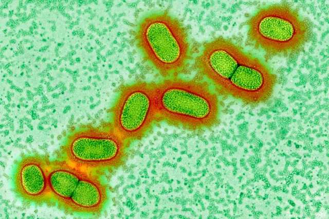
We may finally have found a long-elusive cause of Alzheimer’s disease: Porphyromonas gingivalis, the key bacteria in chronic gum disease. That’s bad, as gum disease affects around a third of all people. But the good news is that a drug that blocks the main toxins of P. gingivalis is entering major clinical trials this year, and research published this week shows it might stop and even reverse Alzheimer’s. There could even be a vaccine.

Alzheimer’s is one of the biggest mysteries in medicine. As populations have aged, dementia has skyrocketed to become the fifth biggest cause of death worldwide. Alzheimer’s constitutes some 70 per cent of these cases and yet, we don’t know what causes it. The disease often involves the accumulation of proteins called amyloid and tau in the brain, and the leading hypothesis has been that the disease arises from defective control of these two proteins. But research in recent years has revealed that people can have amyloid plaques without having dementia. So many efforts to treat Alzheimer’s by moderating these proteins have failed, and the hypothesis has now been seriously questioned.

Indeed, evidence has been growing that the function of amyloid proteins may be as a defence against bacteria, leading to a spate of recent studies looking at bacteria in Alzheimer’s, particularly those that cause gum disease, which is known to be a major risk factor for the condition.

Bacteria involved in gum disease and other illnesses have been found after death in the brains of people who had Alzheimer’s, but until now, it hasn’t been clear whether these bacteria caused the disease or simply got in via brain damage caused by the condition.

Gum disease link

Multiple research teams have been investigating P. gingivalis, and have so far found that it invades and inflames brain regions affected by Alzheimer’s; that gum infections can worsen symptoms in mice genetically engineered to have Alzheimer’s; and that it can cause Alzheimer’s-like brain inflammation, neural damage, and amyloid plaques in healthy mice.

“When science converges from multiple independent laboratories like this, it is very compelling,” says Casey Lynch of Cortexyme, a pharmaceutical firm in San Francisco, California.

In the new study, Cortexyme have now reported finding the toxic enzymes – called gingipains – that P. gingivalis uses to feed on human tissue in 96 per cent of the 54 Alzheimer’s brain samples they looked at, and found the bacteria themselves in all three Alzheimer’s brains whose DNA they examined.

“This is the first report showing P. gingivalis DNA in human brains, and the associated gingipains, co-lococalising with plaques,” says Sim Singhrao, of the University of Central Lancashire, UK. Her team previously found that P. gingivalis actively invades the brains of mice with gum infections. She adds that the new study is also the first to show that gingipains slice up tau protein in ways that could allow it to kill neurons, causing dementia.

The bacteria and its enzymes were found at higher levels in those who had experienced worse cognitive decline, and had more amyloid and tau accumulations. The team also found the bacteria in the spinal fluid of living people with Alzheimer’s, suggesting that this technique may provide a long-sought after method of diagnosing the disease.

When the team gave P. gingivalis gum disease to mice, it led to brain infection, amyloid production, tangles of tau protein, and neural damage in the regions and nerves normally affected by Alzheimer’s.

Cortexyme had previously developed molecules that block gingipains. Giving some of these to mice reduced their infections, halted amyloid production, lowered brain inflammation and even rescued damaged neurons.

The team found that an antibiotic that killed P. gingivalis did this too, but less effectively, and the bacteria rapidly developed resistance. They did not resist the gingipain blockers. “This provides hope of treating or preventing Alzheimer’s disease one day,” says Singhrao.

New treatment hope

Some brain samples from people without Alzheimer’s also had P. gingivalis and protein accumulations, but at lower levels. We already know that amyloid and tau can accumulate in the brain for 10 to 20 years before Alzheimer’s symptoms begin. This, say the researchers, shows P. gingivalis could be a cause of Alzheimer’s, but it is not a result.

Gum disease is far more common than Alzheimer’s. But “Alzheimer’s strikes people who accumulate gingipains and damage in the brain fast enough to develop symptoms during their lifetimes,” says Lynch. “We believe this is a universal hypothesis of pathogenesis.”

Cortexyme reported in October that the best of their gingipain blockers had passed initial safety tests in people, and entered the brain. It also seemed to improve participants with Alzheimer’s. Later this year the firm will launch a larger trial of the drug, looking for P. gingivalis in spinal fluid, and cognitive improvements, before and after.

They also plan to test it against gum disease itself. Efforts to fight that have led a team in Melbourne to develop a vaccine for P. gingivalis that started tests in 2018. A vaccine for gum disease would be welcome – but if it also stops Alzheimer’s the impact could be enormous.

Journal reference: Science Advances

https://www.newscientist.com/article/2191814-we-may-finally-know-what-causes-alzheimers-and-how-to-stop-it/

Protein Changes Detected in Blood Years Before Alzheimer’s Onset

Levels of a protein called neurofilament light chain increase in the blood and spinal fluid of some Alzheimer’s patients 16 years before they develop symptoms, according to a study published January 21 in Nature Medicine.

The results suggest that neurofilament light chain (NfL), which is part of the cytoskeleton of neurons and has previously been tied to brain damage in mice, could serve as a biomarker to noninvasively track the progression of the disease. “This is something that would be easy to incorporate into a screening test in a neurology clinic,” coauthor Brian Gordon, an assistant professor of radiology at Washington University, says in a press release.

Gordon and his colleagues measured NfL in nearly 250 people carrying an Alzheimer’s-risk allele and more than 160 of their relatives who did not carry the variant. They found that those at risk of developing the disease had higher levels of the protein early on, and that NfL levels in both the blood and spinal fluid were on the rise well before the patients began to show signs of neurodegeneration, more than 16 years before disease onset.

Examining a subset of the patients more closely, the team saw that the rate of increase in NfL correlated with the shrinkage of a brain region called the precuneus, and patients whose NfL levels were rising rapidly tested worse on cognitive tests. “It is not necessarily the absolute levels which tell you your neurodegeneration is ongoing, it is the rate of change,” coauthor Mathias Jucker, a professor of cellular neurology at the German Center for Neurodegenerative Diseases in Tübingen, tells The Guardian.

The Alzheimer’s-linked mutation carried by patients examined in this study only affects about 1 percent of people who get the neurodegenerative disease, so the approach must be validated in a broader patient population, James Pickett, the head of research at the Alzheimer’s Society, tells The Guardian.

“We validated it in people with Alzheimer’s disease because we know their brains undergo lots of neurodegeneration, but this marker isn’t specific for Alzheimer’s,” Gordon says in the release. “I could see this being used in the clinic in a few years to identify signs of brain damage in individual patients.”

Meanwhile, a research team at Seoul National University in South Korea described another potential blood test for Alzheimer’s, focusing on the tau and amyloid proteins known to be associated with the disease. According to their study published today in Brain, blood levels of tau and amyloid correlate with how much tau has accumulated in the brain, as well as other markers of neurodegeneration such as hippocampal volume. “These results indicate that combination of plasma tau and amyloid-β1–42 levels might be potential biomarkers for predicting brain tau pathology and neurodegeneration,” the researchers write in their report.

https://www.the-scientist.com/news-opinion/protein-changes-detected-in-blood-years-before-alzheimers-onset-65347

Case Western Reserve researchers cure drug-resistant infections without antibiotics


Case Western Reserve researchers cure drug-resistant infections without antibiotics

Biochemists, microbiologists, drug discovery experts and infectious disease doctors have teamed up in a new study that shows antibiotics are not always necessary to cure sepsis in mice. Instead of killing causative bacteria with antibiotics, researchers treated infected mice with molecules that block toxin formation in bacteria. Every treated mouse survived. The breakthrough study, published in Scientific Reports, suggests infections in humans might be cured the same way.

The molecules cling to a toxin-making protein found across Gram-positive bacterial species, called AgrA, rendering it ineffective. Treating mice with the therapeutic molecules effectively cured infections caused by methicillin-resistant Staphylococcus aureus (MRSA). S. aureus is notorious for its ability to overcome even the most potent antibiotics. Its resistance arsenal is broad, limiting therapeutic options to treat infections.

In a mouse model of S. aureus sepsis, treatment with small molecules alone resulted in 100 percent survival, while 70 percent of untreated animals died. The small molecules were as effective in promoting survival as antibiotics currently used to treat S. aureus infections. The molecules also appear to give antibiotics a boost. Septic mice treated with a combination of the small molecules and antibiotics had 10x fewer bacteria in their bloodstream than mice treated with antibiotic alone.

“For relatively healthy patients, such as athletes suffering from a MRSA infection, these molecules may be enough to clear an infection,” said Menachem Shoham, associate professor of biochemistry at Case Western Reserve University School of Medicine and senior author on the study. “For immunocompromised patients, combination therapy with the molecules and a low-dose antibiotic may be in order. The antibiotic in the combination could be one to which the bacteria are resistant in monotherapy, because our small molecules enhance the activity of conventional antibiotics, such as penicillin.”

With support from the small molecules, previously obsolete antibiotics could reenter the clinic.

Said Shoham: “This could provide a partial solution to the looming, global threat of antibiotic resistance.”

If available, antibiotics kill most bacteria, but a small number of bacteria with natural resistance survive. Over time, antibiotic-resistant bacteria multiply and spread. By Centers for Disease Control and Prevention estimates, at least two million Americans get an antibiotic-resistant infection annually. For some infections, effective antibiotics are no longer available. Disarming bacteria of disease-causing toxins represents a promising alternative to dwindling antibiotics.

Eliminating toxins frees up the immune system to eliminate bacterial pathogens instead of antibiotics, said Shoham, who also is affiliated with Q2 Pharma, Ltd., Haifa, Israel. “Without the toxins the bacteria become harmless. And since they don’t need the toxins to survive, there is less pressure to develop resistance.”

The small molecules work against multiple bacterial species. The new study included preliminary experiments showing the molecules prevent three other bacterial species from killing immune cells.

“These results indicate broad-spectrum efficacy against Gram-positive pathogens,” wrote the authors.

Added Shoham: “We have proven efficacy not only against MRSA but also against Staphylococcus epidermidis, which is notorious for clogging catheters, Streptococcus pyogenes that causes strep throat, Streptococcus pneumoniae, and other pathogens.”

Shoham led the study in collaboration with colleagues from the departments of biochemistry and dermatology and the Center for RNA and Therapeutics at Case Western Reserve University. The researchers developed two small molecules, F12 and F19, both of which potentiate antibiotic efficacy in the mouse models. The researchers are now working to commercialize both potential drugs. Case Western Reserve University has issued a license to Q2Pharma, Ltd., a biopharmaceutical startup company in Israel, to perform additional preclinical studies and develop F12 and F19 for clinical trials. Their initial trials will focus on patients suffering from systemic multi-drug resistant infections.

This research was supported by a Transformational Award to Menachem Shoham by the Dr. Ralph and Marian Falk Medical Research Trust Bank of America, N.A., Trustee. Some in vitro studies were supported by NIH/NIAID Preclinical Services under contract numbers HHSN272201100012I and HHSN27200007.

Greenberg, M, et al. “Small-molecule AgrA inhibitors F12 and F19 act as antivirulence agents against Gram-positive pathogens.” Scientific Reports. 2018 Oct 1;8(1):14578. doi: 10.1038/s41598-018-32829-w. PMID: 30275455.

A new 3-D printed ‘sponge’ sops up excess chemo drugs

Bringing the filtering abilities of a fuel cell into the blood vessels of living organisms, a new device could cut down on toxic effects of cancer treatment.

At the heart of this approach — recently tested in pigs — is a tiny, cylindrical “sponge” created by 3-D printing. Wedged inside a vein near a tumor being treated with chemotherapy, the sponge could absorb excess drug before it spreads through the body — thus lessening chemotherapy’s harmful side effects, including vomiting, immune suppression or even heart failure.

A human study could launch “in a couple of years, if all the stars are aligned,” says Steve Hetts, a neuroradiologist at the University of California, San Francisco who came up with the drug-capture concept. He worked with engineers at UC Berkeley and elsewhere to create and test prototypes.

A test of the most recent prototype showed that the absorber captured nearly two-thirds of a common chemotherapy drug infused into a nearby vein, without triggering blood clots or other obvious problems in the pig, Hetts and his colleagues report January 9 in ACS Central Science.

The study addresses a major need, says Eleni Liapi, a radiologist at Johns Hopkins University School of Medicine not involved with the new work. Existing methods for controlling chemotherapy delivery do not fully block drug escape, she notes. “A technological advancement to reduce unwanted circulating drug is always welcome.”


This image shows a cross-sectional view of a new 3-D printed cylindrical device that could cut down on toxic side effects from cancer treatment. Resin coatings (gold) bind to a chemo drug used to treat liver cancer, experiments show.

Chemo is often delivered intravenously in the hope that some treatment reaches the cancer site. In a more localized form of chemotherapy used to treat hard-to-remove tumors, the drug travels through catheter wires snaked into arteries going straight to the tumor. Although this technique, known as transarterial chemo embolization, or TACE, is given to tens of thousands of people each year, typically some of the injected drug bypasses the tumor site and slips into general circulation where it can wreak havoc elsewhere.

Hetts uses the transarterial method to treat babies with a rare eye tumor called retinoblastoma – and it was those experiences that birthed the “sponge” idea in the first place. After the chemotherapy ran its course through transarterial catheters, the infants’ eye tumors shrank. However, several weeks later, their blood cell counts tanked, suggesting to Hetts that some of the chemo drugs were escaping the eye and affecting other cells. Those observations eight years ago led Hetts to think that “if only I had a device I could put into the vein to bind up the excess drug, then maybe these little babies wouldn’t get the side effect” of immune suppression.

Heart surgeons use a similar “filter” to remove bits of cholesterol plaque from arteries of people with atherosclerosis, a disease characterized by the clogging and hardening of arteries. Hetts envisioned a similar device for chemotherapy treatment — “but not just a dumb, inert membrane to capture debris,” he says. “I wanted a ‘smart’ membrane that chemically binds to a drug.”

Instead of trying to develop a drug-trap device for a super rare tumor — retinoblastoma has just 300 new cases per year in the United States — Hetts’ team focused on a chemo drug for liver cancer, which is estimated to strike more than 40,000 Americans this year and kill three-quarters of them.

Anand Patel, a trainee in the Hetts’ lab with a bioengineering background, tested a batch of resins and found several that could bind to this drug, known as doxorubicin. To optimize the resins and get them onto the tips of guide wires, Patel sought help with “cold call” e-mails to local professors. Nitash Balsara — a UC Berkeley chemical engineer with expertise in polymer chemistry and membranes — “was actually crazy enough to return my e-mail with interest,” says Patel, who now works as an interventional radiologist in the Los Angeles area.

Balsara’s lab develops materials to regulate ion flow in batteries and fuel cells. As it turns out, these filtration processes are “very similar to those that we needed to capture excess chemotherapy drugs from the blood,” Patel says. The team worked with Carbon, Inc., a 3-D printing company in the San Francisco Bay area, to get the drug-binding material onto a 30-millimeter-long, cylinder-shaped “sponge” about as wide as a drinking straw. Hee Jeung Oh of UC Berkeley spent more than a year working out how to attach the drug-binding material to the 3-D printed cylinder with crisscrossing struts.

In experiments, the team injected the liver cancer drug through the pigs’ leg and pelvic veins — which are similar in width to human liver veins, Hetts says. Before infusing the chemotherapy drug, the researchers inserted the 3-D printed sponge a few centimeters from the infusion site — as well as catheters above and below the sponge for collecting blood samples to measure drug absorption over time. Within a half hour, the device absorbed, on average, 64 percent of the liver cancer drug.

The next round of studies will monitor the capture of doxorubicin by drug sponges inserted directly into the pigs’ liver veins.

A new 3-D printed ‘sponge’ sops up excess chemo drugs