Daily step counts between 5,000 to 10,000 or more reduced depression symptoms across 33 studies.
The associations may be due to several mechanisms, like improvement in sleep quality and inflammation.
Daily step counts of 5,000 or more corresponded with fewer depressive symptoms among adults, results of a systematic review and meta-analysis published in JAMA Network Open suggested.
The results are consistent with previous studies linking exercise to various risk reductions for mental health disorders and show that setting step goals “may be a promising and inclusive public health strategy for the prevention of depression,” the researchers wrote.
According to Bruno Bizzozero-Peroni, PhD, MPH, from Universidad De Castilla-La Mancha in Spain, and colleagues, daily step counts are a “simple and intuitive objective measure” of physical activity, while tracking such counts has become increasingly feasible for the general population thanks to the availability of fitness trackers.
“To our knowledge, the association between the number of daily steps measured
with wearable trackers and depression has not been previously examined through a meta-analytic approach,” they wrote.
The researchers searched multiple databases for analyses assessing the effects of daily step counts on depressive symptoms, ultimately including a total of 27 cross-sectional studies and six longitudinal studies comprising 96,173 adults aged 18 years or older.
They found that in the cross-sectional studies, daily step counts of 10,000 or more (standardized mean difference [SMD] = 0.26; 95% CI, 0.38 to 0.14), 7,500 to 9,999 (SMD = 0.27; 95% CI, 0.43 to 0.11) and 5,000 to 7,499 (SMD = 0.17; 95% CI, 0.3 to 0.04) corresponded with reduced depressive symptoms vs. daily step counts less than 5,000.
In the prospective cohort studies, people with 7,000 or more steps a day had a reduced risk for depression vs. with people with fewer than 7,000 daily steps (RR = 0.69; 95% CI, 0.62-0.77), whereas an increase of 1,000 steps a day suggested an association with a lower risk for depression (RR = 0.91; 95% CI, 0.87-0.94).
There were a couple study limitations. The researchers noted that reverse associations are possible, while they could not rule out residual confounders.
They also pointed out that there are some remaining questions, such as whether there is a ceiling limit after which further step counts would no longer reduce the risk for depression.
Bizzozero-Peroni and colleagues highlighted several possible biological and psychosocial mechanisms behind the associations, like changes in sleep quality, inflammation, social support, self-esteem, neuroplasticity and self-efficacy.
They concluded that “a daily active lifestyle may be a crucial factor in regulating and reinforcing these pathways” regardless of the exact combination of mechanisms responsible for the positive link.
“Specifically designed experimental studies are still needed to explore whether there are optimal and maximal step counts for specific population subgroups,” they wrote.
Ever since the time of the ancient Greeks over 2,000 years ago, human beings have known the Earth is a globe.
Despite this, some people are still convinced that we live on a giant floating disc in space, known as ‘Flat Earth’.
Now, one of the internet’s most famous ‘Flat Earthers’ has finally cottoned on to the truth.
Jeran Campanella, who runs the popular Flat Earth YouTube show ‘Jeransim’, has travelled to Antarctica as part of a trip dubbed ‘The Final Experiment’.
Mr Campanella witnessed first hand that the sun doesn’t set during the southern hemisphere’s summer.
This debunks the belief held by Flat Earthers that Antarctica is an ice wall where the sun rises and sets every day.
Stationed in Antarctica, he says to the camera: ‘Sometimes you are wrong in life and I thought there was no 24-hour sun. In fact I was pretty sure of it. And it’s a fact – the sun does circle you in the south. So what does that mean? You guys are going to have to find that out for yourself.’
Campanella thanked the organiser of the trip, which cost $35,000 (£27,500) – although he stopped short of saying that the Earth is spherical.
Jeran Campanella, who runs the popular Flat Earth YouTube channel ‘Jeransim’, has travelled to Antarctica as part of a trip dubbed ‘The Final Experiment’
Mr Campanella was featured in the 2018 documentary ‘Behind the Curve’ as he unintentionally debunked his own theory with a light experiment.
A co-creator of the GlobeBusters YouTube channel, which has 73,000 subscribers, he describes himself as an ‘open-minded True Earth’ proponent and crypto enthusiast.
His website Jeransim.com flogs everything from ‘anti-NASA’ t-shirts and beenies to crypto consultations, leaf powder medicinal capsules and private Zoom dinner parties.
When he agreed to travel to Antarctica on the proviso he wouldn’t have to cover the trip costs, he expected the sun to rise and fall out of view from the horizon.
But in the new clip, he bashfully admits to the truth and acknowledges people may see him as a ‘shill’ – a trickster who takes part in the supposed cover-up that the Earth is round.
‘I realise that I’ll be called a shill for just saying that and you know what, if you’re a shill for being honest so be it,’ he said.
‘I honestly believed there was no 24-hour sun – I honestly now believe there is.’
As any well-informed person will know, Antarctica is an island continent at the southernmost part of our planet.
A co-creator of the GlobeBusters YouTube channel, Campanella has 73,000 subscribers. He describes himself as an ‘open-minded True Earth’ proponent and crypto enthusiastCampanella agreed to travel to Antarctica on the proviso he wouldn’t have to cover the trip costsIn the new clip, he bashfully admits to the truth and acknowledges people may see him as a ‘shill’ – a trickster who takes part in the supposed cover-up that the Earth is roundDuring the southern hemisphere’s and northern hemisphere’s summer, the sun remains visible all day, including at midnight – a phenomenon dubbed ‘the midnight sun’. Pictured, multiple exposure of midnight sun on Lake Ozhogino in Yakutia, Russia
What do Flat Earthers believe?
People who believe the idea that the Earth is disc-shaped rather than round are called ‘Flat Earthers’.
Because Earth’s surface looks and feels flat when we walk around it, the conspiracy theorists denounce all evidence to the contrary.
The leading theory suggests Earth is a disc with the Arctic Circle in the centre and Antarctica, a 150-foot-tall (45-metre) wall of ice, around the rim.
Proponents of the bizarre theory also claim the Earth is stationary in space rather than orbiting the sun.
Due to the tilt of the Earth, the sun does not set during the southern hemisphere’s (or northern hemisphere’s) summer – it just moves in a circle in the sky.
It means the sun remains visible all day, including at midnight – a phenomenon dubbed ‘the midnight sun’.
But Flat Earthers, or ‘Flerfs’, believe that Antarctica is an ice wall that encircles all the other continents and holds in all the world’s oceans.
Therefore, they think the sun rises and sets each day, every day, regardless of whether it’s summer or not.
To account for night and day, most theorists believe the sun moves in circles around the North Pole, with the sun’s light acting like a spotlight over Earth.
About three years ago, Will Duffy, a pastor from Colorado, was made aware that people still believe the Earth is flat when a friend of his posted about it on Facebook.
Since Flat Earthers said for years that Antarctica will show the world the truth about the shape of the Earth, Mr Duffy decided the easiest solution would be to just go to Antarctica.
‘After we go to Antarctica, no one has to waste any more time debating the shape of the Earth,’ he said.
Mr Campanella was featured in the 2018 documentary ‘Behind the Curve’ as he unintentionally debunked his own theory with a light experimentThe Flat Earther claimed that if the light can be seen with a camera, the holes in the fence and the torch all at equal differences above the ground, then he could draw a positive conclusion that the planet is flat
‘I created The Final Experiment to end this debate, once and for all,’ he said.
On December 14, Mr Duffy flew with four Flat Earthers (including Mr Campanella) and four people who already know the Earth is round (‘globe earthers’) to Antarctica.
Mr Duffy said he filmed the sun for 25 hours straight, using SpaceX’s Starlink satellite internet network for a livestream.
Stunning footage from the frozen landscape shows the gleaming midnight sun with ice-peaked mountains in the background.
‘That is a midnight sun – so the sun has never set the entire time we’ve been here,’ the pastor said to the camera.
‘I put sunscreen on at midnight so that I would not get burned at midnight while I was doing this livestream.’
He passes over to Mr Campanella, who humbly admits he was wrong, having witnessed the sun travel in a circle above his head.
However, another famous Flat Earther on the trip, Austin Witsit, has more trouble letting go of the theory.
Mr Campanella humbly admitted he was wrong, having witnessed the sun travel in a circle above his head
Witsit said there’s ‘clearly’ a 24-hour sun, but denied that this proves the Earth is a globe.
‘I don’t think it falsifies plane [flat] Earth, I don’t think it proves a globe – I think it’s a singular data point,’ he said.
Likewise, some YouTuber conspiracy theorists have been left unconvinced, with many claiming the group were standing in front of a green screen.
In the live chat, YouTube user Nandy posted: ‘THIS IS A GREEN SCREEN, HOLD UP SNOW AND PUT UP THREE FINGERS IF IT’S REAL, the earth will always be flat’, while user Fozzy_Foster said: ‘move the camera green screen.’
Another user, Oz Riv, called Mr Duffy a ‘shill’, adding: ‘You are a pastor advocating for earth that isn’t biblical. Annd you work with Nasa.’
Scientists have know for millennia that Earth is a sphere due to the simple fact that the sun sets at different times in different locations.
If the Earth were flat, then shadows would be the same length, regardless of location.
For space travelers and robotic probes adorned with cameras, the curvature of the Earth has also long been clearly visible.
It was Greek philosopher Pythagoras who first proposed that the Earth was round around 500 BC, but it was about 350 BC that Aristotle declared Earth was a sphere.
This was based on observations Aristotle had made about which constellations you could see in the sky as you travelled further and further away from the equator.
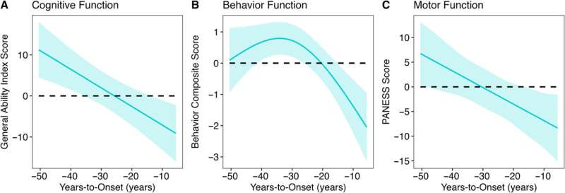
The genetic mutation that causes Huntington’s disease (HD)—a devastating brain disease that disrupts mobility and diminishes cognitive ability—may also enhance early brain development and play a role in promoting human intelligence.
This revelation comes from more than 10 years of brain imaging and brain function data, including motor, cognitive, and behavioral assessments, collected from a unique population—children and young adults who carry the gene for HD. While an HD mutation will eventually cause fatal brain disease in adulthood, the study finds that early in life, children with the HD mutation have bigger brains and higher IQ than children without the mutation.
“The finding suggests that early in life, the gene mutation is actually beneficial to brain development, but that early benefit later becomes a liability,” says Peg Nopoulos, MD, professor and head of psychiatry at the UI Carver College of Medicine, and senior author on the study published in The Annals of Neurology.
The finding may also have implications for developing effective treatments for HD. If the gene’s early action is beneficial, then simply aiming to knock out the gene might result in loss of the developmental benefit, too. Creating therapies that can disrupt the gene’s activity later in the patient’s lifetime might be more useful.
The new data about the gene’s positive effect on early brain development is also exciting to Nopoulos for another reason.
“We are very interested in the fact that this appears to be a gene that drives IQ,” she says. “No previous study has found any gene of significant effect on IQ, even though we know intelligence is heritable.”
HD gene linked to better brain development in early life
Huntington’s disease is caused by a mutation in the huntingtin (HTT) gene. The protein produced by the HTT gene is necessary for normal development, but variations within a segment of the protein have a profound effect on the brain.
The segment in question is a long repeat of one amino acid called glutamine. More repeats are associated with bigger, more complex brains. For example, species such as sea urchins or fish have no repeats, but these repeats start to appear higher up the evolutionary ladder. Rodents have a few repeats, while apes (our closest relatives) have even more repeats; and humans have the most.
Most people have repeats in the range of 10–26, but if a person has 40 or more repeats, then they develop HD. Although the gene expansion is present before birth, HD symptoms do not appear until middle age. Nopoulos’s team at the University of Iowa has a long history of studying how the HTT gene expansion affects brain development in the decades before disease onset.
“We know that the expanded gene causes a horrible degenerative disease later in life, but we also know it is a gene that is crucial for general development,” she says.
“We were surprised to find that it does have a positive effect on brain development early in life. Those who have the gene expansion have an enhanced brain with larger volumes of the cerebrum and higher IQ compared to those who don’t.”
In particular, the study found that decades before HD symptoms appeared, children with the HD gene expansion showed significantly better cognitive, behavioral, and motor scores compared to children with repeats within the normal range. Children with the expanded gene also had larger cerebral volumes and greater cortical surface area and folding. After this initial peak, a prolonged deterioration was seen in both brain function and structure.
The study gathered this data by following almost 200 participants in the Kids-HD study, the only longitudinal study of children and young adults at risk for HD due to having a parent or grandparent with the disease.
Evolutionary benefit comes at a cost
Although surprising, the findings are in line with studies by evolutionary biologists who believe that genes like HTT may have been “positively selected” for human brain evolution. This theory, known as antagonistic pleiotropy, suggests that certain genes can produce a beneficial effect early in life, but come at a cost later in life.
The finding also challenges the idea that the protein produced by the HD gene is solely a toxic protein that causes brain degeneration.
“Overall, our study suggests that we should rethink the notion of the toxic protein theory,” says Nopoulos, who is also a member of the Iowa Neuroscience Institute.
“Instead, we should consider the theory of antagonistic pleiotropy—a theory that suggests that genes like HTT build a better brain early in life, but the cost of the superior brain is that it isn’t built to last and may be prone to premature or accelerating aging.
“This means that instead of knocking down the gene for therapy, drugs that slow the aging process may be more effective.”
Next steps
Nopoulos’s team is already making progress extending the research from the Kids-HD program. Nopoulos has established the Children to Adult Neurodevelopment in Gene-Expanded Huntington’s Disease (ChANGE-HD), a multi-site study that aims to recruit hundreds of participants for a total of over 1,200 assessments to validate the key findings from the Kids-HD study and to enhance future research on HD.
A primary area of focus will be understanding how an enlarged brain can later lead to degeneration. One hypothesis Nopoulos and her team will explore involves the idea that an enlarged cortex might produce excess glutamate (an important neurotransmitter), which is beneficial in early brain development, but later leads to neurotoxicity and brain degeneration.
In addition to Nopoulos, the UI team included Mohit Neema, MD, UI research scientist and first author of the study; Jordan Schultz, PharmD; Douglas Langbehn, MD, Ph.D.; Amy Conrad, Ph.D.; Eric Epping, MD, Ph.D.; and Vincent Magnotta, Ph.D.
More information: Mohit Neema et al, Mutant Huntingtin Drives Development of an Advantageous Brain Early in Life: Evidence in Support of Antagonistic Pleiotropy, Annals of Neurology (2024). DOI: 10.1002/ana.27046
Scientists at the University of Copenhagen have discovered a new weight loss drug target that reduces appetite, increases energy expenditure, and improves insulin sensitivity without causing nausea or loss of muscle mass. The discovery was reported in the journal Nature and could lead to a new therapy for millions of people with both obesity and type 2 diabetes who do not respond well to current treatments.
Millions of people around the world benefit from weight-loss drugs based on the incretin hormone GLP-1. These drugs also improve kidney function, reduce the risk of fatal cardiac events, and are linked to protection against neurodegeneration.
However, many people stop taking the drugs due to common side effects, including nausea and vomiting. Studies also show that incretin-based therapies like Wegovy and Mounjaro are much less effective at lowering weight in people living with both obesity and type 2 diabetes—a group numbering more than 380 million people globally.
In the study, scientists from the University of Copenhagen describe a powerful new drug candidate that lowers appetite without loss of muscle mass or side effects like nausea and vomiting. And, unlike the current generation of treatments, the drug also increases the body’s energy expenditure—the capacity of the body to burn calories.
“While GLP-1-based therapies have revolutionized patient care for obesity and type 2 diabetes, safely harnessing energy expenditure and controlling appetite without nausea remain two Holy Grails in this field. By addressing these needs, we believe our discovery will propel current approaches to make more tolerable, effective treatments accessible to millions more individuals,” says Associate Professor Zach Gerhart-Hines from the NNF Foundation Center for Basic Metabolic Research (CBMR) at the University of Copenhagen.
NK2R activation lowers body weight and reverses diabetes
Our weight is largely determined by the balance between the energy we consume and the amount of energy we expend. Eating more and burning less creates a positive energy balance leading to weight gain, while eating less and burning more creates a negative balance, resulting in weight loss.
The current generation of incretin-based therapies tip the scales toward a negative energy balance by lowering appetite and the total calories a person consumes. But scientists have also recognized the potential on the other side of the equation—increasing the calories the body burns.
This approach is especially relevant, given recent research that has shown that our bodies seem to be burning fewer calories at rest than they did a few decades ago. However, there are currently no clinically approved ways to safely increase energy expenditure, and few options are in development.
This was the starting point when scientists at the University of Copenhagen decided to test the effect of activating the neurokinin 2 receptor (NK2R) in mice. The Gerhart-Hines Group identified the receptor through genetic screens that suggested NK2R played a role in maintaining energy balance and glucose control.
They were astonished by the results of the studies—not only did activating the receptor safely increase calorie-burning, it also lowered appetite without any signs of nausea.
Further studies in non-human primates with type 2 diabetes and obesity showed that NK2R activation lowered body weight and reversed their diabetes by increasing insulin sensitivity and lowering blood sugar, triglycerides, and cholesterol.
“One of the biggest hurdles in drug development is translation between mice and humans. This is why we were excited that the benefits of NK2R agonism translated to diabetic and obese nonhuman primates, which represents a big step towards clinical translation,” says Ph.D. Student Frederike Sass from CBMR at the University of Copenhagen, and first author of the study.
The discovery could result in the next generation of drug therapies that bring more efficacious and tolerable treatments for the almost 400 million people globally who live with both type 2 diabetes and obesity.
The University of Copenhagen holds the patent rights for targeting NK2R. To date, research by the Gerhart-Hines lab has led to the creation of three biotech companies—Embark Biotech, Embark Laboratories, and Incipiam Pharma.
In 2023, Embark Biotech was acquired by Novo Nordisk to develop next generation therapeutics for cardiometabolic disease.
Researchers have developed an AI-powered model that—in 10 seconds—can determine during surgery if any part of a cancerous brain tumor that could be removed remains, a study published in Nature suggests.
The technology, called FastGlioma, outperformed conventional methods for identifying what remains of a tumor by a wide margin, according to the research team led by University of Michigan and University of California San Francisco.
“FastGlioma is an artificial intelligence-based diagnostic system that has the potential to change the field of neurosurgery by immediately improving comprehensive management of patients with diffuse gliomas,” said senior author Todd Hollon, M.D., a neurosurgeon at University of Michigan Health and assistant professor of neurosurgery at U-M Medical School.
“The technology works faster and more accurately than current standard of care methods for tumor detection and could be generalized to other pediatric and adult brain tumor diagnoses. It could serve as a foundational model for guiding brain tumor surgery.”
When a neurosurgeon removes a life threatening tumor from a patient’s brain, they are rarely able to remove the entire mass.
Commonly, the tumor is missed during the operation because surgeons are not able to differentiate between healthy brain and residual tumor in the cavity where the mass was removed. Residual tumor’s ability to resemble healthy brain tissue remains a major challenge in surgery.
Neurosurgical teams employ different methods to locate that residual tumor during a procedure.
They may get MRI imaging, which requires intraoperative machinery that is not available everywhere. The surgeon might also use a fluorescent imaging agent to identify tumor tissue, which is not applicable for all tumor types. These limitations prevent their widespread use.
In this international study of the AI-driven technology, neurosurgical teams analyzed fresh, unprocessed specimens sampled from 220 patients who had operations for low- or high-grade diffuse glioma.
FastGlioma detected and calculated how much tumor remained with an average accuracy of approximately 92%.
In a comparison of surgeries guided by FastGlioma predictions or image- and fluorescent-guided methods, the AI technology missed high-risk, residual tumor just 3.8% of the time—compared to a nearly 25% miss rate for conventional methods.
“This model is an innovative departure from existing surgical techniques by rapidly identifying tumor infiltration at microscopic resolution using AI, greatly reducing the risk of missing residual tumor in the area where a glioma is resected,” said co-senior author Shawn Hervey-Jumper, M.D., professor of neurosurgery at University of California San Francisco and a former neurosurgery resident at U-M Health.
“The development of FastGlioma can minimize the reliance on radiographic imaging, contrast enhancement or fluorescent labels to achieve maximal tumor removal.”
How it works
To assess what remains of a brain tumor, FastGlioma combines microscopic optical imaging with a type of artificial intelligence called foundation models. These are AI models, such as GPT-4 and DALL·E 3, trained on massive, diverse datasets that can be adapted to a wide range of tasks.
After large scale training, foundation models can classify images, act as chatbots, reply to emails and generate images from text descriptions.
To build FastGlioma, investigators pre-trained the visual foundation model using over 11,000 surgical specimens and 4 million unique microscopic fields of view.
The tumor specimens are imaged through stimulated Raman histology, a method of rapid, high resolution optical imaging developed at U-M. The same technology was used to train DeepGlioma, an AI based diagnostic screening system that detects a brain tumor’s genetic mutations in under 90 seconds.
“FastGlioma can detect residual tumor tissue without relying on time-consuming histology procedures and large, labeled datasets in medical AI, which are scarce,” said Honglak Lee, Ph.D., co-author and professor of computer science and engineering at U-M.
Full resolution images take around 100 seconds to acquire using stimulated Raman histology; a “fast mode” lower resolution image takes just 10 seconds.
Researchers found that the full resolution model achieved accuracy up to 92%, with the fast mode slightly lower at approximately 90%.
“This means that we can detect tumor infiltration in seconds with extremely high accuracy, which could inform surgeons if more resection is needed during an operation,” Hollon said.
AI’s future in cancer
Over the last 20 years, the rates of residual tumor after neurosurgery have not improved.
Not only is FastGlioma an accessible and affordable tool for neurosurgical teams operating on gliomas, but researchers say, it can also accurately detect residual tumor for several non-glioma tumor diagnoses, including pediatric brain tumors, such as medulloblastoma and ependymoma, and meningiomas.
“These results demonstrate the advantage of visual foundation models such as FastGlioma for medical AI applications and the potential to generalize to other human cancers without requiring extensive model retraining or fine-tuning,” said co-author Aditya S. Pandey, M.D., chair of the Department of Neurosurgery at U-M Health.
“In future studies, we will focus on applying the FastGlioma workflow to other cancers, including lung, prostate, breast, and head and neck cancers.”
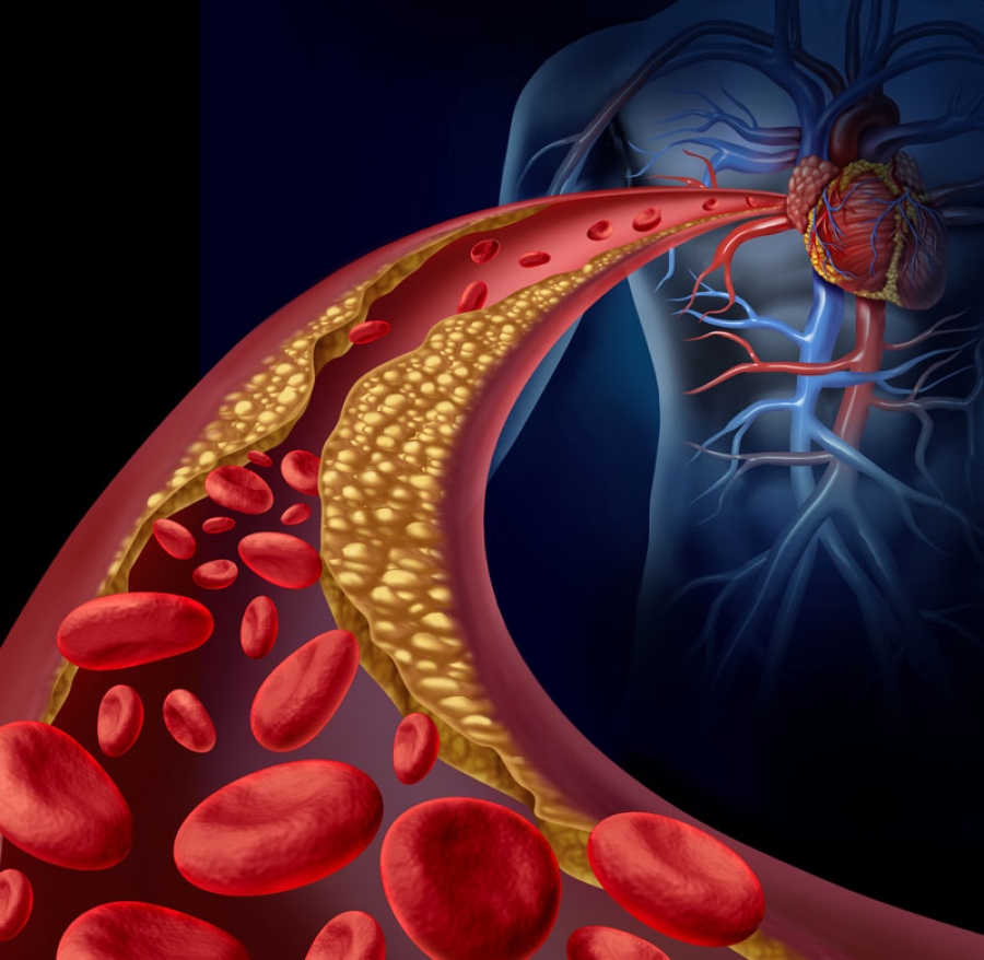
Researchers at Case Western Reserve University have identified a new target to treat atherosclerosis, a condition where plaque clogs arteries and causes major cardiac issues, including stroke and heart attack.
In a new study, published in the journal Cell Reports, the team identified an inflammation-reducing molecule—called itaconate (ITA)—that could be the foundation of a new approach to treat such a common and deadly disease.
Heart disease is the leading cause of death for men, women and people of most racial and ethnic groups, according to the U.S. Centers for Disease Control and Prevention.
Medications help but don’t completely protect patients from cardiovascular risk. So, doctors also recommend lifestyle changes, such as a low-cholesterol/low-fat diet (LCLFD), to further reduce plaque and inflammation that increase the risk of cardiovascular disease. Yet many patients find it challenging to follow diet restrictions long-term.
Identifying the role ITA plays in diet and heart disease may help address this.
“We’ve found that itaconate is crucial to the diet’s ability to stabilize plaques and reduce inflammation, which has been a mystery until now,” said Andrei Maiseyeu, associate professor at the Cardiovascular Research Institute and Department of Biomedical Engineering at Case Western Reserve’s School of Medicine.
“This discovery marks a major leap forward in the understanding of how diet-induced plaque resolution occurs at a molecular level.”
Based on their discovery, Maiseyeu and his team have developed a new treatment: ITA-conjugated lipid nanoparticles. This new therapeutic approach allows ITA to accumulate in plaque and bone marrow, where it reduces inflammation and mimics the beneficial effects of LCLFD without requiring drastic lifestyle changes.
“We have already seen its effectiveness in multiple models of atherosclerosis,” Maiseyeu said. “We are optimistic that this will result in better treatments that will greatly lower the long-term risk of heart attacks and strokes while also improving patients’ quality of life.”
Maiseyeu and his team are now taking steps to translate ITA-LNP to the clinic, including engineering a pill form of the treatment, which they believe will not only be convenient for patients, but also transformative.
More information: Natalie E. Hong et al, Nanoparticle-based itaconate treatment recapitulates low-cholesterol/low-fat diet-induced atherosclerotic plaque resolution, Cell Reports (2024). DOI: 10.1016/j.celrep.2024.114911
Scientists wielding a minute hollow needle — and a bike pump — have managed to implant bacteria into a larger cell, creating a relationship similar to those that sparked the evolution of complex life.
The feat — described1 in Nature on 2 October — could help researchers to understand the origins of pairings that gave rise to specialized organelles called mitochondria and chloroplasts more than one billion years ago.
Endosymbiotic relationships — in which a microbial partner lives harmoniously within the cells of another organism — are found in numerous life forms, including insects and fungi. Scientists think that mitochondria, the organelles that are responsible for cells’ energy production, evolved when a bacterium took up residence inside an ancestor of eukaryotic cells. Chloroplasts emerged when an ancestor of plants swallowed a photosynthetic microorganism.
Determining the factors that formed and sustained these couplings is difficult because they occurred so long ago. To get around this problem, a team led by microbiologist Julia Vorholt, at the Swiss Federal Institute of Technology in Zurich (ETH Zurich), has spent the past few years engineering endosymbioses in the laboratory. Their approach uses a 500-1000 nanometre wide needle to puncture host cells and then deliver bacterial cells one at a time.
Sparking symbiosis
Even with this technical wizardry, initial pairings tended to fail; for instance, because the would-be symbiont divided too fast and killed its host2. The team’s luck changed when they recreated a natural symbiosis that occurs between some strains of a fungal plant pathogen, Rhizopus microsporus, and the bacterium Mycetohabitans rhizoxinica, which produces a toxin that protects the fungus from predation.
Yet delivering bacterial cells into the fungi, which have thick cell walls that maintain a high internal pressure, was a challenge. After piercing the wall with the needle, the researchers used a bicycle pump — and later an air compressor — to maintain enough pressure to deliver the bacteria.
After overcoming the initial shock of surgery, the fungi continued their life cycles and produced spores, a fraction of which contained bacteria. When these spores germinated, bacteria were also present in the cells of the next generation of fungi. This showed that the new endosymbiosis could be passed onto offspring — a key finding.
Vanishing bacteria
But the germination success of the bacteria-containing spores was low. In a mixed population of spores (some with bacteria and some without), those with bacteria vanished after two generations. To see whether relations could be improved, the researchers used a fluorescent cell sorter to select spores containing bacteria — which had been labelled with a glowing protein — and propagated only these spores in future rounds of reproduction. By ten generations, the bacteria-containing spores germinated nearly as efficiently as those without bacteria.
The basis of this adaptation isn’t clear. Genome sequencing identified a handful of mutations associated with improved germination success in the fungus — which was a strain of R. microsporus not known to carry endosymbionts naturally — and found no changes in the bacteria.
The line that germinated most efficiently tended to limit the number of bacteria in each spore, says study co-author Gabriel Giger, a microbiologist at ETH Zurich. “There are ways for these two partners to make a better, easier living with each other. That’s something that’s really important for us to understand.”
Fungal immune system
Researchers don’t know much about the genetics of R. microsporus. But Thomas Richards, an evolutionary biologist at the University of Oxford, UK, wonders whether a fungal immune system is preventing symbiosis — and whether mutations to this system could be easing relations. “I’m a big fan of this work,” he adds.
Eva Nowack, a microbiologist at Heinrich Heine University Düsseldorf in Germany, was surprised at how quickly adaptations to symbiotic life seemed to evolve. In the future, she would love to see what happens after even longer time periods; for example, more than 1,000 generations.
Engineering such symbioses could lead to the development of novel organisms with useful traits, such as the ability to consume carbon dioxide or atmospheric nitrogen, says Vorholt. “That’s the idea: to bring in new traits that an organism doesn’t have, and that would be difficult to implement otherwise.”
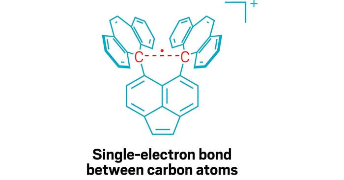
For a little more than a century, chemists have believed that strong atomic links called covalent bonds are formed when atoms share one or more electron pairs. Now, researchers have made the first observations of single-electron covalent bonds between two carbon atoms.
This unusual bonding behaviour has been seen between a few other atoms, but scientists are particularly excited to see it in carbon, the basic building block of life on Earth and the key component of industrial chemicals including drugs, plastics, sugars and proteins. The discovery was published1 in Nature on 25 September.
“The covalent bond is one of the most important concepts in chemistry, and discovery of new types of chemical bonds holds great promise for expanding vast areas of chemical space,” says University of Tokyo chemist Takuya Shimajiri, who was part of the carbon bonding research team.
Most chemical bonds in molecules are made up of a single pair of electrons, shared between atoms. These are called covalent single bonds. In particularly strong bonds, atoms might share two electron pairs in a double bond, or three pairs — a triple bond. But chemists know that atoms interact in many other ways, and by studying more unusual bond types at the boundaries of the possible, they hope to better understand what a chemical bond is in the first place.
Pauling’s proposal
The concept of single-electron covalent bonds dates to 1931, when chemist Linus Pauling proposed them. But at the time, chemists didn’t have the tools to observe such bonds, says Marc-Etienne Moret, a chemist at Utrecht University in the Netherlands. Even with modern analytical techniques, these bonds are challenging to observe. “The situation in which only one electron makes a bond is very unstable,” says Moret. “This means the bond will break easily and have a strong tendency to either release or capture an electron to restore an even number of electrons.”
In 1998, scientists observed2 a single-electron bond between two phosphorus atoms; Moret was part of a group that created3 one between copper and boron in 2013. Chemists have theorized that these unusual bonds might occur between carbon atoms in short-lived intermediate structures that appear during chemical reactions. But to observe these fickle bonds, chemists have to stabilize a compound that contains them. A stable compound that contains a one-electron C–C bond had eluded chemists.
Shimajiri says the key to observing the single-electron carbon bond was carefully designing a molecule that would stabilize it. The research team, which included Hokkaido University chemist Yusuke Ishigaki, created a molecule that provides a stable ‘shell’ of fused carbon rings that helps hold together the carbon–carbon bond in its centre. That central bond is stretched out to a relatively long length for a C–C bond, which makes it susceptible to losing one electron in an oxidation reaction, creating the elusive single-electron bond.
Stable bond
To capture this compound in a stable, observable form, they crystallized it. When the oxidation is performed in the presence of iodine, the reaction yields a purple salt, with the stable shell of the molecule holding together the single-electron C–C bond inside. They then used various analytical techniques to characterize the molecule and the bond. Shimajiri says the compound is extremely stable under ambient conditions.
“In several chemical reactions, the involvement of one-electron bonds has been proposed, but so far, they have remained hypothetical,” says Shimajiri. Creating stable compounds containing these bonds could help researchers to better understand what happens during these reactions.
Guy Bertrand, a chemist at the University of California, Santa Barbara, was part of the team that created the phosphorus single-electron bond. He says it’s significant to see it in carbon. “Anytime you do something with carbon, the impact is greater than with any other element,” he says. Carbon is the stuff of organic chemistry. But he says it’s not so easy to say whether this work will have any applications. “This is a curiosity,” he says. “But it will be in the textbooks.”
Shimajiri hopes that the description of the single-electron carbon bond will help chemists to better understand the basic nature of chemical bonds. “We aim to clarify what a covalent bond is — specifically, at what point does a bond qualify as covalent, and at what point does it not?”
The NIH announced that Eliezer Masliah, MD, now former director of the division of neuroscience at the National Institute on Aging, engaged in research misconduct while serving in the agency.
The NIH said in a statement that Masliah committed falsification and/or fabrication involving repeated use and relabeling of “figure panels representing different experimental results in two publications.”
The NIH further stated it will notify the two journals in which the panels appeared of its findings so that appropriate action can be taken.
The agency initiated a research misconduct review process in May 2023 after it was notified of allegations from the HHS Office of Research Integrity. An investigation was subsequently initiated in December 2023 and concluded on Sept. 15.
Masliah joined the NIH in 2016 as director of the division of neuroscience at the National Institute on Aging (NIA) and an intramural researcher studying synaptic damage in patients with neurodegenerative disorders. Per the NIH, he no longer serves as director. NIA Deputy Director Amy Kelley, MD, has assumed the role of acting director.
The NIH declined to comment further when asked about details surrounding its decision.
Life and death are traditionally viewed as opposites. But the emergence of new multicellular life-forms from the cells of a dead organism introduces a “third state” that lies beyond the traditional boundaries of life and death.
Usually, scientists consider death to be the irreversible halt of functioning of an organism as a whole. However, practices such as organ donation highlight how organs, tissues and cells can continue to function even after an organism’s demise. This resilience raises the question: What mechanisms allow certain cells to keep working after an organism has died?
The third state challenges how scientists typically understand cell behavior. While caterpillars metamorphosing into butterflies, or tadpoles evolving into frogs, may be familiar developmental transformations, there are few instances where organisms change in ways that are not predetermined. Tumors, organoids and cell lines that can indefinitely divide in a petri dish, like HeLa cells, are not considered part of the third state because they do not develop new functions.
However, researchers found that skin cells extracted from deceased frog embryos were able to adapt to the new conditions of a petri dish in a lab, spontaneously reorganizing into multicellular organisms called xenobots. These organisms exhibited behaviors that extend far beyond their original biological roles. Specifically, these xenobots use their cilia – small, hair-like structures – to navigate and move through their surroundings, whereas in a living frog embryo, cilia are typically used to move mucus.
Xenobots are also able to perform kinematic self-replication, meaning they can physically replicate their structure and function without growing. This differs from more common replication processes that involve growth within or on the organism’s body.
Researchers have also found that solitary human lung cells can self-assemble into miniature multicellular organisms that can move around. These anthrobots behave and are structured in new ways. They are not only able to navigate their surroundings but also repair both themselves and injured neuron cells placed nearby.
Taken together, these findings demonstrate the inherent plasticity of cellular systems and challenge the idea that cells and organisms can evolve only in predetermined ways. The third state suggests that organismal death may play a significant role in how life transforms over time.
Postmortem conditions
Several factors influence whether certain cells and tissues can survive and function after an organism dies. These include environmental conditions, metabolic activity and preservation techniques.
Different cell types have varying survival times. For example, in humans, white blood cells die between 60 and 86 hours after organismal death. In mice, skeletal muscle cells can be regrown after 14 days postmortem, while fibroblast cells from sheepandgoats can be cultured up to a month or so postmortem.
Metabolic activity plays an important role in whether cells can continue to survive and function. Active cells that require a continuous and substantial supply of energy to maintain their function are more difficult to culture than cells with lower energy requirements. Preservation techniques such as cryopreservation can allow tissue samples such as bone marrow to function similarly to that of living donor sources.
Factors such as age, health, sex and type of species further shape the postmortem landscape. This is seen in the challenge of culturing and transplanting metabolically active islet cells, which produce insulin in the pancreas, from donors to recipients. Researchers believe that autoimmune processes, high energy costs and the degradation of protective mechanisms could be the reason behind many islet transplant failures.
How the interplay of these variables allows certain cells to continue functioning after an organism dies remains unclear. One hypothesis is that specialized channels and pumps embedded in the outer membranes of cells serve as intricate electrical circuits. These channels and pumps generate electrical signals that allow cells to communicate with each other and execute specific functions such as growth and movement, shaping the structure of the organism they form.
The extent to which different types of cells can undergo transformation after death is also uncertain. Previous research has found that specific genes involved in stress, immunity and epigenetic regulation are activated after death in mice, zebrafishand people, suggesting widespread potential for transformation among diverse cell types.
Implications for biology and medicine
The third state not only offers new insights into the adaptability of cells. It also offers prospects for new treatments.
For example, anthrobots could be sourced from an individual’s living tissue to deliver drugs without triggering an unwanted immune response. Engineered anthrobots injected into the body could potentially dissolve arterial plaque in atherosclerosis patients and remove excess mucus in cystic fibrosis patients.
Importantly, these multicellular organisms have a finite life span, naturally degrading after four to six weeks. This “kill switch” prevents the growth of potentially invasive cells.
A better understanding of how some cells continue to function and metamorphose into multicellular entities some time after an organism’s demise holds promise for advancing personalized and preventive medicine.