Researchers 3D Print on Skin for Breakthrough Applications


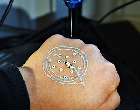
Researchers at the University of Minnesota use a customized 3D printer to print electronics on a real hand. Image: McAlpine group, University of Minnesota

Soldiers are commonly thrust into situations where the danger is the unknown: Where is the enemy, how many are there, what weaponry is being used? The military already uses a mix of technology to help answer those questions quickly, and another may be on its way. Researchers at the University of Minnesota have developed a low-cost 3D printer that prints sensors and electronics directly on skin. The development could allow soldiers to directly print temporary, disposable sensors on their hands to detect such things as chemical or biological agents in the field.

The technology also could be used in medicine. The Minnesota researchers successfully used bioink with the device to print cells directly on the wounds of a mouse. Researchers believe it could eventually provide new methods of faster and more efficient treatment, or direct printing of grafts for skin wounds or conditions.

“The concept was to go beyond smart materials, to integrate them directly on to skin,” says Michael McAlpine, professor of mechanical engineering whose research group focuses on 3D printing functional materials and devices. “It is a biological merger with electronics. We wanted to push the limits of what a 3D printer can do.”

McAlpine calls it a very simple idea, “One of those ideas so simple, it turns out no one has done it.”

Others have used 3D printers to print electronics and biological cells. But printing on skin presented a few challenges. No matter how hard a person tries to remain still, there always will be some movement during the printing process. “If you put a hand under the printer, it is going to move,” he says.

To adjust for that, the printer the Minnesota team developed uses a machine vision algorithm written by Ph.D. student Zhijie Zhu to track the motion of the hand in real time while printing. Temporary markers are placed on the skin, which then is scanned. The printer tracks the hand using the markers and adjusts in real time to any movement. That allows the printed electronics to maintain a circuit shape. The printed device can be peeled off the skin when it is no longer needed.

The team also needed to develop a special ink that could not only be conductive but print and cure at room temperature. Standard 3D printing inks cure at high temperatures of 212 °F and would burn skin.

In a paper recently published in Advanced Materals, the team identified three criteria for conductive inks: The viscosity of the ink should be tunable while maintaining self-supporting structures; the ink solvent should evaporate quickly so the device becomes functional on the same timescale as the printing process; and the printed electrodes should become highly conductive under ambient conditions.

The solution was an ink using silver flakes to provide conductivity rather than particles more commonly used in other applications. Fibers were found to be too large, and cure at high temperatures. The flakes are aligned by their shear forces during printing, and the addition of ethanol to the mix increases speed of evaporation, allowing the ink to cure quickly at room temperature.

“Printing electronics directly on skin would have been a breakthrough in itself, but when you add all of these other components, this is big,” McAlpine says.

The printer is portable, lightweight and cost less than $400. It consists of a delta robot, monitor cameras for long-distance observation of printing states and tracking cameras mounted for precise localization of the surface. The team added a syringe-type nozzle to squeeze and deliver the ink

Furthering the printer’s versatility, McAlpine’s team worked with staff from the university’s medical school and hospital to print skin cells directly on a skin wound of a mouse. The mouse was anesthetized, but still moved slightly during the procedure, he says. The initial success makes the team optimistic that it could open up a new method of treating skin diseases.

“Think about what the applications could be,” McAlpine says. “A soldier in the field could take the printer out of a pack and print a solar panel. On the cellular side, you could bring a printer to the site of an accident and print cells directly on wounds, speeding the treatment. Eventually, you may be able to print biomedical devices within the body.”

In its paper, the team suggests that devices can be “autonomously fabricated without the need for microfabrication facilities in freeform geometries that are actively adaptive to target surfaces in real time, driven by advances in multifunctional 3D printing technologies.”

Besides the ability to print directly on skin, McAlpine says the work may offer advantages over other skin electronic devices. For example, soft, thin, stretchable patches that stick to the skin have been fitted with off-the-shelf chip-based electronics for monitoring a patient’s health. They stick to skin like a temporary tattoo and send updates wirelessly to a computer.

“The advantage of our approach is that you don’t have to start with electronic wafers made in a clean room,” McAlpine says. “This is a completely new paradigm for printing electronics using 3D printing.”

http://www.asme.org/engineering-topics/articles/bioengineering/researchers-3d-print-skin-breakthrough

Bursts of brain activity linked to memory reactivation

By Hilary Hurd Anyaso

Leading theories propose that sleep presents an opportune time for important, new memories to become stabilized. And it’s long been known which brain waves are produced during sleep. But in a new study, researchers set out to better understand the brain mechanisms that secure memory storage.

The team from Northwestern and Princeton universities set out to find more direct and precisely timed evidence for the involvement of one particular sleep wave — known as the “sleep spindle.”

In the study, sleep spindles, described as bursts of brain activity typically lasting around one second, were linked to memory reactivation. The paper, “Sleep spindle refractoriness segregates periods of memory reactivation,” published today in the journal Current Biology.

“The most novel aspect of our study is that we found these spindles occur rhythmically — about every three to six seconds — and this rhythm is related to memory,” said James W. Antony, first author of the study and a postdoctoral fellow in Princeton’s Computational Memory Lab.

Three experiments explored how recent memories are reactivated during sleep. While volunteers took an afternoon nap, sound cues were surreptitiously played. Each was linked to a specific memory. The researchers’ final experiment showed that if cues were presented at opportune times such that spindles could follow them, the linked memories were more likely to be retained. If they were presented when a spindle was unlikely to follow, the linked memories were more likely to be forgotten.

“One particularly remarkable aspect of the study was that we were able to monitor spindles moment by moment while people slept,” said Ken A. Paller, senior author of the study and professor of psychology at Northwestern’s Weinberg College of Arts and Sciences. “Therefore, we could know when the brain was most ready for us to prompt memory reactivation.”
If the researchers reminded people of a recently learned fact, a spindle would likely be evident in the cerebral cortex, and memory for that information would be improved, added Paller, also director of Northwestern’s Cognitive Neuroscience Program.

“In memory research, we know it’s important to segregate experiences while you’re awake so that everything doesn’t just blend together,” said Antony, who worked in Paller’s lab at Northwestern as a doctoral student. “If that happens, you may have difficulty retrieving information because so many things will come to mind at once. We believe the spindle rhythmicity shown here might play a role in segregating successive memory reactivations from each other, preventing overlap that might cause later interference between memories.”

Ultimately, the researchers’ goal is to understand how sleep affects memory under natural conditions and how aging or disease can impact these functions.

“With that goal in mind, we’ve helped elucidate the importance of sleep spindles more generally,” Antony said.

Paller said they are on the trail of the physiology of memory reactivation.

“Future work will be needed to see how spindles fit together with other aspects of the physiology of memory and will involve other types of memory testing and other species,” Paller said.

In addition to Antony and Paller, co-authors are Luis Piloto, Margaret Wang, Paula Pacheco and Kenneth A. Norman, all of Princeton.

https://news.northwestern.edu/stories/2018/may/bursts-of-brain-activity-linked-to-memory-reactivation/

How brown fat keeps us warm


Adipose Connective Tissue Stores Fat in Our Body. Credit: Berkshire Community College Bioscience Image Library

A new technique to study fat stores in the body could aid efforts to find treatments to tackle obesity.

The approach focuses on energy-burning tissues found deep inside the body – called brown fat – that help to keep us warm when temperatures drop.

Experts are aiming to find it this calorie-burning power can be harnessed to stop weight gain, but little is known about how the process works.

Previous studies have mainly relied on a medical imaging technique called PET/CT to watch brown fat in action deep inside the body. But the method is unable to directly measure the chemical factors in the tissue.

Scientists at the University of Edinburgh developed a technique called microdialysis to measure how brown fat generates heat in people.

The approach involves inserting a small tube into an area of brown fat in the body and flushing it with fluid to collect a snapshot of the tissues’ chemical make-up.

The team tested the technique in six healthy volunteers, using PET/CT to guide the tube to the right location.

They discovered that in cold conditions, brown fat uses its own energy stores and other substances to generate heat.

Brown fat was active under warm conditions too, when the body does not need to generate its own heat, an outcome that had not been seen before.

Researchers hope the technique will help them to analyse the specific chemicals involved, so that they can better understand how brown fat works.

Most of the fat in our body is white fat, which is found under the skin and surrounding internal organs. It stores excess energy when we consume more calories than we burn.

Brown fat is mainly found in babies and helps them to stay warm. Levels can decrease with age but adults can still have substantial amounts of it, mainly in the neck and upper back region. People who are lean tend to have more brown fat.

The study, published in Cell Metabolism, was funded by the Medical Research Council and Wellcome.

Lead researcher Dr Roland Stimson, of the British Heart Foundation Centre for Cardiovascular Science at the University of Edinburgh, said: “Understanding how brown fat is activated could reveal potential targets for therapies that boost its energy-burning power, which could help with weight loss.”

This article has been republished from materials provided by the University of Edinburgh. Note: material may have been edited for length and content. For further information, please contact the cited source.

Reference: Weir, G., Ramage, L. E., Akyol, M., Rhodes, J. K., Kyle, C. J., Fletcher, A. M., … Stimson, R. H. (2018). Substantial Metabolic Activity of Human Brown Adipose Tissue during Warm Conditions and Cold-Induced Lipolysis of Local Triglycerides. Cell Metabolism, 0(0). https://doi.org/10.1016/j.cmet.2018.04.020

https://www.technologynetworks.com/proteomics/news/how-brown-fat-keeps-us-warm-304351?utm_campaign=Newsletter_TN_BreakingScienceNews&utm_source=hs_email&utm_medium=email&utm_content=63228690&_hsenc=p2ANqtz-9oqDIw3te1NPoj51s94kxnA1ClK8Oiecfela6I4WiITEbm_-SWdmw6pjMTwm2YP24gqSzRaBvUK1kkb2kZEJKPcL5JtQ&_hsmi=63228690

UMass Amherst Chemists Develop New Blood Test to Detect Liver Damage in Under an Hour

Chemist Vincent Rotello at the University of Massachusetts Amherst, with colleagues at University College London (UCL), U.K., announce today that they have developed a “quick and robust” blood test that can detect liver damage before symptoms appear, offering what they hope is a significant advance in early detection of liver disease. Details appear in Advanced Materials.

Their new method can detect liver fibrosis, the first stage of liver scarring that can lead to fatal disease if left unchecked, from a blood sample in 30-45 minutes, the authors note. They point out that liver disease is a leading cause of premature mortality in the United States and U.K., and is rising. It often goes unnoticed until late stages of the disease when the damage is irreversible.

For this work, Rotello and his team at UMass Amherst’s Institute of Applied Life Sciences (IALS) designed a sensor that uses polymers coated with fluorescent dyes that bind to blood proteins based on their chemical processes. The dyes change in brightness and color, offering a different signature or blood protein pattern.

He says, “This platform provides a simple and inexpensive way of diagnosing disease with potential for both personal health monitoring and applications in developing parts of the world.” Rotello and colleagues hope the new test can be used routinely in medical offices, clinics and hospitals to screen people with elevated liver disease risk so they can be treated “before it’s too late.”

The UCL team tested the sensor by comparing results from small blood samples equivalent to finger-prick checks from 65 people, in three balanced groups of healthy patients and among those with early-stage and late-stage fibrosis. This was determined using the Enhanced Liver Fibrosis (ELF) test, the existing benchmark for liver fibrosis detection. They found that the sensor identified different protein-level patterns in the blood of people in the three groups. The ELF test requires samples to be sent away to a lab.

Co-author William Peveler, a chemist now at the University of Glasgow, adds, “By comparing the different samples, the sensor array identified a ‘fingerprint’ of liver damage. It’s the first time this approach has been validated in something as complex as blood, to detect something as important as liver disease.”

The investigators report that the test distinguished fibrotic samples from healthy blood 80 percent of the time, reaching the standard threshold of clinical relevance on a widely-used metric and comparable to existing methods of diagnosing and monitoring fibrosis. The test distinguished between mild-moderate fibrosis and severe fibrosis 60 percent of the time. The researchers plan further tests with larger samples to refine the method’s effectiveness.

Peter Reinhart, director of UMass Amherst’s IALS says, “These exciting findings epitomize the mission of IALS to translate excellent basic science into diagnostics, therapeutic candidates and personalized health monitoring devices to improve human health and well-being.”

Peveler adds, “This may open the door to a cost-effective regular screening program thanks to its simplicity, low cost and robustness. We’re addressing a vital need for point-of-care diagnostics and monitoring, which could help millions of people access the care they need to prevent fatal liver disease.”

Rotello explains that the sensing strategy uses a “signature-based” approach that is highly versatile and should be useful in other areas. “A key feature of this sensing strategy is that it is not disease-specific, so it is applicable to a wide spectrum of conditions, which opens up the possibility of diagnostic systems that can track health status, providing both disease detection and monitoring wellness.”

In addition to UMass Amherst, UCL and the University of Glasgow, the U.K.-based research and development firm iQur Ltd. took part in the study. The work was supported by the U.K. Royal Society, the U.K. Engineering and Physical Sciences Research Council, the U.S. National Institutes of Health and the U.K. National Institute for Health Research UCLH Biomedical Research Centre.

http://www.umass.edu/newsoffice/article/umass-amherst-chemists-international-team

Depression speeds up brain ageing

Psychologists at the University of Sussex have found a link between depression and an acceleration of the rate at which the brain ages. Although scientists have previously reported that people with depression or anxiety have an increased risk of dementia in later life, this is the first study that provides comprehensive evidence for the effect of depression on decline in overall cognitive function (also referred to as cognitive state), in a general population.

For the study, published today, Thursday 24 May 2018, in the journal Psychological Medicine, researchers conducted a robust systematic review of 34 longitudinal studies, with the focus on the link between depression or anxiety and decline in cognitive function over time. Evidence from more than 71,000 participants was combined and reviewed. Including people who presented with symptoms of depression as well as those that were diagnosed as clinically depressed, the study looked at the rate of decline of overall cognitive state – encompassing memory loss, executive function (such as decision making) and information processing speed – in older adults.

Importantly, any studies of participants who were diagnosed with dementia at the start of study were excluded from the analysis. This was done in order to assess more broadly the impact of depression on cognitive ageing in the general population. The study found that people with depression experienced a greater decline in cognitive state in older adulthood than those without depression. As there is a long pre-clinical period of several decades before dementia may be diagnosed, the findings are important for early interventions as currently there is no cure for the disease.

Lead authors of the paper, Dr Darya Gaysina and Amber John from the EDGE (Environment, Development, Genetics and Epigenetics in Psychology and Psychiatry) Lab at the University of Sussex, are calling for greater awareness of the importance of supporting mental health to protect brain health in later life.

Dr Gaysina, a Lecturer in Psychology and EDGE Lab Lead, comments: “This study is of great importance – our populations are ageing at a rapid rate and the number of people living with decreasing cognitive abilities and dementia is expected to grow substantially over the next thirty years.

“Our findings should give the government even more reason to take mental health issues seriously and to ensure that health provisions are properly resourced. We need to protect the mental wellbeing of our older adults and to provide robust support services to those experiencing depression and anxiety in order to safeguard brain function in later life.”

Researcher Amber John, who carried out this research for her PhD at the University of Sussex adds: “Depression is a common mental health problem – each year, at least 1 in 5 people in the UK experience symptoms. But people living with depression shouldn’t despair – it’s not inevitable that you will see a greater decline in cognitive abilities and taking preventative measures such as exercising, practicing mindfulness and undertaking recommended therapeutic treatments, such as Cognitive Behaviour Therapy, have all been shown to be helpful in supporting wellbeing, which in turn may help to protect cognitive health in older age.”

The research paper, ‘Affective problems and decline in cognitive state in older adults’ will be available at: https:// doi.org/10.1017/S0033291718001137 from Thursday 24 May 2018.

http://www.sussex.ac.uk/broadcast/read/44977

Ultrasound Fires Up the Auditory Cortex—Even Though Animals Can’t Hear It


Ultrasound activates auditory pathways in the rodent brain (red arrows) regardless of where in the brain the ultrasound-generating transducer is placed.

By Abby Olena

Activating or suppressing neuronal activity with ultrasound has shown promise both in the lab and the clinic, based on the ability to focus noninvasive, high-frequency sound waves on specific brain areas. But in mice and guinea pigs, it appears that the technique has effects that scientists didn’t expect. In two studies published today (May 24) in Neuron, researchers demonstrate that ultrasound activates the brains of rodents by stimulating an auditory response—not, as researchers had presumed, only the specific neurons where the ultrasound is focused.

“These papers are a very good warning to folks who are trying to use ultrasound as a tool to manipulate brain activity,” says Raag Airan, a neuroradiologist and researcher at Stanford University Medical Center who did not participate in either study, but coauthored an accompanying commentary. “In doing these experiments going forward [the hearing component] is something that every single experimenter is going to have to think about and control,” he adds.

Over the past decade, researchers have used ultrasound to elicit electrical responses from cells in culture and motor and sensory responses from the brains of rodents and primates. Clinicians have also used so-called ultrasonic neuromodulation to treat movement disorders. But the mechanism by which high frequency sound waves work to exert their influence is not well understood.

The University of Minnesota’s Hubert Lim studies ways to restore hearing, but many of the strategies that his group uses are invasive, such as cochlear implants, which require surgery to insert a device inside the ear. He says that he and his colleagues were excited by the prospect of using noninvasive and precise ultrasound to activate the parts of the brain responsible for hearing.

Lim’s team started by stimulating the brains of guinea pigs with audible noise or with pulsed ultrasound directly over the auditory cortex. They were surprised to observe similar neuronal responses to the two different stimuli because ultrasound is outside the spectrum that the guinea pigs—and humans—can hear. The researchers also found that the rodents’ neurons showed comparable electrical activity in the auditory cortex regardless of where in the brain the researchers directed the ultrasound. This raised the question: are the animals’ brains responding directly to the ultrasound or to responses of the auditory system?

When the authors cut the guinea pigs’ auditory nerves or removed their cochlear fluid, the guinea pigs stopped responding to the ultrasound and to audible noise. Lim’s team concluded that what must be happening is ultrasound moves through brain tissue and vibrates the cochlear fluid. This vibration then triggers auditory signaling and indirectly activates the auditory cortex and other brain regions, rather than ultrasound having a direct effect on the activity of the neurons.

“I am actually very hopeful that ultrasound can be a powerful tool that can not only modulate but also treat different neurologic and psychiatric disorders, and that it can achieve a noninvasive yet localized activation,” says Lim. “But what we’re trying to show in this paper is that there are many confounding effects that are actually happening with ultrasound, and we have to remove those effects to really see how it’s activating the brain.”

A coauthor on the companion study, Mikhail Shapiro of Caltech, says that previous work showing that it is possible to apply ultrasound to the brains of mice and rats to elicit electrical activity and movement in their limbs left him and his colleagues curious about how it works. To determine where and when neural activation happens, they applied ultrasonic pulses to the brains of transgenic mice that have neurons that light up when stimulated. As with guinea pigs, ultrasound is inaudible to mice.

“To our surprise, we found that the main activation pattern that we were seeing was not in the region where we were applying the ultrasound directly, but actually in the auditory areas of the brain, those responsible for processing information about sound,” Shapiro tells The Scientist.

Consistent with the findings of Lim and colleagues, Shapiro and his coauthors determined that the mouse brains lit up across the cortex, starting from the auditory cortex. And as in the guinea pigs, the mouse neurons responded similarly to ultrasound and audible sounds. The researchers also showed that both ultrasound and audible noise elicited motor movements that decreased when they used chemicals to deafen the mice.

“We’re not trying to imply that [the effects of ultrasound observed in previous studies are] due to this auditory side effect,” says Shapiro. “We’re very optimistic that now that we know that it’s there, we will be able to design ways to get around it and still be able to use this technology scientifically.”

Shy Shoham, a neuroscientist and biomedical engineer at New York University Langone Medical Center who did not participate in the studies, tells The Scientist that these papers highlight how careful researchers must be in the future when using ultrasound to modify neuronal function. “In the field of neural stimulation in general, we should always be very concerned about off-target effects,” he says. We must “delineate what is real and what isn’t.”

“The big take home point here is that we need to take care of the auditory effects,” says Kim Butts Pauly, who studies ultrasound neuromodulation at Stanford University Medical Center and who coauthored the accompanying commentary with Airan. “There’s been very compelling data from other studies that ultrasound can stimulate the brain and change recordings from the brain that are completely separate from any auditory effects. As we get rid of the auditory effects, then the more subtle effects may become apparent.”

H. Guo et al., “Ultrasound produces extensive brain activation via a cochlear pathway,” Neuron, doi:10.1016/j.neuron.2018.04.036, 2018.

T. Sato et al., “Ultrasonic neuromodulation causes widespread cortical activation via an indirect auditory mechanism,” Neuron, doi:10.1016/j.neuron.2018.05.009, 2018.

https://www.the-scientist.com/?articles.view/articleNo/54652/title/Ultrasound-Fires-Up-the-Auditory-Cortex-Even-Though-Animals-Can-t-Hear-It/

A Hangover Pill? Tests on drunk mice show promise

“Civilization begins with distillation,” said William Faulkner, a writer and drinker. Although our thirst for alcohol dates back to the Stone Age, nobody has figured out a good way to deal with the ensuing hangover after getting drunk.

As a chemical engineering professor and wine enthusiast, I felt I needed to find a solution. As frivolous as this project may sound, it has serious implications. Between 8 and 10 percent of emergency room visits in America are due to acute alcohol poisoning. Alcohol is the leading risk factor for premature deaths and disability among people aged 15-49 and its abuse leads to serious health problems, including cardiovascular and liver cancer. Despite these sobering facts, current treatments for alcohol overdose largely rely on the body’s own enzymes to break down this drug.

I decided to design an antidote that could help people enjoy wine or cocktails or beer without a hangover, and at the same time create a lifesaving therapy to treat intoxication and overdose victims in the ER. I chose to create capsules filled with natural enzymes usually found in liver cells to help the body process the alcohol faster.

Together with professor Cheng Ji, an expert in liver diseases from Keck School of Medicine at the University of Southern California, and my graduate student Duo Xu, we developed an antidote and tested it in mice.

Inspired by the body’s approach for breaking down alcohol, we chose three natural enzymes that convert alcohol into harmless molecules that are then excreted. That might sound simple, because these enzymes were not new, but the tricky part was to figure out a safe, effective way to deliver them to the liver.

To protect the enzymes, we wrapped each of them in a shell, using a material the U.S. Food and Drug Administration had already approved for pills. We then injected these nanocapsules into the veins of drunk mice where they hurtled through the circulatory system, eventually arriving in the liver where they entered the cells and served as mini–reactors to digest alcohol.

We showed that in inebriated mice (which fall asleep much faster than drunk humans), the treatment decreased the blood alcohol level by 45 percent in just four hours compared to mice that didn’t receive any. Meanwhile, the blood concentration of acetaldehyde – a highly toxic compound that is carcinogenic, causes headaches and vomiting, makes people blush after drinking, and is produced during the normal alcohol metabolism – remained extremely low. The animals given the drug woke from their alcohol-induced slumber faster than their untreated counterparts – something all college students would appreciate.

The ability to efficiently break down alcohol quickly should help patients wake up earlier and prevent alcohol poisoning. It should also protect their liver from alcohol–associated stress and damage.

We are currently completing tests to ensure that our nanocapsules are safe and don’t trigger unexpected or dangerous side effects. If our treatments prove effective in animals, we could begin human clinical trials in as early as one year.

This sort of antidote won’t stop people from going too far when consuming alcohol, but it could help them recover quicker.

https://www.technologynetworks.com/neuroscience/articles/a-hangover-pill-tests-on-drunk-mice-show-promise-302970?utm_campaign=NEWSLETTER_TN_Neuroscience_2017&utm_source=hs_email&utm_medium=email&utm_content=63148685&_hsenc=p2ANqtz-_9-CBGC564lH1Jr5Fxrauf8vQZ42sDx9gSSQj_dPJTj3gm3QDvY74R4WiynR1vM5L7tdtTLBIV40iEWBKcEB7JzwFUnQ&_hsmi=63148685

Scientists plan DNA testing of Loch Ness lake

Prof Neil Gemmell, a New Zealand scientist leading the project, said he did not believe in Nessie, but was confident of finding genetic codes for other creatures.

He said a “biological explanation” might be found to explain some of the stories about the Loch Ness Monster.

The team will collect tiny fragments of skin and scales for two weeks in June.

Prof Gemmell, from the University of Otago in Dunedin, said: “I don’t believe in the idea of a monster, but I’m open to the idea that there are things yet to be discovered and not fully understood.

“Maybe there’s a biological explanation for some of the stories.”

The University of the Highlands and Islands’ UHI Rivers and Lochs Institute in Inverness is assisting in the project.

Other organisms

After the research team’s trip to Loch Ness, the samples will be sent to laboratories in New Zealand, Australia, Denmark and France to be analysed against a genetic database.

Prof Gemmell said: “There’s absolutely no doubt that we will find new stuff. And that’s very exciting.

“While the prospect of looking for evidence of the Loch Ness monster is the hook to this project, there is an extraordinary amount of new knowledge that we will gain from the work about organisms that inhabit Loch Ness – the UK’s largest freshwater body.”

The scientist said the team expected to find sequences of DNA from plants, fish and other organisms.

He said it would be possible to identify these plants and animals by comparing the sequences of their DNA against sequences held on a large, international database.

Prof Gemmell added: “There is this idea that an ancient Jurassic Age reptile might be in Loch Ness.

“If we find any reptilian DNA sequences in Loch Ness, that would be surprising and would be very, very interesting.”

The Loch Ness Monster is one of Scotland’s oldest and most enduring myths. It inspires books, TV shows and films, and sustains a major tourism industry around its home.

The story of the monster can be traced back 1,500 years when Irish missionary St Columba is said to have encountered a beast in the River Ness in 565AD.

Later, in the 1930s, The Inverness Courier reported the first modern sighting of Nessie.

Whale-like creature

In 1933, the newspaper’s Fort Augustus correspondent, Alec Campbell, reported a sighting by Aldie Mackay of what she believed to be Nessie.

Mr Campbell’s report described a whale-like creature and the loch’s water “cascading and churning”.

The editor at the time, Evan Barron, suggested the beast be described as a “monster”, kick starting the modern myth of the Loch Ness Monster.

Over the years various efforts have tried and failed to find the beast.

In tourism terms, there are two exhibitions dedicated to the monster and there is not a tourist shop in the Highlands, and even more widely across Scotland, where a cuddly toy of Nessie cannot be found.

In 2016, the inaugural Inverness Loch Ness International Knitting Festival exhibited knitted Nessie’s made from all parts of the world.

‘Record high’

In popular culture, the Loch Ness Monster has reared its head many times, including in 1975’s four-part Doctor Who – Terror of the Zygons, the 1980s cartoon The Family-Ness as well as The Simpsons and 1996’s Loch Ness starring Ted Danson.

In 2014, it was reported that for the first time in almost 90 years no “confirmed sightings” had been made of the Loch Ness Monster.

Gary Campbell, who keeps a register of sightings, said no-one had come forward in 18 months to say they had seen the monster.

But last year, sightings hit a record high.

http://www.bbc.com/news/uk-scotland-highlands-islands-44223259

Biomaterial developed at UCLA helps regrow brain tissue after stroke in mice

by Leigh Hopper

Tnew stroke-healing gel created by UCLA researchers helped regrow neurons and blood vessels in mice whose brains had been damaged by strokes. The finding is reported May 21 in Nature Materials.

“We tested this in laboratory mice to determine if it would repair the brain and lead to recovery in a model of stroke,” said Dr. S. Thomas Carmichael, professor of neurology at the David Geffen School of Medicine at UCLA. “The study indicated that new brain tissue can be regenerated in what was previously just an inactive brain scar after stroke.”

The results suggest that such an approach could some day be used to treat people who have had a stroke, said Tatiana Segura, a former professor of chemical and biomolecular engineering at UCLA who collaborated on the research. Segura is now a professor at Duke University.

The brain has a limited capacity for recovery after stroke. Unlike the liver, skin and some other organs, the brain does not regenerate new connections, blood vessels or tissue structures after it is damaged. Instead, dead brain tissue is absorbed, which leaves a cavity devoid of blood vessels, neurons or axons — the thin nerve fibers that project from neurons.

To see if healthy tissue surrounding the cavity could be coaxed into healing the stroke injury, Segura engineered a hydrogel that, when injected into the cavity, thickens to create a scaffolding into which blood vessels and neurons can grow. The gel is infused with medications that stimulate blood vessel growth and suppress inflammation, since inflammation results in scars and impedes functional tissue from regrowing.

After 16 weeks, the stroke cavities contained regenerated brain tissue, including new neuronal connections — a result that had not been seen before. The mice’s ability to reach for food improved, a sign of improved motor behavior, although the exact mechanism for the improvement wasn’t clear.

“The new axons could actually be working,” Segura said. “Or the new tissue could be improving the performance of the surrounding, unharmed brain tissue.”

The gel was eventually absorbed by the body, leaving behind only new tissue.

The research was designed to explore recovery in acute stroke, the period immediately following a stroke — in mice, that period lasts five days; in humans, it’s two months. Next, Carmichael and Segura plan to investigate whether brain tissue can be regenerated in mice long after the stroke injury. More than 6 million Americans are living with long-term effects of stroke, which is known as chronic stroke.

The other authors of the paper are Lina Nih and Shiva Gojgini, both of UCLA.

The study was supported by the National Institutes of Health.

http://newsroom.ucla.edu/releases/biomaterial-ucla-regrow-brain-tissue-after-stroke-mice

Molecular link between long-term memory and neurodegenerative disease discovered

Scientists have just discovered that a small region of a cellular protein that helps long-term memories form also drives the neurodegeneration seen in motor neuron disease (MND). This small part of the Ataxin-2 protein thus works for good and for bad. When a version of the protein lacking this region was substituted for the normal form in fruit flies (model organisms), the animals could not form long-term memories – but, surprisingly, the same flies showed a remarkable resistance to neurodegeneration.

The popular “ice bucket challenge” highlighted the social significance of MND, as well as the need to better understand and treat neurodegenerative conditions. This new research identifies a very specific basic mechanism that facilitates progression of neuronal loss in an animal model of MND, and, by shedding light on a potential way to protect against cell death in MND, it should inform strategies for the development of therapeutics to treat or manage these devastating conditions, which are currently incurable.

The Science Foundation Ireland-funded research, involving scientists from the Trinity College Institute of Neuroscience, NCBS Bangalore and HMMI, University of Colorado, Boulder, has just been published in the leading international journal Neuron.

Professor of Neurogenetics at Trinity College Dublin, Mani Ramaswami, said: “This work, by collaborating young researchers based in Irish, Indian and American laboratories, provides a great example of the ability of fundamental research in model organisms to produce biologically and clinically interesting information.”

A common feature of neurodegenerative diseases is the presence of specific protein aggregates in nerve cells, which accumulate and clump together — usually as protein fibres called amyloid filaments. Such aggregates are believed to trigger processes that cause the neuronal death associated with these debilitating diseases. For example, amyloid-beta (Aβ) aggregates are associated with Alzheimer’s disease, while TDP-43, FUS and Ataxin-2 proteins are commonly found in MND patients.

The scientists behind the current study set out to test this “amyloid hypothesis” to see whether it may explain how MND develops. The scientists genetically engineered fruit flies with mutations designed to reduce Ataxin-2 protein assembly into aggregates without affecting other functions of the protein.

Arnas Petrauskas, Trinity, said: “The flies with this altered, non-aggregating version of the protein showed a striking resistance to neurodegeneration. This suggests the normal Ataxin-2 protein and its ability to form aggregates is required for the progression of at least some forms of MND, which means these results provide support for the amyloid hypothesis.”

“What really surprised us though was that this same protein region seems to be required for the flies to develop long-term memory, as those with the altered version of Ataxin-2 showed normal short-term but defective long-term memories.”

Fruit flies normally respond strongly to new odorants, but weakly to familiar odorants through a process called habituation. This memory of the familiar can be of the short-term kind – to an odorant encountered for half-an-hour, or of the long-term kind, to odorants encountered for days (think of it as remembering a phone number of a new acquaintance versus remembering your own phone number). Flies lacking this small domain of Ataxin-2 showed greatly reduced long-term memory.

So how is long-term memory formation and disease progression connected? It turns out that proteins like the TDP-43, FUS and Ataxin-2 found in MND are also involved in the natural control and management of protein expression in the cell. The very same region of Ataxin-2 is needed to form RNP granules that store RNAs (essentially blueprints, or recipes for specific proteins) in a silent form until they are unpackaged by a signal and used to produce molecules when they are required. This local control of RNAs is required for long-term changes at neuronal synapses that underlie long-term memory.

The new discovery shows that Ataxin-2 concentrates several RNA-binding proteins used in the process of memory storing, but in doing so, it creates a biological environment that can help these proteins aggregate into disease-causing amyloids. A “trade-off” therefore exists in nature where the Ataxin-2 gene increases the danger of neurodegeneration, but helps our cells control RNA and form long-term memories.

In a commentary on the research published in the same issue of the journal Neuron, Aaron Gitler, Professor of Genetics in the Stanford Neuroscience Institute, an independent expert in MND research said: “This data suggest that manipulating RNP granule formation by genetically manipulating ataxin-2’s IDRs, or by other means could be therapeutic in ALS. Beyond ataxin-2, the race is now on to discover additional proteins that help build RNP granules.”

https://www.tcd.ie/news_events/articles/link-between-long-term-memory-and-neurodegenerative-disease/8941