The neurobiological basis of leadership rests in low aversion to responsibility


Low responsibility aversion is an important determinant of the decision to lead.

Leaders are more willing to take responsibility for making decisions that affect the welfare of others. In a new study, researchers at the University of Zurich identified the cognitive and neurobiological processes that influence whether someone is more likely to take on leadership or to delegate decision-making.

Parents, company bosses and army generals, as well as teachers and heads of state, all have to make decisions that affect not only themselves, but also influence the welfare of others. Sometimes, the consequences will be borne by individuals, but sometimes by whole organizations or even countries.

Researchers from the Department of Economics investigated what distinguishes people with high leadership abilities. In the study, which has just been published in the journal Science, they identify a common decision process that may characterize followers: Responsibility aversion, or the unwillingness to make decisions that also affect others.

Controlled experiments and brain imaging

In the study, leaders of groups could either make a decision themselves or delegate it to the group. A distinction was drawn between “self” trials, in which the decision only affected the decision-makers themselves, and “group” trials, in which there were consequences for the whole group. The neurobiological processes taking place in the brains of the participants as they were making the decisions were examined using functional magnetic resonance imaging (fMRI).

The scientists tested several common intuitive beliefs, such as the notion that individuals who are less afraid of potential losses or taking risks, or who like being in control, will be more willing to take on responsibility for others. These characteristics, however, did not explain the differing extent of responsibility aversion found in the study participants. Instead, they found that responsibility aversion was driven by a greater need for certainty about the best course of action when the decision also had an effect on others. This shift in the need for certainty was particularly pronounced in people with a strong aversion to responsibility.

“Because this framework highlights the change in the amount of certainty required to make a decision, and not the individual’s general tendency for assuming control, it can account for many different leadership types,” says lead author Micah Edelson. “These can include authoritarian leaders who make most decisions themselves, and egalitarian leaders who frequently seek a group consensus.”

More information: Computational and neurobiological foundations of leadership decisions. Science: August 2, 2018. DOI: 10.1126/science.aat0036

Machine learning links dimensions of mental illness to abnormalities of brain networks

A new study using machine learning has identified brain-based dimensions of mental health disorders, an advance towards much-needed biomarkers to more accurately diagnose and treat patients. A team at Penn Medicine led by Theodore D. Satterthwaite, MD, an assistant professor in the department of Psychiatry, mapped abnormalities in brain networks to four dimensions of psychopathology: mood, psychosis, fear, and disruptive externalizing behavior. The research is published in Nature Communications this week.

Currently, psychiatry relies on patient reporting and physician observations alone for clinical decision making, while other branches of medicine have incorporated biomarkers to aid in diagnosis, determination of prognosis, and selection of treatment for patients. While previous studies using standard clinical diagnostic categories have found evidence for brain abnormalities, the high level of diversity within disorders and comorbidity between disorders has limited how this kind of research may lead to improvements in clinical care.

“Psychiatry is behind the rest of medicine when it comes to diagnosing illness,” said Satterthwaite. “For example, when a patient comes in to see a doctor with most problems, in addition to talking to the patient, the physician will recommend lab tests and imaging studies to help diagnose their condition. Right now, that is not how things work in psychiatry. In most cases, all psychiatric diagnoses rely on just talking to the patient. One of the reasons for this is that we don’t understand how abnormalities in the brain lead to psychiatric symptoms. This research effort aims to link mental health issues and their associated brain network abnormalities to psychiatric symptoms using a data-driven approach.”

To uncover the brain networks associated with psychiatric disorders, the team studied a large sample of adolescents and young adults (999 participants, ages 8 to 22). All participants completed both functional MRI scans and a comprehensive evaluation of psychiatric symptoms as part of the Philadelphia Neurodevelopmental Cohort (PNC), an effort lead by Raquel E. Gur, MD, Ph.D., professor of Psychiatry, Neurology, and Radiology, that was funded by the National Institute of Mental Health. The brain and symptom data were then jointly analyzed using a machine learning method called sparse canonical correlation analysis.

This analysis revealed patterns of changes in brain networks that were strongly related to psychiatric symptoms. In particular, the findings highlighted four distinct dimensions of psychopathology—mood, psychosis, fear, and disruptive behavior—all of which were associated with a distinct pattern of abnormal connectivity across the brain.

The researchers found that each brain-guided dimension contained symptoms from several different clinical diagnostic categories. For example, the mood dimension was comprised of symptoms from three categories, e.g. depression (feeling sad), mania (irritability), and obsessive-compulsive disorder (recurrent thoughts of self-harm). Similarly, the disruptive externalizing behavior dimension was driven primarily by symptoms of both Attention Deficit Hyperactivity Disorder(ADHD) and Oppositional Defiant Disorder (ODD), but also included the irritability item from the depression domain. These findings suggest that when both brain and symptomatic data are taken into consideration, psychiatric symptoms do not neatly fall into established categories. Instead, groups of symptoms emerge from diverse clinical domains to form dimensions that are linked to specific patterns of abnormal connectivity in the brain.

“In addition to these specific brain patterns in each dimension, we also found common brain connectivity abnormalities that are shared across dimensions,” said Cedric Xia, a MD-Ph.D. candidate and the paper’s lead author. “Specifically, a pair of brain networks called default mode network and frontal-parietal network, whose connections usually grow apart during brain development, become abnormally integrated in all dimensions.”

These two brain networks have long intrigued psychiatrists and neuroscientists because of their crucial role in complex mental processes such as self-control, memory, and social interactions. The findings in this study support the theory that many types of psychiatric illness are related to abnormalities of brain development.

The team also examined how psychopathology differed across age and sex. They found that patterns associated with both mood and psychosis became significantly more prominent with age. Additionally, brain connectivity patterns linked to mood and fear were both stronger in female participants than males.

“This study shows that we can start to use the brain to guide our understanding of psychiatric disorders in a way that’s fundamentally different than grouping symptoms into clinical diagnostic categories. By moving away from clinical labels developed decades ago, perhaps we can let the biology speak for itself,” said Satterthwaite. “Our ultimate hope is that understanding the biology of mental illnesses will allow us to develop better treatments for our patients.”

More information: Cedric Huchuan Xia et al, Linked dimensions of psychopathology and connectivity in functional brain networks, Nature Communications (2018). DOI: 10.1038/s41467-018-05317-y

https://medicalxpress.com/news/2018-08-machine-links-dimensions-mental-illness.html

pH imbalance in brain cells may contribute to Alzheimer’s disease


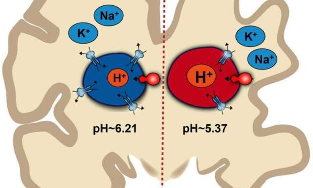
Illustration of how pH imbalance inside endosomes may contribute to Alzheimer’s disease

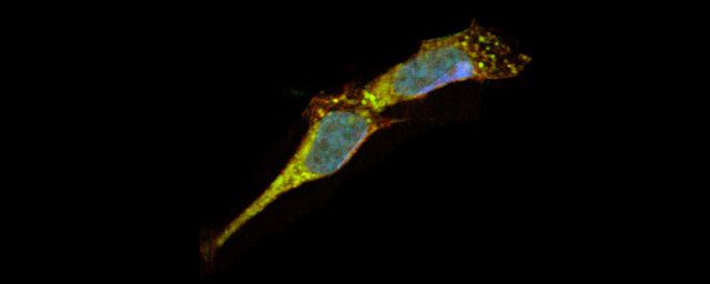
Johns Hopkins Medicine scientists say they have found new evidence in lab-grown mouse brain cells, called astrocytes, that one root of Alzheimer’s disease may be a simple imbalance in acid-alkaline—or pH—chemistry inside endosomes, the nutrient and chemical cargo shuttles in cells.

Astrocytes work to clear so-called amyloid beta proteins from the spaces between neurons, but decades of evidence has shown that if the clearing process goes awry, amyloid proteins pile up around neurons, leading to the characteristic amyloid plaques and nerve cell degeneration that are the hallmarks of memory-destroying Alzheimer’s disease.

The new study, described online June 26 in Proceedings of the National Academy of Sciences, also reports that the scientists gave drugs called histone deacetylase (HDAC) inhibitors to pH-imbalanced mice cells engineered with a common Alzheimer’s gene variant. The experiment successfully reversed the pH problem and improved the capacity for amyloid beta clearance.

HDAC inhibitors are approved by the U.S. Food and Drug Administration for use in people with certain types of blood cancers, but not in people with Alzheimer’s. They cautioned that most HDAC inhibitors cannot cross the blood-brain barrier, a significant challenge to the direct use of the drugs for brain disorders. The scientists say they are planning additional experiments to see if HDAC inhibitors have a similar effect in lab-grown astrocytes from Alzheimer’s patients, and that there is the potential to design HDAC inhibitors that can cross the barrier.

However, the scientists caution that even before those experiments can happen, far more research is needed to verify and explain the precise relationship between amyloid proteins and Alzheimer’s disease, which affects an estimated 50 million people worldwide. To date, there is no cure and no drugs that can predictably or demonstrably prevent or reverse Alzheimer’s disease symptoms.

“By the time Alzheimer’s disease is diagnosed, most of the neurological damage is done, and it’s likely too late to reverse the disease’s progression,” says Rajini Rao, Ph.D., professor of physiology at the Johns Hopkins University School of Medicine. “That’s why we need to focus on the earliest pathological symptoms or markers of Alzheimer’s disease, and we know that the biology and chemistry of endosomes is an important factor long before cognitive decline sets in.”

Nearly 20 years ago, scientists at Johns Hopkins and New York University discovered that endosomes, circular compartments that ferry cargo within cells, are larger and far more abundant in brain cells of people destined to develop Alzheimer’s disease. This hinted at an underlying problem with endosomes that could lead to an accumulation of amyloid protein in spaces around neurons, says Rao.

To shuttle their cargo from place to place, endosomes use chaperones—proteins that bind to specific cargo and bring them back and forth from the cell’s surface. Whether and how well this binding occurs depends on the proper pH level inside the endosome, a delicate balance of acidity and alkalinity, or acid and base, that makes endosomes float to the surface and slip back down into the cell.

Embedded in the endosome membrane are proteins that shuttle charged hydrogen atoms, known as protons, in and out of endosomes. The amount of protons inside the endosome determines its pH.

When fluids in the endosome become too acidic, the cargo is trapped within the endosome deep inside the cell. When the endosome contents are more alkaline, the cargo lingers at the cell’s surface for too long.

To help determine whether such pH imbalances occur in Alzheimer’s disease, Johns Hopkins graduate student Hari Prasad scoured scientific studies of Alzheimer’s disease looking for genes that were dialed down in diseased brains compared with normal ones. Comparing a dataset of 15 brains of Alzheimer’s disease patients with 12 normal ones, he found that 10 of the 100 most frequently down-regulated genes were related to the proton flow in the cell.

In another set of brain tissue samples from 96 people with Alzheimer’s disease and 82 without it, gene expression of the proton shuttle in endosomes, known as NHE6, was approximately 50 percent lower in people with Alzheimer’s disease compared with those with normal brains. In cells grown from people with Alzheimer’s disease and in mouse astrocytes engineered to carry a human Alzheimer’s disease gene variant, the amount of NHE6 was about half the amount found in normal cells.

To measure the pH balance within endosomes without breaking open the astrocyte, Prasad and Rao used pH sensitive probes that are absorbed by endosomes and emit light based on pH levels. They found that mouse cell lines containing the Alzheimer’s disease gene variant had more acidic endosomes (average of 5.37 pH) than cell lines without the gene variant (average of 6.21 pH).

“Without properly functioning NHE6, endosomes become too acidic and linger inside astrocytes, avoiding their duties to clear amyloid beta proteins,” says Rao.

While it’s likely that changes in NHE6 happen over time in people who develop sporadic Alzheimer’s disease, people who have inherited mutations in NHE6 develop what’s known as Christianson syndrome in infancy and have rapid brain degeneration.

Prasad and Rao also found that a protein called LRP1, which picks up amyloid beta proteins outside the astrocyte and delivers them to endosomes, was half as abundant on the surface of lab grown mouse astrocytes engineered with a human gene variant called APOE4, commonly linked to Alzheimer’s disease.

Looking for ways to restore the function of NHE6, Prasad searched databases of yeast studies to find that HDAC inhibitors tend to increase expression of the NHE6 gene in yeast. This gene is very similar across species, including flies, mice and humans.

Prasad and Rao tested nine types of HDAC inhibitors on cell cultures of mouse astrocytes engineered with the APOE4 gene variant. Broad-spectrum HDAC inhibitors increased NHE6 expression to levels associated with mouse astrocytes that did not have the Alzheimer’s gene variant. They also found that HDAC inhibitors corrected the pH imbalance inside endosomes and restored LRP1 to the astrocyte surface, resulting in efficient clearance of amyloid beta protein.

More information: Hari Prasad et al. Amyloid clearance defect in ApoE4 astrocytes is reversed by epigenetic correction of endosomal pH, Proceedings of the National Academy of Sciences (2018). DOI: 10.1073/pnas.1801612115

https://medicalxpress.com/news/2018-08-ph-imbalance-brain-cells-contribute.html

Magnetic particle mapped in the human brain

Researchers at Ludwing Maximilliams Universitat Muchen have for the first time mapped the distribution of magnetic particles in the human brain. The study reveals that the particles are primarily located in the cerebellum and the brainstem, which are the more ancient parts of the brain.

Many living organisms, such as migratory birds, are thought to possess a magnetotactic sense, which enables them to respond to the Earth’s magnetic field. Whether or not humans are capable of sensing magnetism is the subject of debate. However, several studies have already shown that one of the preconditions required for such a magnetic sensory system is indeed met: magnetic particles exist in the human brain. Now a team led by Stuart A. Gilder (a professor at LMU‘s Department of Earth and Environmental Sciences) and Christoph Schmitz (a professor at LMU’s Department of Neuroanatomy) has systematically mapped the distribution of magnetic particles in human post mortem brains. Their findings were published in the journal Scientific Reports (Nature Publishing Group)

In their study, the LMU researchers confirmed the presence of magnetic particles in human brains. The particles were found primarily in the cerebellum and the brainstem, and there was striking asymmetry in the distribution between the left and right hemispheres of the brain. “The human brain exploits asymmetries in sensory responses for spatial orientation, and also for sound-source localization,” Schmitz explains. The asymmetric distribution of the magnetic particles is therefore compatible with the idea that humans might have a magnetic sensor. But in all probability, this sensor is much too insensitive to serve any useful biological function, he adds. Furthermore, the chemical nature of the magnetic particles remains unknown. “We assume that they are all made of magnetite (Fe3O4), but it is not yet possible to be sure,” says Gilder.

The study was funded by the Volkswagen Foundation’s “Experiment!” program, which is designed specifically to get daring new research projects, whose ultimate outcome is uncertain, off the ground. This is in contrast to traditional NIH-style support, which largely supports research that has already been conducted and for which the outcome is almost certain. The data were obtained from seven human post mortem brains, which had been donated for use in medical research. In all, a total of 822 tissue samples were subjected to magnetometry. The measurements were performed under the supervision of Stuart Gilder in a magnetically shielded laboratory located in a forest 80 km from Munich which is largely free from pervasive magnetic pollution that is characteristic of urban settings nowadays.

In further experiments, the LMU team plans to characterize the properties of the magnetic particles found in human brains. In collaboration with Professor Patrick R. Hof (Fishberg Department of Neuroscience, Icahn School of Medicine at Mount Sinai, New York), they also hope to perform analogous localization studies on far larger mammals – whales. These huge marine mammals are known to migrate between feeding and breeding grounds across great distances in the world’s oceans. “We want determine whether we can detect magnetic particles in the brains of whales, and if so whether they are also asymmetrically distributed“ says Schmitz. “It goes without saying that such studies will be carried out on animals that have died of natural causes.”

https://www.en.uni-muenchen.de/news/newsarchiv/2018/schmitz_magnetite.html

Distribution of magnetic remanence carriers in the human brain
Stuart A. Gilder, Michael Wack, Leon Kaub, Sophie C. Roud, Nikolai Petersen, Helmut Heinsen, Peter Hillenbrand, Stefan Milz & Christoph Schmitz
Scientific Reportsvolume 8, Article number: 11363 (2018)

LRRK2 enzyme is implicated in even more Parkinson’s disease cases than originally realized, suggesting that inhibitors of the enzyme could treat more patients.

by SUKANYA CHARUCHANDRA

Even when Parkinson’s patients don’t have mutations in a gene called LRRK2, more of the active enzyme the gene generates is present in their brains than in healthy brains, researchers reported last week (July 25) in Science Translational Medicine. The finding suggests that LRRK2 inhibitors could help to reduce harmful effects of the enzyme in the vast majority of Parkinson’s patients.

“This is the really interesting bit of data … the demonstration that when you look in the brains of individuals with idiopathic Parkinson’s [where the cause is unknown], that there’s evidence that LRRK2 is activated,” says Patrick Lewis, who studies Parkinson’s disease at University College London and the University of Reading in the UK. He has collaborated with one of the paper’s coauthors but was not involved in this study.

Ten percent of Parkinson’s cases have known genetic causes. Three percent of cases are due to a mutation in LRRK2, the gene encoding the LRRK2 enzyme. The enzyme is highly active in Parkinson’s patients with a mutated LRRK2 gene, and the increased enzyme activity has been linked to the development of the disease.

In the new study, Timothy Greenamyre, a professor of neurology at the University of Pittsburgh, and his team wanted to look at the level of active LRRK2 in patients without an LRRK2 mutation. “Because [LRRK2] is a low-abundance protein, people have had difficulty detecting it,” Greenamyre says. To spot active LRRK2, the researchers first developed two versions of an assay: the first detects the active enzyme and the second, the inactive enzyme. In the first detection method, researchers used two different antibodies, one that binds to a specific subunit that acts as a known indicator of the active enzyme and another that binds to a different proximal portion of it. When both antibodies bind successfully, their close contact generates a fluorescent signal—a sign of active LRRK2. The second method detects a protein known to regulate LRRK2 activity. Higher levels of this protein indicate lower levels of available active LRRK2.

The team used the assay on postmortem brain tissue from Parkinson’s disease patients and from healthy individuals. The researchers observed higher levels of the active LRRK2 enzyme in substantia nigra dopamine-producing neurons—the death of which indicate neurodegenerative disease—in the brain tissue of Parkinson’s patients’ with no mutation in the LRRK2 gene than in healthy brain tissue.

“We have been wondering for a very long time whether LRRK2 plays a role in sporadic Parkinson’s disease,” says Mark Cookson, who studies the neurodegenerative disorder in the National Institutes of Health’s Laboratory of Neurogenetics. He has collaborated with Greenamyre before but was not involved in this work. According to Cookson, this study provides “defensive evidence” of LRRK2’s role in the disease, even in patients without a mutation in the gene.

In the next set of experiments, Greenamyre and his colleagues wanted to see if active LRRK2 turned up in two rat models of Parkinson’s disease. In the first rodent model, the animals were given the toxin rotenone to induce symptoms of the disease. Even without a mutation in the LRRK2 gene, the rats had higher levels of active LRRK2 protein. In the rats’ brains, the active LRRK2 enzymes were linked with clumps of another protein, α-synuclein. The clumps eventually help form Lewy bodies, a characteristic feature of Parkinson’s brains. In the second rodent model, the researchers overexpressed wildtype α-synuclein in the rats’ substantia nigra, which caused levels of active LRRK2 to rise. When the group treated the rotenone-rodent model with a drug that inhibited the LRRK2 protein, the number of clumps and Lewy bodies dropped.

The team also observed higher levels of reactive oxygen species (ROS)—chemically responsive molecules such as peroxides—in the brains of both rat models of Parkinson’s disease. As a result, Greenamyre and his colleagues wanted to see if directly increasing ROS led to more active LRRK2. In a third set of experiments, the team dosed healthy human cell lines with hydrogen peroxide and found the addition of the ROS increased the levels of LRRK2. A spike in ROS levels, the researchers suggest, activates LRRK2, which in turn aids in the development of some classic Parkinson’s features. Blocking the production of ROS resulted in a drop in active LRRK2. The result gives clues to an environmental cause for Parkinson’s disease.

Pharmaceutical companies are already developing LRRK2 inhibitors that can help the small percentage of Parkinson’s patients that have a mutation in the LRRK2 gene. “The inhibitors may benefit patients not only with the mutation but also patients who have idiopathic diseases—they’re much more common,” says coauthor Dario Alessi, a professor who studies signaling pathways in neurodegenerative disorders at the University of Dundee in the UK.

LRRK2 inhibitors, the researchers note, cause mild, yet reversible side effects, in the lungs and kidneys.

R.D. Maio et al., “LRRK2 activation in idiopathic Parkinson’s disease,” Science Translational Medicine, doi:10.1126/scitranslmed.aar5429, 2018.

https://www.the-scientist.com/news-opinion/key-enzyme-active-in-brains-of-patients-without-parkinsons-mutation-64599

Ketamine shows promise as an effective treatment for adolescents with depression

BY ARISTOS GEORGIOU

Young people suffering from treatment-resistant depression (TRD) showed a significant reduction of their symptoms after being administered ketamine injections, according to a study published in the Journal of Child and Adolescent Psychopharmacology.

Researchers from the University of Minnesota (UM) and the nonprofit Mayo Clinic found that ketamine caused an average decrease of 42 percent on the Children’s Depression Rating Scale (CDRS)—the most widely used rating scale in research trials for assessing the severity of depression and change in depressive symptoms among adolescents.

Ketamine is perhaps best known for being a popular recreational drug and a useful medical anesthetic, but a growing body of research is indicating that the compound could be an effective treatment for depression. Several recent studies have shown that even a single dose in adults can lead to rapid reductions in depressive symptoms. However, relatively little research has been conducted into ketamine’s antidepressant effects in adolescents.

“Adolescence is a very important time for studying depression, first because depression often starts during these years, and second because it is an important time for brain development,” Kathryn Cullen, from the Department of Psychiatry at UM, told Newsweek.

“When adolescent depression persists without successful treatment, it can interfere with achieving important developmental milestones. Finding the right treatment is critical to allow the restoration of healthy brain development and prevent negative outcomes like chronic depression, disability and suicide.”

Unfortunately, about 40 percent of adolescents do not respond to their first intervention and only half of nonresponders respond to the second treatment, according to the researchers.

“Standard antidepressant treatments do not work for everyone and take weeks to months to take effect, a time period when patients are at risk for continued suffering and suicide attempts,” Cullen said. “The field is in need of new treatment options. Ketamine has a very different mechanism of action than standard treatments.”

The latest study involved 13 young people ages 12 to 18 who had failed two previous trials of antidepressants. During a two-week period, the researchers gave them six ketamine infusions.

They found that the treatment was well tolerated, with the participants showing an average decrease in CDRS scores of 42.5 percent. Five of the participants met the criteria for clinical response and remission. Of these, three were still in remission after six weeks, while the remaining two relapsed within two weeks.

According to the scientists, the results demonstrate the potential role for ketamine in treating adolescents with TRD. However, they note that the study was limited by its small sample size, so future research will be needed to confirm these results.

“The purpose of our study was to investigate the effects of ketamine for TRD in younger patients for whom this indication for ketamine administration is not well studied,” Mark Roback, a professor of pediatrics at the University of Minnesota, told Newsweek.

“I think our results show promise for this population, however this study is just a beginning. The study serves to point out the need for further, rigorous, study designed to answer the many questions that remain about ketamine for TRD, such as optimal dosing and route of administration, dosing interval and treatment length, and long-term effects—just to name a few.”

James Stone, a clinical senior lecturer from the Institute of Psychiatry, Psychology and Neuroscience at King’s College London, who was not involved in the study, told Newsweek that there is “a lot of potential for the use of ketamine as a second or third line antidepressant where other treatments have failed.”

“Although ketamine is potentially a huge breakthrough in the treatment of depression, we still don’t know about the long-term safety, or about how to keep people well from depression without requiring regular ketamine dosing,” Stone added. “Further studies are needed to address these questions.”

https://www.newsweek.com/ketamine-shows-promise-treatment-adolescents-depression-1054021

New research shows a mechanism by which tau may be toxic in Alzheimer’s disease

New evidence suggests a mechanism by which progressive accumulation of Tau protein in brain cells may lead to Alzheimer’s disease. Scientists studied more than 600 human brains and fruit fly models of Alzheimer’s disease and found the first evidence of a strong link between Tau protein within neurons and the activity of particular DNA sequences called transposable elements, which might trigger neurodegeneration. The study appears in the journal Cell Reports.

“One of the key characteristics of Alzheimer’s disease is the accumulation of Tau protein within brain cells, in combination with progressive cell death,” said corresponding author Dr. Joshua Shulman, associate professor of neurology, neuroscience and molecular and human genetics at Baylor College of Medicine and investigator at the Jan and Dan Duncan Neurological Research Institute at Texas Children’s Hospital. “In this study we provide novel insights into how accumulation of Tau protein may contribute to the development of Alzheimer’s disease.”

Although scientists have studied for years what happens when Tau forms aggregates inside neurons, it still is not clear why brain cells ultimately die. One thing that scientists have noticed is that neurons affected by Tau accumulation also appear to have genomic instability.

“Genomic instability refers to an increased tendency to have alterations in the genetic material, DNA, such as mutations or other impairments. This means that the genome is not functioning correctly. Genomic instability is known to be a major driving force behind other diseases such as cancer,” Shulman said. “Our study focused on a new possible causal connection between Tau accumulation within neurons and the resulting genomic instability in Alzheimer’s disease.”

Enter transposable elements
Previous studies of brain tissues from patients with other neurologic diseases and of animal models have suggested that the neurons not only present with genomic instability, but also with activation of transposable elements.

“Transposable elements are short pieces of DNA that do not seem to contribute to the production of proteins that make cells function. They behave in a way similar to viruses; they can make copies of themselves that are inserted within the genome and this can create mutations that lead to disease,” Shulman said. “Although most transposable elements are dormant or dysfunctional, some may become active in human brains late in life or in disease. That’s what led us to look specifically at Alzheimer’s disease and the possible association between Tau accumulation and activated transposable elements.”

Shulman and his colleagues began their investigations by studying more than 600 human brains from a population study run by co-author Dr. David Bennett at Rush University Medical Center in Chicago. This population study follows participants throughout their lives and at death, allowing the researchers to examine their brains in detail postmortem. One of the evaluations is the amount of Tau accumulation across many brain regions. In addition, co-author Dr. Philip De Jager at the Broad Institute and Columbia University comprehensively profiled gene expression in the same brains.

“With this large amount of data, we looked to identify signatures of active transposable elements, but this was not easy,” Shulman said. “We therefore reached out to Dr. Zhandong Liu, a co-author in this study, and together we developed a new software tool to detect signatures of active transposable elements from postmortem human brains. Then we conducted a statistical analysis in which we compared the amount of active transposable elements signatures with the amount of Tau accumulation, brain by brain.” Liu also is assistant professor of pediatrics – neurology at Baylor and a member of the Dan L Duncan Comprehensive Cancer Center.

The researchers found a strong link between the amount of Tau accumulation in neurons and detectable activity of transposable elements.

“We identified individual transposable elements that were active when Tau aggregates were present. Surprisingly, we also found evidence that the activation of transposable elements was quite broad across the genome,” Shulman said.

Other research has shown that Tau may disrupt the tightly packed architecture of the genome. It is believed that tightly packed DNA limits gene activation, while opening up the DNA may promote it. Keeping the DNA tightly packed may be an important mechanism to suppress the activity of transposable elements that lead to disease.

“The fact that Tau aggregates can affect that architecture of the genome may be one possible mechanism by which transposable elements are activated in Alzheimer’s disease,” Shulman said. “However, our studies in human brains only establish an association between Tau accumulation and activation of transposable elements. To determine whether Tau accumulation could in fact cause transposable element activation, we conducted studies with a fruit fly model of Alzheimer’s disease.”

In this fruit fly model of the disease, the researchers found that triggering Tau changes similar to those observed in human brains resulted in the activation of fruit fly transposable elements, strongly suggesting that Tau aggregates that disrupt the architecture of the genome can potentially mediate the activation of transposable elements and ultimately cause neurodegeneration.

“We think our experiments reveal new and potentially important insights relevant for understanding Alzheimer’s disease mechanisms,” Shulman said. “There is still a lot of work to be done, but by presenting our results we hope we can stimulate others in the research community to help work on this problem.”

https://www.bcm.edu/news/neurology/research-links-tau-aggregates-cell-death

Our brains need silence to make sense of things

by CHRISTIAN COTRONEO

The mind may seem to thrive on stimuli — the honking horns, the pixels percolating on this screen at this very moment, or even the way the keyboard feels under your fingers at any given time of day.

But, in fact, it may be what lies between — the time between honks, if you will — when the brain focuses on encoding the information, according to a new study from Neuroscience Research Australia (NeuRA) and the University of New South Wales.

Of course, we’ve long known that silence is golden — especially when it comes to mental health and dealing with stress. But the new research points to the absence of stimulation as a window when the brain has a chance to learn from its environment.

Think of it as a micro-breather for the mind, allowing it to grasp and distill what it’s experiencing.

To reach that conclusion, researchers Ingvars Birznieks and Richard Vickery developed a unique way to control the neural information that’s presented to the brain. Essentially, they delivered short mechanical taps to the fingertips of study subjects.

Birznieks and Vickery ensured that each tap generated a corresponding nerve impulse to a neuron in the brain. By triggering the sense of touch — which the brain registers from vibrations along the ridge of our fingertips — the scientists were able to monitor how nerve impulses encoded the information.

The thing is, the frequency of those neuron bursts didn’t match the frequency of taps.

“Instead, it was the silent period between bursts that best explained the subjects’ experiences,” Birznieks noted in the NeuRA blog.

Prevailing theories had it that every vibration or tap would have a corresponding nerve impulse, or the brain would be able to detect a periodic regularity in the impulse patterns.

“We were hoping to disprove one of the two competing theories, but showing they were both incorrect and finding a completely new coding strategy totally surprised us,” Birznieks added.

The brain just kept ticking along to its own beat, independent of how often those fingertips were stimulated.

For neuroscience, the findings could be a game-changer. A better understanding of how the brain fields daily neural impulses could pave the way for more efficient interfaces between brain and machine.

And for the rest of us, it suggests that in a increasingly noise-addled society — where every sense seems in danger of over-stimulation — it may do a body good to give the brain a breather.

https://www.mnn.com/green-tech/research-innovations/stories/silence-brain-study-touch-stimulation

Scientists are now able to make human neurons from white blood cells


Fresh or frozen human blood samples can be directly transformed into patient-specific neurons to study disorders such as schizophrenia and autism, Stanford researcher Marius Wernig has found.

Human immune cells in blood can be converted directly into functional neurons in the laboratory in about three weeks with the addition of just four proteins, researchers at the Stanford University School of Medicine have found.

The dramatic transformation does not require the cells to first enter a state called pluripotency but instead occurs through a more direct process called transdifferentiation.

The conversion occurs with relatively high efficiency — generating as many as 50,000 neurons from 1 milliliter of blood — and it can be achieved with fresh or previously frozen and stored blood samples, which vastly enhances opportunities for the study of neurological disorders such as schizophrenia and autism.

“Blood is one of the easiest biological samples to obtain,” said Marius Wernig, MD, associate professor of pathology and a member of Stanford’s Institute for Stem Cell Biology and Regenerative Medicine. “Nearly every patient who walks into a hospital leaves a blood sample, and often these samples are frozen and stored for future study. This technique is a breakthrough that opens the possibility to learn about complex disease processes by studying large numbers of patients.”

A paper describing the findings was published online June 4 in the Proceedings of the National Academy of Sciences. Wernig is the senior author. Former postdoctoral scholar Koji Tanabe, PhD, and graduate student Cheen Ang are the lead authors.

Dogged by challenges

The transdifferentiation technique was first developed in Wernig’s laboratory in 2010 when he and his colleagues showed that they could convert mouse skin cells into mouse neurons without first inducing the cells to become pluripotent — a developmentally flexible stage from which the cells can become nearly any type of tissue. They went on to show the technique could also be used on human skin and liver cells.

But each approach has been dogged by challenges, particularly for researchers wishing to study genetically complex mental disorders, such as autism or schizophrenia, for which many hundreds of individual, patient-specific samples are needed in order to suss out the relative contributions of dozens or more disease-associated mutations.

“Generating induced pluripotent stem cells from large numbers of patients is expensive and laborious. Moreover, obtaining skin cells involves an invasive and painful procedure,” Wernig said. “The prospect of generating iPS cells from hundreds of patients is daunting and would require automation of the complex reprogramming process.”

Although it’s possible to directly convert skin cells to neurons, the biopsied skin cells first have to be grown in the laboratory for a period of time until their numbers increase — a process likely to introduce genetic mutations not found in the person from whom the cells were obtained.

The researchers wondered if there was an easier, more efficient way to generate patient-specific neurons.

‘Somewhat mind-boggling’
In the new study, Wernig and his colleague focused on highly specialized immune cells called T cells that circulate in the blood. T cells protect us from disease by recognizing and killing infected or cancerous cells. In contrast, neurons are long and skinny cells capable of conducting electrical impulses along their length and passing them from cell to cell. But despite the cells’ vastly different shapes, locations and biological missions, the researchers found it unexpectedly easy to complete their quest.

“It’s kind of shocking how simple it is to convert T cells into functional neurons in just a few days,” Wernig said. “T cells are very specialized immune cells with a simple round shape, so the rapid transformation is somewhat mind-boggling.”

The resulting human neurons aren’t perfect. They lack the ability to form mature synapses, or connections, with one another. But they are able to carry out the main fundamental functions of neurons, and Wernig and his colleague are hopeful they will be able to further optimize the technique in the future. In the meantime, they’ve started to collect blood samples from children with autism.

“We now have a way to directly study the neuronal function of, in principle, hundreds of people with schizophrenia and autism,” Wernig said. “For decades we’ve had very few clues about the origins of these disorders or how to treat them. Now we can start to answer so many questions.”

Other Stanford co-authors are postdoctoral scholars Soham Chanda, PhD, and Daniel Haag, PhD; undergraduate student Victor Olmos; professor of psychiatry and behavioral sciences Douglas Levinson, MD; and professor of molecular and cellular physiology Thomas Südhof, MD.

The research was supported by the National Institutes of Health (grants MH092931 and MH104172), the California Institute for Regenerative Medicine, the New York Stem Cell Foundation, the Howard Hughes Medical Institute, the Siebel Foundation and the Stanford Schizophrenia Genetics Research Fund.

http://med.stanford.edu/news/all-news/2018/06/human-blood-cells-transformed-into-functional-neurons.html

Researchers discovery an independent role for astrocytes in cognition

The majority of the cells in the brain are no neurons, but Glia (from “glue”) cells, that support the structure and function of the brain. Astrocytes (“start cells”) are star-shaped glial cells providing many supportive functions for the neurons surrounding them, such as the provision of nutrients and the regulation of their chemical environment. Newer studies showed that astrocytes also monitor and modulate neuronal activity. For example, these studies have shown that astrocytes are necessary for the ability of neurons to change the strength of the connections between them, the process underlying learning and memory, and indeed astrocytes are also necessary for normal cognitive function. However, it is still unknown whether astrocytic activity is only necessary, or is it may also be sufficient to induce synaptic potentiation and enhance cognitive performance.

In a new study published in Cell, two graduate students, Adar Adamsky and Adi Kol, from Inbal Goshen’s lab, employed chemogenetic and optogenetic tools that allow specific activation of astrocytes in behaving mice, to explore their role in synaptic activity and memory performance. They found that astrocytic activation in the hippocampus, a brain region that plays an important role in memory acquisition and consolidation, potentiated the synaptic connections in this region, measured in brain slices. Moreover, in the intact brain, astrocytic activation enhanced hippocampal neuronal activity in a task-dependent way: i.e. only during when it was combined with memory acquisition, but not when mice were at their home cage with no meaningful stimuli. The ability of astrocytes to increase neuronal activity during memory acquisition had a significant effect on cognitive function: Specifically, astrocytic activation during learning resulted in enhanced memory in two memory tests. In contrast, direct neuronal activation in the hippocampus induced a non-selective increase in activity (during learning or in the home cage), and thus resulted in drastic memory impairment.

The results suggest that the memory enhancement induced by astrocytic activation during learning is not simply a result of a general increase in hippocampal neuronal activity. Rather, the astrocytes, which sense and respond to changes in the surrounding neuronal activity, can detect and specifically enhance only the neuronal activity involved in learning, without affecting the general activity. This may explain why general astrocytic activation improves memory performance, whereas a similar activation of neurons impairs it.

Memory is not a binary process (remember/don’t remember); the strength of a memory can vary greatly, either for the same memory or between different memories. Here, we show that activating astrocytes in mice with intact cognition improves their memory performance. This finding has important clinical implications for cognitive augmentation treatments. Furthermore, the ability of astrocytes to strengthen neuronal communication and improve memory performance supports the claim that astrocytes are able to take an active part in the neuronal processes underlying cognitive function. This perspective expands the definition of the role of astrocytes, from passive support cells to active cells that can modulate neural activity and thus shape behavior.

Link: https://www.cell.com/cell/pdf/S0092-8674(18)30575-0.pdf

https://elsc.huji.ac.il/content/article-month-june-2018-goshens-lab