Neuroscientists Translate Brain Waves Into Recognizable Speech

by George Dvorsky

Using brain-scanning technology, artificial intelligence, and speech synthesizers, scientists have converted brain patterns into intelligible verbal speech—an advance that could eventually give voice to those without.

It’s a shame Stephen Hawking isn’t alive to see this, as he may have gotten a real kick out of it. The new speech system, developed by researchers at the ​Neural Acoustic Processing Lab at Columbia University in New York City, is something the late physicist might have benefited from.

Hawking had amyotrophic lateral sclerosis (ALS), a motor neuron disease that took away his verbal speech, but he continued to communicate using a computer and a speech synthesizer. By using a cheek switch affixed to his glasses, Hawking was able to pre-select words on a computer, which were read out by a voice synthesizer. It was a bit tedious, but it allowed Hawking to produce around a dozen words per minute.

But imagine if Hawking didn’t have to manually select and trigger the words. Indeed, some individuals, whether they have ALS, locked-in syndrome, or are recovering from a stroke, may not have the motor skills required to control a computer, even by just a tweak of the cheek. Ideally, an artificial voice system would capture an individual’s thoughts directly to produce speech, eliminating the need to control a computer.

New research published today in Scientific Advances takes us an important step closer to that goal, but instead of capturing an individual’s internal thoughts to reconstruct speech, it uses the brain patterns produced while listening to speech.

To devise such a speech neuroprosthesis, neuroscientist Nima Mesgarani and his colleagues combined recent advances in deep learning with speech synthesis technologies. Their resulting brain-computer interface, though still rudimentary, captured brain patterns directly from the auditory cortex, which were then decoded by an AI-powered vocoder, or speech synthesizer, to produce intelligible speech. The speech was very robotic sounding, but nearly three in four listeners were able to discern the content. It’s an exciting advance—one that could eventually help people who have lost the capacity for speech.

To be clear, Mesgarani’s neuroprosthetic device isn’t translating an individual’s covert speech—that is, the thoughts in our heads, also called imagined speech—directly into words. Unfortunately, we’re not quite there yet in terms of the science. Instead, the system captured an individual’s distinctive cognitive responses as they listened to recordings of people speaking. A deep neural network was then able to decode, or translate, these patterns, allowing the system to reconstruct speech.

“This study continues a recent trend in applying deep learning techniques to decode neural signals,” Andrew Jackson, a professor of neural interfaces at Newcastle University who wasn’t involved in the new study, told Gizmodo. “In this case, the neural signals are recorded from the brain surface of humans during epilepsy surgery. The participants listen to different words and sentences which are read by actors. Neural networks are trained to learn the relationship between brain signals and sounds, and as a result can then reconstruct intelligible reproductions of the words/sentences based only on the brain signals.”

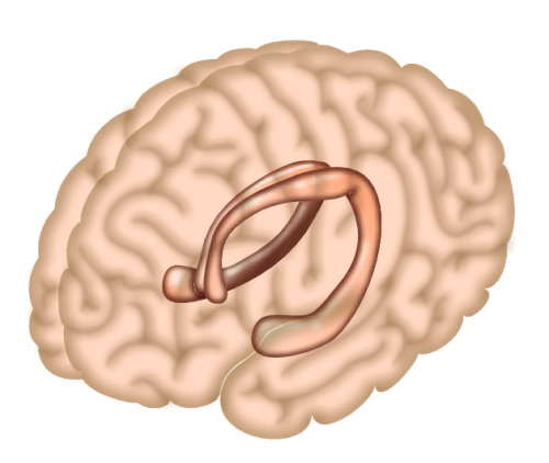
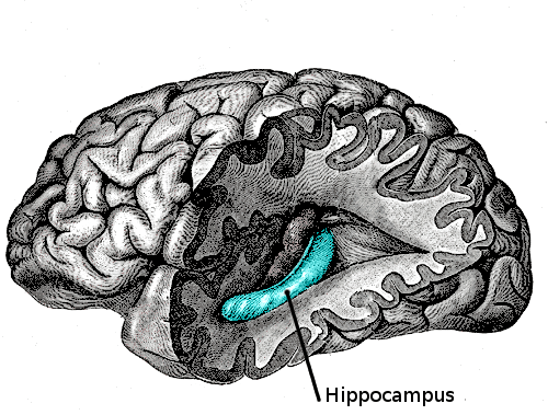
Epilepsy patients were chosen for the study because they often have to undergo brain surgery. Mesgarani, with the help of Ashesh Dinesh Mehta, a neurosurgeon at Northwell Health Physician Partners Neuroscience Institute and a co-author of the new study, recruited five volunteers for the experiment. The team used invasive electrocorticography (ECoG) to measure neural activity as the patients listened to continuous speech sounds. The patients listened, for example, to speakers reciting digits from zero to nine. Their brain patterns were then fed into the AI-enabled vocoder, resulting in the synthesized speech.

The results were very robotic-sounding, but fairly intelligible. In tests, listeners could correctly identify spoken digits around 75 percent of the time. They could even tell if the speaker was male or female. Not bad, and a result that even came as “a surprise” to Mesgaran, as he told Gizmodo in an email.

Recordings of the speech synthesizer can be found here (the researchers tested various techniques, but the best result came from the combination of deep neural networks with the vocoder).

The use of a voice synthesizer in this context, as opposed to a system that can match and recite pre-recorded words, was important to Mesgarani. As he explained to Gizmodo, there’s more to speech than just putting the right words together.

“Since the goal of this work is to restore speech communication in those who have lost the ability to talk, we aimed to learn the direct mapping from the brain signal to the speech sound itself,” he told Gizmodo. “It is possible to also decode phonemes [distinct units of sound] or words, however, speech has a lot more information than just the content—such as the speaker [with their distinct voice and style], intonation, emotional tone, and so on. Therefore, our goal in this particular paper has been to recover the sound itself.”

Looking ahead, Mesgarani would like to synthesize more complicated words and sentences, and collect brain signals of people who are simply thinking or imagining the act of speaking.

Jackson was impressed with the new study, but he said it’s still not clear if this approach will apply directly to brain-computer interfaces.

“In the paper, the decoded signals reflect actual words heard by the brain. To be useful, a communication device would have to decode words that are imagined by the user,” Jackson told Gizmodo. “Although there is often some overlap between brain areas involved in hearing, speaking, and imagining speech, we don’t yet know exactly how similar the associated brain signals will be.”

William Tatum, a neurologist at the Mayo Clinic who was also not involved in the new study, said the research is important in that it’s the first to use artificial intelligence to reconstruct speech from the brain waves involved in generating known acoustic stimuli. The significance is notable, “because it advances application of deep learning in the next generation of better designed speech-producing systems,” he told Gizmodo. That said, he felt the sample size of participants was too small, and that the use of data extracted directly from the human brain during surgery is not ideal.

Another limitation of the study is that the neural networks, in order for them do more than just reproduce words from zero to nine, would have to be trained on a large number of brain signals from each participant. The system is patient-specific, as we all produce different brain patterns when we listen to speech.

“It will be interesting in future to see how well decoders trained for one person generalize to other individuals,” said Jackson. “It’s a bit like early speech recognition systems that needed to be individually trained by the user, as opposed to today’s technology, such as Siri and Alexa, that can make sense of anyone’s voice, again using neural networks. Only time will tell whether these technologies could one day do the same for brain signals.”

No doubt, there’s still lots of work to do. But the new paper is an encouraging step toward the achievement of implantable speech neuroprosthetics.

https://gizmodo.com/neuroscientists-translate-brain-waves-into-recognizable-1832155006

https://www.nature.com/articles/s41598-018-37359-z

Protein Changes Detected in Blood Years Before Alzheimer’s Onset

Levels of a protein called neurofilament light chain increase in the blood and spinal fluid of some Alzheimer’s patients 16 years before they develop symptoms, according to a study published January 21 in Nature Medicine.

The results suggest that neurofilament light chain (NfL), which is part of the cytoskeleton of neurons and has previously been tied to brain damage in mice, could serve as a biomarker to noninvasively track the progression of the disease. “This is something that would be easy to incorporate into a screening test in a neurology clinic,” coauthor Brian Gordon, an assistant professor of radiology at Washington University, says in a press release.

Gordon and his colleagues measured NfL in nearly 250 people carrying an Alzheimer’s-risk allele and more than 160 of their relatives who did not carry the variant. They found that those at risk of developing the disease had higher levels of the protein early on, and that NfL levels in both the blood and spinal fluid were on the rise well before the patients began to show signs of neurodegeneration, more than 16 years before disease onset.

Examining a subset of the patients more closely, the team saw that the rate of increase in NfL correlated with the shrinkage of a brain region called the precuneus, and patients whose NfL levels were rising rapidly tested worse on cognitive tests. “It is not necessarily the absolute levels which tell you your neurodegeneration is ongoing, it is the rate of change,” coauthor Mathias Jucker, a professor of cellular neurology at the German Center for Neurodegenerative Diseases in Tübingen, tells The Guardian.

The Alzheimer’s-linked mutation carried by patients examined in this study only affects about 1 percent of people who get the neurodegenerative disease, so the approach must be validated in a broader patient population, James Pickett, the head of research at the Alzheimer’s Society, tells The Guardian.

“We validated it in people with Alzheimer’s disease because we know their brains undergo lots of neurodegeneration, but this marker isn’t specific for Alzheimer’s,” Gordon says in the release. “I could see this being used in the clinic in a few years to identify signs of brain damage in individual patients.”

Meanwhile, a research team at Seoul National University in South Korea described another potential blood test for Alzheimer’s, focusing on the tau and amyloid proteins known to be associated with the disease. According to their study published today in Brain, blood levels of tau and amyloid correlate with how much tau has accumulated in the brain, as well as other markers of neurodegeneration such as hippocampal volume. “These results indicate that combination of plasma tau and amyloid-β1–42 levels might be potential biomarkers for predicting brain tau pathology and neurodegeneration,” the researchers write in their report.

https://www.the-scientist.com/news-opinion/protein-changes-detected-in-blood-years-before-alzheimers-onset-65347

Lewis Judd, psychiatrist who probed the science of the brain, dies at 88


Dr. Lewis L. Judd led the National Institute of Mental Health from 1988 to 1990. (National Library of Medicine)

By Emily Langer

Lewis L. Judd, a nationally known psychiatrist who helped turn the focus of his profession from psychoanalysis to neuroscience, an approach that sought to destigmatize mental illness by treating it as cancer, heart disease or any other medical problem, died Dec. 16 in La Jolla, Calif. He was 88.

The cause was cardiac arrest, said his wife, Pat Judd.

For decades, psychiatrists were schooled in the theories of Sigmund Freud, the founder of psychoanalysis, who posited that mental disturbances could be treated through dialogue with a therapist. Practitioners sought to interpret their patients’ dreams, giving little attention to the physical functioning of the brain or the chemicals that regulate it.

Dr. Judd agreed, he once told the Associated Press, that a physician must look at patients as a “whole individual,” with all their “worries, concerns, aspirations and needs,” and not resort to simply “popping a pill in their mouth.” But he found the long-prevailing psychoanalytic approach too limiting to explain or treat afflictions such as depression, bipolar disorder, severe anxiety and schizophrenia — “these serious mental disorders that have defied our understanding for centuries,” he once told the Chicago Tribune.

Instead, he advocated a biological approach, starting at the molecular level of the brain. As director of the National Institute of Mental Health in Bethesda, Md. — a post he held from 1988 to 1990, during a hiatus from his decades-long chairmanship of the psychiatry department at the University of California at San Diego — he helped launch a federal research initiative known as the “Decade of the Brain.”

“He was obsessed with educating the public and the profession . . . that mental illnesses were biological illnesses, that schizophrenia and depression were diseases of the brain,” Alan I. Leshner, Dr. Judd’s deputy at NIMH and later chief executive of the American Association for the Advancement of Science, said in an interview. “At the time, that was a heretical thought.”

Today, the biological component of many mental illnesses is widely accepted. When Dr. Judd led NIMH, it was not; he once cited a survey in which 71 percent of respondents said mental illness was a result of personal weakness and a third attributed it to sinful behavior. Poor parenting was another common alleged culprit.

Dr. Judd argued that the biological approach to psychiatry held the promise not only of deepening understanding of the body’s most complex organ but of improving lives: If mental disorders could be shown to be a result of brain chemistry or of physical dysfunction, patients might feel less stigmatized and therefore more willing to seek treatment.

“We look at the homeless and feel that if they only got their act together, they could lift themselves up,” Dr. Judd told the Los Angeles Times in 1988, discussing the prevalence of mental illness among homeless people. “We would never believe that about someone who has cancer or some other physical disease.”

As head of NIMH, which is an arm of the National Institutes of Health and the chief federal agency for research on mental illness, Dr. Judd oversaw more than $500 million in research money. He described the Decade of the Brain — a designation conferred by Congress and President George H.W. Bush — as a “research plan designed to bring a precise and detailed understanding of all the elements of brain function within our own lifetimes.”

During his tenure at NIMH, scientists for the first time successfully grew brain tissue in a laboratory. Dr. Judd was among those scientists who touted the potential of medical imaging, such as MRIs and PET scans, to reveal the inner workings of the brain and the potential causes of diseases such as schizophrenia.

Almost 30 years after the Decade of the Brain began, much about the organ remains elusive. Leshner credited the initiative with helping bring attention to the importance of brain research as well as inspiring the Brain Initiative, a public-private research effort advanced by the Obama administration.

“The brain is really the last frontier for scientists,” Dr. Judd said.

Lewis Lund Judd was born in Los Angeles on Feb. 10, 1930. His father was an obstetrician-gynecologist, and his mother was a homemaker. Dr. Judd’s brother, Howard Judd, also became an OB/GYN and a noted researcher in women’s health at the University of California at Los Angeles.

Dr. Judd received a bachelor’s degree in psychology from the University of Utah in 1954 and a medical degree from UCLA in 1958. In the early years of his career, he served in the Air Force as a base psychiatrist.

He joined UC-San Diego in 1970 and became department chairman in 1977, helping grow his faculty into one of the most respected the country. He stepped down as chairman in 2013 and retired in 2015.

Dr. Judd’s first marriage, to Anne Nealy, ended in divorce. Survivors include his wife of 45 years, the former Patricia Hoffman, who is also a psychiatry professor at UC-San Diego, of La Jolla; three daughters from his first marriage, Allison Fee of Whidbey Island, Wash., Catherine Judd of Miami and Stephanie Judd of Chevy Chase, Md.; and four grandchildren.

Ever exploring the outer reaches of his field, Dr. Judd participated in a dialogue with the Dalai Lama in 1989 about life and the mind.

“Our model of mental health is mostly defined in terms of the absence of mental illness,” Dr. Judd told the New York Times, reflecting on the Tibetan Buddhist leader’s discussion of wisdom and compassion. “They may have more positive ones that might be worth our study.”

https://www.washingtonpost.com/local/obituaries/lewis-judd-psychiatrist-who-probed-the-science-of-the-brain-dies-at-88/2019/01/11/271e1f48-1549-11e9-b6ad-9cfd62dbb0a8_story.html?noredirect=on&utm_term=.18ed788ae8b3

Monogamy may have a telltale signature of gene activity


Patterns of gene expression unite the prairie vole Microtus ochrogaster with other monogamous species, including certain frogs, fish, and birds. YVA MOMATIUK AND JOHN EASTCOTT/MINDEN PICTURES

By Kelly Servick

In the animal world, monogamy has some clear perks. Living in pairs can give animals some stability and certainty in the constant struggle to reproduce and protect their young—which may be why it has evolved independently in various species. Now, an analysis of gene activity within the brains of frogs, rodents, fish, and birds suggests there may be a pattern common to monogamous creatures. Despite very different brain structures and evolutionary histories, these animals all seem to have developed monogamy by turning on and off some of the same sets of genes.

“It is quite surprising,” says Harvard University evolutionary biologist Hopi Hoekstra, who was not involved in the new work. “It suggests that there’s a sort of genomic strategy to becoming monogamous that evolution has repeatedly tapped into.”

Evolutionary biologists have proposed various benefits to so-called social monogamy, where mates pair up for at least a breeding season to care for their young and defend their territory. When potential mates are scarce or widely dispersed, for example, forming a single-pair bond can ensure they get to keep reproducing.

Neuroscientist Hans Hofmann and evolutionary biologist Rebecca Young at the University of Texas in Austin wanted to explore how the regulation of genes in the brain might have changed when a nonmonogamous species evolved to become monogamous. For example, the complex set of genes that underlie the ability to tolerate the presence of another member of one’s species presumably exists in nonmonogamous animals, but might be activated in different patterns to allow prolonged partnerships in monogamous ones.

“We wanted to be bold—and maybe a little bit crazy” in the new experiment, Hofmann says. Instead of doing a relatively straightforward genetic comparison between closely related species on either side of the monogamy divide, he and colleagues wanted to hunt down a gene activity signature associated with monogamy in males across a wide variety of species—frogs, mice, voles, birds, and fish. So in each of these groups, they selected two species, one monogamous and one nonmonogamous.

Rounding up the brains of those animals took an international team and years of effort. Hostile regional authorities and a complicated permitting system confronted the team in Romania as they tried to capture two types of a native songbird. Hofmann donned scuba gear and plunged into Africa’s Lake Tanganyika to chase finger-length cichlid fish into nets. Delicately debraining them while aboard a rocking boat, he says, was a struggle.

Back the lab, the researchers then grouped roughly comparable genes across all 10 species based on similarities in their sequences. For each of these cross-species gene groups, they measured activity based on how much the cells in the brain transcribed the DNA’s proteinmaking instructions into strands of RNA.

Among the monogamous animals, a pattern emerged. The researchers found certain sets of genes were more likely to be “turned up” or “turned down” in those creatures than in the nonmonogamous species. And they ruled out other reasons why these monogamous animals might have similar gene expression patterns, including similar environments or close evolutionary relationships.

Among the genes with increased activity in monogamous species were those involved in neural development, signaling between cells, learning, and memory, the researchers report online today in the Proceedings of the National Academy of Sciences. They speculate that genes that make the brain more adaptable—and better able to remember—might also help animals recognize their mates and find their presence rewarding.

It makes sense that genes involved in brain development and function would underlie a complex behavior like monogamy, says behavioral neuroscientist Claudio Mello of Oregon Health & Science University in Portland. But because the researchers didn’t dissect out specific brain regions and analyze their RNA production independently, they can’t describe the finely tuned patterns of gene expression in areas that are key to reproductive behavior. “It seems to me unlikely that by themselves these genes will be able to ‘explain’ this behavior,” he says.

“The fact that they got any common genes at all is interesting,” adds Lisa Stubbs, a developmental geneticist at the University of Illinois in Urbana. “It is a superb data set and an expert analysis,” she says, “[but] the authors have not actually uncovered many important biological insights into monogamy.”

The study did turn up a curious outlier. Some of the genes with decreased expression in most of the monogamous species showed increased expression in one of them—the poison dart frog Ranitomeya imitator. Young notes that in this species’s evolutionary history, fathers cared for the young before cooperative parenting evolved. As a result, these frogs may have had a different evolutionary starting point than other animals in the study, later tapping into different genes to become monogamous.

Hoekstra, who has studied the genetics of monogamy in mice, sees “a lot of exciting next steps.” There are likely mutations in other regions of DNA that regulate the expression of the genes this study identified. But it will take more work to show a causal relationship between any particular genetic sequence and monogamous behavior.

People also often opt for monogamy, albeit for a complicated set of social and cultural reasons. So, do we share the gene activity signature common to monogamous birds, fish, and frogs? “We don’t know that,” says Hofmann, but “we certainly would speculate that the kind of gene expression patterns … might [show up] in humans as well.”

http://www.sciencemag.org/news/2019/01/monogamy-may-have-telltale-signature-gene-activity

Chronic Bullying Could Actually Reshape The Brains of Teens

by Carly Cassella

Sticks and stones may break your bones, but name-calling could actually change the structure of your brain.

A new study has found that persistent bullying in high school is not just psychologically traumatising, it could also cause real and lasting damage to the developing brain.

The findings are drawn from a long-term study on teenage brain development and mental health, which collected brain scans and mental health questionnaires from European teenagers between the ages of 14 and 19.

Following 682 young people in England, Ireland, France and Germany, the researchers tallied 36 in total who reported experiencing chronic bullying during these years.

When the researchers compared the bullied participants to those who had experienced less intense bullying, they noticed that their brains looked different.

Across the length of the study, in certain regions, the brains of the bullied participants appeared to have actually shrunk in size.

In particular, the pattern of shrinking was observed in two parts of the brain called the putamen and the caudate, a change oddly reminiscent of adults who have experienced early life stress, such as childhood maltreatment.

Sure enough, the researchers found that they could partly explain these changes using the relationship between extreme bullying and higher levels of general anxiety at age 19. And this was true even when controlling for other types of stress and co-morbid depressive symptoms.

The connection is further supported by previous functional MRI studies that found differences in the connectivity and activation of the caudate and putamen activation in those with anxiety.

“Although not classically considered relevant to anxiety, the importance of structural changes in the putamen and caudate to the development of anxiety most likely lies in their contribution to related behaviours such as reward sensitivity, motivation, conditioning, attention, and emotional processing,” explains lead author Erin Burke Quinlan from King’s College London.

In other words, the authors think all of this shrinking could be a mark of mental illness, or at least help explain why these 19-year-olds are experiencing such unusually high anxiety.

But while numerous past studies have already linked childhood and adolescent bullying to mental illness, this is the very first study to show that unrelenting victimisation could impact a teenager’s mental health by actually reshaping their brain.

The results are cause for worry. During adolescence, a young person’s brain is absolutely exploding with growth, expanding at an incredible place.

And even though it’s normal for the brain to prune back some of this overabundance, in the brains of those who experienced chronic bullying, the whole pruning process appears to have spiralled out of control.

The teenage years are an extremely important and formative period in a person’s life, and these sorts of significant changes do not bode well. The authors suspect that as these children age, they might even begin to experience greater shrinkage in the brain.

But an even longer long-term study will need to be done if we want to verify that hunch. In the meantime, the authors are recommending that every effort be made to limit bullying before it can cause damage to a teenager’s brain and their mental health.

This study has been published in Molecular Psychiatry.

https://www.sciencealert.com/chronic-bullying-could-actually-reshape-the-brains-of-teens

Bravery-associated brain cells identified in the hippocampus

hippocampus
The hippocampus is a region of the brain largely responsible for memory formation.

Why can some people comfortably walk between skyscrapers on a high-wire or fearlessly raft Niagara Falls in a wooden barrel, whereas others freeze at the mere thought of climbing off escalators in a shopping mall? In a new study, scientists have found that a certain type of cell in the hippocampus plays a key role.

People differ when it comes to trying dangerous or exhilarating activities. Even siblings can show dramatic differences in risk-taking behaviour. The neural mechanisms that drive risk-taking behaviour are largely unknown. However, scientists from the Department of Neuroscience of Uppsala University in Sweden and the Brain Institute of the Federal University of Rio Grande do Norte in Brazil have found that some cells in the hippocampus play a key role in risk-taking behaviour and anxiety.

In an article published in the journal Nature Communications, the authors show that neurons known as OLM cells, when stimulated, produce a brain rhythm that is present when animals feel safe in a threatening environment (for example, when they are hiding from a predator but aware of the predator’s proximity). The study, produced by Drs. Sanja Mikulovic, Ernesto Restrepo, Klas Kullander and Richardson Leao, among others, showed that anxiety and risk-taking behaviour can be controlled by the manipulation of OLM cells. To find a pathway that quickly and robustly modulates risk-taking behaviour is very important for treatment of pathological anxiety, since reduced risk-taking behaviour is a trait in people with high anxiety levels.

Adaptive (or normal) anxiety is essential for survival because it protects us from harm. Unfortunately, in a large number of people, anxiety can be dysfunctional and severely interfere with daily life. In these cases, doctors often rely on antidepressants to help patients recover from the dysfunctional state. However, these drugs act in the entire brain and not only in the areas where it is needed, and may therefore cause severe side-effects. Thus, drugs that affect a single brain region or a very specific group of cells may be a major breakthrough in treating anxiety and associated disorders like depression. Another interesting finding in the study is that OLM cells can be controlled by pharmacological agents. In the past, the same group of scientists found that OLM cells were the gatekeepers of memories in the hippocampus, and that these cells were very sensitive to nicotine.

“This finding may explain why people binge-smoke when they are anxious,” says Dr. Richardson Leao, researcher at the Brain Institute of the Federal University of Rio Grande do Norte.

The participation of the hippocampus in emotions is much less studied than its role in memory and cognition. In 2014, for example, the Nobel prize was awarded for the discovery of “place cells” that represent a biological GPS and underlie the memories of where we are located in our surroundings. In the past decade, scientists have also started to appreciate the role of the hippocampus in regulating emotions.

“It is fascinating how different regions of the same brain structure control distinct behaviours and how they interact with each other. Identifying specific circuits that underlie either cognitive or emotional processes is crucial for the general understanding of brain function and for more specific drug development to treat disorders,” says Dr. Sanja Mikulovic, Uppsala University.

The discovery of these neurons and their role in anxiety and risk-taking may open a path for the development of highly efficient anxiolytics and antidepressants without common side-effects, such as apathy.

Sanja Mikulovic et al, Ventral hippocampal OLM cells control type 2 theta oscillations and response to predator odor, Nature Communications (2018). DOI: 10.1038/s41467-018-05907-w

https://medicalxpress.com/news/2018-09-bravery-associated-cells-hippocampus.html

Scientists have found a previously unknown mechanism in which the protein tau, which is implicated in Alzheimer’s disease, damages brain cells by interfering with their internal communications.

The discovery sheds new light on the origins of this most common cause of dementia, a hallmark of which is the buildup of tangled tau protein filaments in the brain.

The finding could also lead to new treatments for Alzheimer’s and other diseases that progressively destroy brain tissue, conclude the researchers in a paper about their work that now features in the journal Neuron.

Scientists from Massachusetts General Hospital (MGH) in Charlestown and the Johns Hopkins School of Medicine in Baltimore, MD, led the study, which set out to investigate how tau protein might contribute to brain cell damage.

Alzheimer’s disease does not go away and gets worse over time. It is the sixth most common cause of death in adults in the United States, where an estimated 5.7 million people have the disease.

Exact causes of Alzheimer’s still unknown

Exactly what causes Alzheimer’s and other forms of dementia is still a mystery to science. Evidence suggests that a combination of environment, genes, and lifestyle is involved, with different factors having different amounts of influence in different people.

Most cases of Alzheimer’s do not show symptoms until people are in their 60s and older. The risk of getting the disease rises rapidly with age after this.

Brain studies of people with the disease — together with postmortem analyses of brain tissue — have revealed much about how Alzheimer’s changes and harms the brain.

“Age-related changes” include: inflammation; shrinkage in some brain regions; creation of unstable, short-lived molecules known as free radicals; and disruption of cellular energy production.

The brain of a person with Alzheimer’s disease also has two distinguishing features: plaques of amyloid protein that form between cells, and tangles of tau protein that form inside cells. The recent study concerns the latter.

Changes to tau behavior

Brain cells, or neurons, have internal structures known as microtubules that support the cell and its function. They are highly active cell components that help carry substances from the body of the cell out to the parts that connect it to other cells.

In healthy brain cells, tau protein normally “binds to and stabilizes” the microtubules. Tau behaves differently, however, in Alzheimer’s disease.

Changes in brain chemistry make tau protein molecules come away from the microtubules and stick to each other instead.

Eventually, the detached tau molecules form long filaments, or neurofibrillary tangles, that disrupt the brain cell’s ability to communicate with other cells.

The new study introduces the possibility that, in Alzheimer’s disease, tau disrupts yet another mechanism that involves communication between the nucleus of the brain cell and its body.

Communication with cell nucleus

The cell nucleus communicates with the rest of the cell using structures called nuclear pores, which comprise more than 400 different proteins and control the movement of molecules.

Studies on the causes of amyotrophic lateral sclerosis, frontotemporal, and other types of dementia have suggested that flaws in these nuclear pores are involved somehow.

The recent study reveals that animal and human cells with Alzheimer’s disease have faulty nuclear pores, and that the fault is linked to tau accumulation in the brain cell.

“Under disease conditions,” explains co-senior study author Bradley T. Hyman, the director of the Alzheimer’s Unit at MGH, “it appears that tau interacts with the nuclear pore and changes its properties.”

He and his colleagues discovered that the presence of tau disrupts the orderly structure of nuclear pores containing the major structural protein Nup98. In Alzheimer’s disease cells, there were fewer of these pores and those that were there tended to be stuck to each other.

‘Mislocalized’ Nup98
They also observed another curious change involving Nup98 inside Alzheimer’s disease brain cells. In cells with aggregated tau, the Nup98 was “mislocalized” instead of staying in the nuclear pore.

They revealed that this feature was more exaggerated in brain tissue of people who had died with more extreme forms of Alzheimer’s disease.

Finally, when they added human tau to living cultures of rodent brain cells, the researchers found that it caused mislocalization of Nup98 in the cell body and disrupted the transport of molecules into the nucleus.

This was evidence of a “functional link” between the presence of tau protein and damage to the nuclear transport mechanism.

The authors note, however, that it is not clear whether the Nup98-tau interaction uncovered in the study just occurs because of disease or whether it is a normal mechanism that behaves in an extreme fashion under disease conditions.

They conclude:

“Taken together, our data provide an unconventional mechanism for tau-induced neurodegeneration.”

https://www.medicalnewstoday.com/articles/322991.php

Cleveland Clinic Researchers Discover Novel Subtype of Multiple Sclerosis


Reprinted from The Lancet Neurology, http://dx.doi.org/10.1016/S1474-4422(18)30245-X, Trapp et al, Cortical neuronal densities and cerebral white matter demyelination in multiple sclerosis: a retrospective study, Copyright (2018), with permission from Elsevier


Bruce Trapp, Ph.D., chair of Cleveland Clinic’s Lerner Research Institute Department of Neurosciences

Cleveland Clinic researchers have discovered a new subtype of multiple sclerosis (MS), providing a better understanding of the individualized nature of the disease.

MS has long been characterized as a disease of the brain’s white matter, where immune cells destroy myelin – the fatty protective covering on nerve cells. The destruction of myelin (called demyelination) was believed to be responsible for nerve cell (neuron) death that leads to irreversible disability in patients with MS.

However, in the new findings, a research team led by Bruce Trapp, Ph.D., identified for the first time a subtype of the disease that features neuronal loss but no demyelination of the brain’s white matter. The findings, published in Lancet Neurology, could potentially lead to more personalized diagnosis and treatments.

The team’s findings support the concept that neurodegeneration and demyelination can occur independently in MS and underscore the need for more sensitive MRI imaging techniques for evaluating brain pathology in real time and monitoring treatment response in patients with the disease. This new subtype of MS, called myelocortical MS (MCMS), was indistinguishable from traditional MS on MRI. The researchers observed that in MCMS, part of the neurons become swollen and look like typical MS lesions indicative of white matter myelin loss on MRI. The disease was only diagnosed in post-mortem tissues.

“This study opens up a new arena in MS research. It is the first to provide pathological evidence that neuronal degeneration can occur without white matter myelin loss in the brains of patients with the disease,” said Trapp, chair of Cleveland Clinic’s Lerner Research Institute Department of Neurosciences. “This information highlights the need for combination therapies to stop disability progression in MS.”

In the study of brain tissue from 100 MS patients who donated their brains after death, the researchers observed that 12 brains did not have white matter demyelination. They compared microscopic tissue characteristics from the brains and spinal cords of 12 MCMS patients, 12 traditional MS patients and also individuals without neurological disease. Although both MCMS and traditional MS patients had typical MS lesions in the spinal cord and cerebral cortex, only the latter group had MS lesions in the brain white matter.

Despite having no typical MS lesions in the white matter, MCMS brains did have reduced neuronal density and cortical thickness, which are hallmarks of brain degeneration also observed in traditional MS. Contrary to previous belief, these observations show that neuronal loss can occur independently of white matter demyelination.

“The importance of this research is two-fold. The identification of this new MS subtype highlights the need to develop more sensitive strategies for properly diagnosing and understanding the pathology of MCMS,” said Daniel Ontaneda, M.D., clinical director of the brain donation program at Cleveland Clinic’s Mellen Center for Treatment and Research in MS. “We are hopeful these findings will lead to new tailored treatment strategies for patients living with different forms of MS.”

Dr. Trapp is internationally known for his work on mechanisms of neurodegeneration and repair in MS and has published more than 240 peer-reviewed articles and 40 book chapters. He also holds the Morris R. and Ruth V. Graham Endowed Chair in Biomedical Research. In 2017 he received the prestigious Outstanding Investigator award by the National Institute of Neurological Disorders and Stroke to examine the biology of MS and to seek treatments that could slow or reverse the disease.

Cleveland Clinic Researchers Discover Novel Subtype of Multiple Sclerosis

New Research Suggests It’s all About the Bass

When we listen to music, we often tap our feet or bob our head along to the beat – but why do we do it? New research led by Western Sydney University’s MARCS Institute suggests the reason could be related to the way our brain processes low-frequency sounds.

The study, published in PNAS, recorded the electrical activity of volunteers’ brains while they listened to rhythmic patterns played at either low or high-pitched tones. The study found that while listening, volunteer’s brain activities and the rhythmic structure of the sound became synchronized – particularly at the frequency of the beat.

Co-author of the paper, Dr Sylvie Nozaradan from the MARCS Institute, say these findings strongly suggest that the bass exploits a neurophysiological mechanism in the brain – essentially forcing it to lock onto the beat.

“There is mounting evidence supporting the hypothesis that selective synchronization of large pools of neurons of the brain to the beat frequency may support perception and movement to the musical beat”, says Dr Nozaradan.

While this research is an important step in answering the mystery of why we ‘dance to the beat of the drum’, according to co-author Dr Peter Keller from the MARCS Institute, these findings could also prove important in clinical rehabilitation.

“Music is increasingly being used in clinical rehabilitation of cognitive and motor disorders caused by brain damage and these findings, and a better understanding of the relationship between music and movement, could help develop such treatments,” says Dr Keller.

The research team – also comprising of co-authors Dr Manuel Varlet and Tomas Lenc – suggests that while this research is an important step in understanding the relationship between bass and movement, there are still many open questions about the mechanisms behind this phenomenon.

“Future research is needed to clarify what networks of brain areas are responsible for this synchronization to the beat and how it develops from early in infancy” says Dr Nozaradan.

https://www.westernsydney.edu.au/newscentre/news_centre/more_news_stories/new_research_suggests_its_all_about_the_bass

New research shows that being forgetful is a sign of unusual intelligence

By Timothy Roberts

Being able to recall memories, whether short-term or long-term is something that we all need in life. It comes in handy when we are studying at school or when we are trying to remember where we left our keys. We also tend to use our memory at work and remembering somebody’s name is certainly a good thing.

Although many of us may consider ourselves to have a good memory, we are all going to forget things from time to time. When it happens, we might feel as if we are slipping but there may be more behind it than you realize.

Imagine this scenario; you go to the grocery store to pick up 3 items and suddenly, you forget why you were there. Even worse, you may walk from one room to another and forget why you got up in the first place!

If you often struggle with these types of problems, you will be happy to learn that there is probably nothing wrong with you. In fact, a study that was done by the Neuron Journal and it has some rather good news. It says that forgetting is part of the brain process that might actually make you smarter by the time the day is over.

Professors took part in a study at the University of Toronto and they discovered that the perfect memory actually doesn’t necessarily reflect your level of intelligence.

You might even be surprised to learn that when you forget details on occasion, it can make you smarter.

Most people would go by the general thought that remembering more means that you are smarter.

According to the study, however, when you forget a detail on occasion, it’s perfectly normal. It has to do with remembering the big picture compared to remembering little details. Remembering the big picture is better for the brain and for our safety.

Our brains are perhaps more of a computer than many of us think. The hippocampus, which is the part of the brain where memories are stored, tends to filter out the unnecessary details.

In other words, it helps us to “optimize intelligent decision making by holding onto what’s important and letting go of what’s not.”

Think about it this way; is it easier to remember somebody’s face or their name? Which is the most important?

In a social setting, it is typically better to remember both but if we were part of the animal kingdom, remembering somebody as being a threat would mean our very lives. Remembering their name would be inconsequential.

The brain doesn’t automatically decide what we should remember and what we shouldn’t. It holds new memories but it sometimes overwrites old memories.

When the brain becomes cluttered with memories, they tend to conflict with each other and that can make it difficult to make important decisions.

That is why the brain tends to hold on to those big picture memories but they are becoming less important with the advent of technology.

As an example, at one time, we would have learned how to spell words but now, we just use Google if we don’t know how to spell them. We also tend to look everything up online, from how to change a showerhead to how to cook meatloaf for dinner.

If you forget everything, you may want to consider having a checkup but if you forget things on occasion, it’s perfectly okay.

The moral of the story is, the next time you forget something, just think of it as your brain doing what it was designed to do.

http://wetpaintlife.com/scientists-say-that-being-forgetful-is-actually-a-sign-you-are-unusually-intelligent/?utm_source=vn&utm_tracking=11&utm_medium=Social