Good Grip, Good Health

Measuring hand grip can help identify youths who could benefit from lifestyle changes, Baylor University researcher says. While other studies have shown that muscle weakness as measured by grip strength is a predictor of unhealthy outcomes — including cardiovascular and metabolic diseases, disability and even early mortality — this is the first to do so for adolescent health over time, a Baylor University researcher said.

“What we know about today’s kids is that because of the prevalence of obesity, they are more at risk for developing pre-diabetes and cardiovascular disease than previous generations,” said senior author Paul M. Gordon, Ph.D., professor and chair of health, human performance and recreation in Baylor’s Robbins College of Health and Human Sciences.

“This study gives multiple snapshots over time that provide more insight about grip strength and future risks for developing diabetes and cardiovascular disease,” he said. “Low grip strength could be used to predict cardiometabolic risk and to identify adolescents who would benefit from lifestyle changes to improve muscular fitness.”

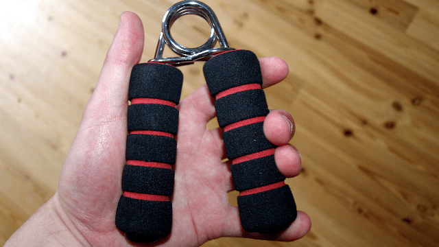
Students tracked in the study were assessed in the fall of their fourth-grade year and at the end of the fifth grade. Using the norms for grip strengths in boys and girls, researchers measured the students’ grips in their dominant and non-dominant hands with an instrument called a handgrip dynamometer.

Researchers found that initially, 27.9 percent of the boys and 20.1 percent of the girls were classified as weak. Over the course of the study, boys and girls with weak grips were more than three times as likely to decline in health or maintain poor health as those who were strong.

Researchers also screened for and analyzed other metabolic risk factor indicators, including physical activity, cardiorespiratory fitness, body composition (the proportion of fat and fat-free mass), blood pressure, family history, fasting blood lipids and glucose levels.

“Even after taking into account other factors like cardiorespiratory fitness, physical activity and lean body mass, we continue to see an independent association between grip strength and both cardiometabolic health maintenance and health improvements,” Gordon said.

While much emphasis has been placed on the benefits of a nutritious diet and aerobic activity, this study suggests that greater emphasis needs to be placed on improving and maintaining muscular strength during adolescence.

If someone with a strong grip develops an even stronger grip, “we don’t necessarily see a drastic improvement in that individual’s health,” Gordon noted. “It’s the low strength that puts you at risk.

“Given that grip strength is a simple indicator for all-cause death, cardiovascular death and cardiovascular disease in adults, future research is certainly warranted to better understand how weakness during childhood tracks into and throughout adulthood,” he said. “Testing grip strength is simple, non-invasive and can easily be done in a health care professional’s office. It has value for adults and children.”

An estimated 17.2 percent of U.S. children and adolescents aged 2 to 19 years are obese and another 16.2 percent are overweight, according to the National Center for Health Statistics. Excess weight carries a greater lifetime risk of diabetes and premature heart disease. While the World Health Organization and the U.S. Department of Health and Human Services recommend that youths perform at least 60 minutes of moderate to vigorous physical activity daily — including vigorous activity at least three days a week — fewer than a quarter of U.S. children do so, according to a report by the nonprofit National Physical Activity Plan Alliance.

Reference: Peterson, M. D., Gordon, P. M., Smeding, S., & Visich, P. (2018). Grip Strength Is Associated with Longitudinal Health Maintenance and Improvement in Adolescents. The Journal of Pediatrics. https://doi.org/10.1016/j.jpeds.2018.07.020

https://www.technologynetworks.com/proteomics/news/good-grip-good-health-307585?utm_campaign=Newsletter_TN_BreakingScienceNews&utm_source=hs_email&utm_medium=email&utm_content=65175478&_hsenc=p2ANqtz-887HvGM-iiCBXuYuQ-OC_o-JSzmK_HOnCxRga2M8gAVZDF4SejOFma20Bb04GZ9F3uhKOjczHVcuNF-Htnak8rN-Hfow&_hsmi=65175479

New research shows that degenerative eye diseases are associated with risk of developing Alzheimer’s disease


Age-related macular degeneration, diabetic retinopathy and glaucoma were all associated with a higher risk of developing Alzheimer’s disease in a new study.

by Rich Haridy

A new study has found an interesting correlation between several degenerative eye diseases and the onset of Alzheimer’s disease. No mechanism explaining the connection has been proposed at this stage but it is thought these eye conditions may help physicians identify patients at risk of developing Alzheimer’s at a stage before major symptoms appear.

The five-year study followed almost 4,000 patients over the age of 65, all without clinically diagnosed Alzheimer’s disease at the time of enrolment. After five years, 792 subjects were officially diagnosed with Alzheimer’s. The study found that those subjects with age-related macular degeneration, diabetic retinopathy or glaucoma, were 40 to 50 percent more likely to develop Alzheimer’s compared to patients without those specific conditions. No correlation between cataracts and an increased risk of Alzheimer’s were found.

“We don’t mean people with these eye conditions will get Alzheimer’s disease,” cautions Cecilia Lee, lead researcher on the study. “The main message from this study is that ophthalmologists should be more aware of the risks of developing dementia for people with these eye conditions and primary care doctors seeing patients with these eye conditions might be more careful on checking on possible dementia or memory loss.”

The researchers are clear that there are no definable causal connections between these eye conditions and Alzheimer’s at this stage, but the study does highlight the potential of using the eye as a way to better understand what is going on in the brain. Intriguingly, this isn’t the first bit of research that has found correlations between signs detected in the eye and the onset of Alzheimer’s disease.

Last year, a team from Cedars-Sinai Medical Center revealed that the same type of amyloid protein deposits found in the brain, and hypothesized as a major pathogenic cause of Alzheimer’s, can also be detected on the retina. That research suggested a possible investigational eye scan could become an effective early screening device for the disease.

While this new study does not at all cross over with last year’s research, and there is no implication that amyloid proteins play a part in these degenerative eye diseases, it does add to a fascinating growing body of work that highlights the eye’s role in helping offer a deeper insight into the cognitive health of our brain.

The research was published in the journal Alzheimer’s & Dementia.

https://newatlas.com/eye-disease-alzheimers-connection/55823/

pH imbalance in brain cells may contribute to Alzheimer’s disease


Illustration of how pH imbalance inside endosomes may contribute to Alzheimer’s disease

Johns Hopkins Medicine scientists say they have found new evidence in lab-grown mouse brain cells, called astrocytes, that one root of Alzheimer’s disease may be a simple imbalance in acid-alkaline—or pH—chemistry inside endosomes, the nutrient and chemical cargo shuttles in cells.

Astrocytes work to clear so-called amyloid beta proteins from the spaces between neurons, but decades of evidence has shown that if the clearing process goes awry, amyloid proteins pile up around neurons, leading to the characteristic amyloid plaques and nerve cell degeneration that are the hallmarks of memory-destroying Alzheimer’s disease.

The new study, described online June 26 in Proceedings of the National Academy of Sciences, also reports that the scientists gave drugs called histone deacetylase (HDAC) inhibitors to pH-imbalanced mice cells engineered with a common Alzheimer’s gene variant. The experiment successfully reversed the pH problem and improved the capacity for amyloid beta clearance.

HDAC inhibitors are approved by the U.S. Food and Drug Administration for use in people with certain types of blood cancers, but not in people with Alzheimer’s. They cautioned that most HDAC inhibitors cannot cross the blood-brain barrier, a significant challenge to the direct use of the drugs for brain disorders. The scientists say they are planning additional experiments to see if HDAC inhibitors have a similar effect in lab-grown astrocytes from Alzheimer’s patients, and that there is the potential to design HDAC inhibitors that can cross the barrier.

However, the scientists caution that even before those experiments can happen, far more research is needed to verify and explain the precise relationship between amyloid proteins and Alzheimer’s disease, which affects an estimated 50 million people worldwide. To date, there is no cure and no drugs that can predictably or demonstrably prevent or reverse Alzheimer’s disease symptoms.

“By the time Alzheimer’s disease is diagnosed, most of the neurological damage is done, and it’s likely too late to reverse the disease’s progression,” says Rajini Rao, Ph.D., professor of physiology at the Johns Hopkins University School of Medicine. “That’s why we need to focus on the earliest pathological symptoms or markers of Alzheimer’s disease, and we know that the biology and chemistry of endosomes is an important factor long before cognitive decline sets in.”

Nearly 20 years ago, scientists at Johns Hopkins and New York University discovered that endosomes, circular compartments that ferry cargo within cells, are larger and far more abundant in brain cells of people destined to develop Alzheimer’s disease. This hinted at an underlying problem with endosomes that could lead to an accumulation of amyloid protein in spaces around neurons, says Rao.

To shuttle their cargo from place to place, endosomes use chaperones—proteins that bind to specific cargo and bring them back and forth from the cell’s surface. Whether and how well this binding occurs depends on the proper pH level inside the endosome, a delicate balance of acidity and alkalinity, or acid and base, that makes endosomes float to the surface and slip back down into the cell.

Embedded in the endosome membrane are proteins that shuttle charged hydrogen atoms, known as protons, in and out of endosomes. The amount of protons inside the endosome determines its pH.

When fluids in the endosome become too acidic, the cargo is trapped within the endosome deep inside the cell. When the endosome contents are more alkaline, the cargo lingers at the cell’s surface for too long.

To help determine whether such pH imbalances occur in Alzheimer’s disease, Johns Hopkins graduate student Hari Prasad scoured scientific studies of Alzheimer’s disease looking for genes that were dialed down in diseased brains compared with normal ones. Comparing a dataset of 15 brains of Alzheimer’s disease patients with 12 normal ones, he found that 10 of the 100 most frequently down-regulated genes were related to the proton flow in the cell.

In another set of brain tissue samples from 96 people with Alzheimer’s disease and 82 without it, gene expression of the proton shuttle in endosomes, known as NHE6, was approximately 50 percent lower in people with Alzheimer’s disease compared with those with normal brains. In cells grown from people with Alzheimer’s disease and in mouse astrocytes engineered to carry a human Alzheimer’s disease gene variant, the amount of NHE6 was about half the amount found in normal cells.

To measure the pH balance within endosomes without breaking open the astrocyte, Prasad and Rao used pH sensitive probes that are absorbed by endosomes and emit light based on pH levels. They found that mouse cell lines containing the Alzheimer’s disease gene variant had more acidic endosomes (average of 5.37 pH) than cell lines without the gene variant (average of 6.21 pH).

“Without properly functioning NHE6, endosomes become too acidic and linger inside astrocytes, avoiding their duties to clear amyloid beta proteins,” says Rao.

While it’s likely that changes in NHE6 happen over time in people who develop sporadic Alzheimer’s disease, people who have inherited mutations in NHE6 develop what’s known as Christianson syndrome in infancy and have rapid brain degeneration.

Prasad and Rao also found that a protein called LRP1, which picks up amyloid beta proteins outside the astrocyte and delivers them to endosomes, was half as abundant on the surface of lab grown mouse astrocytes engineered with a human gene variant called APOE4, commonly linked to Alzheimer’s disease.

Looking for ways to restore the function of NHE6, Prasad searched databases of yeast studies to find that HDAC inhibitors tend to increase expression of the NHE6 gene in yeast. This gene is very similar across species, including flies, mice and humans.

Prasad and Rao tested nine types of HDAC inhibitors on cell cultures of mouse astrocytes engineered with the APOE4 gene variant. Broad-spectrum HDAC inhibitors increased NHE6 expression to levels associated with mouse astrocytes that did not have the Alzheimer’s gene variant. They also found that HDAC inhibitors corrected the pH imbalance inside endosomes and restored LRP1 to the astrocyte surface, resulting in efficient clearance of amyloid beta protein.

More information: Hari Prasad et al. Amyloid clearance defect in ApoE4 astrocytes is reversed by epigenetic correction of endosomal pH, Proceedings of the National Academy of Sciences (2018). DOI: 10.1073/pnas.1801612115

https://medicalxpress.com/news/2018-08-ph-imbalance-brain-cells-contribute.html

A Lack of This One Molecule Might Be The Reason Millions of People Have Depression

By Michelle Star

People who live with depression have low blood levels of a specific molecule, new medical research has revealed. It’s called acetyl-L-carnitine, and those with particularly severe, treatment-resistant or childhood onset depression were found to have the lowest levels.

Naturally produced by the body, acetyl-L-carnitine plays a crucial role in metabolising fat and the production of energy. It’s also widely available as a dietary supplement – not some strange and esoteric thing.

Now researchers from multiple institutions have found a link to depression, noticing a clear correlation between the condition and noticeably low levels of acetyl-L-carnitine.

In recent years, more and more evidence has been building to suggest this link. Since at least 1991, medical researchers have been aware of acetyl-L-carnitine’s potential to treat depression, particularly in geriatric and comorbid patients, with the substance showing greater efficacy than a placebo.

More recently, Carla Nasca of the Rockefeller University led a study on rodents, which found that acetyl-L-carnitine had a fast-acting antidepressant effect on rats, kicking into effect in just a few days, rather than the weeks it takes for drugs like SSRIs.

Now Nasca and colleagues have conducted a study on human patients to see if there’s a basis for a similar trial in people.

“As a clinical psychiatrist, I have treated many people with this disorder in my practice,” said Stanford University School of Medicine psychiatrist Natalie Rasgon.

“It’s the number one reason for absenteeism at work, and one of the leading causes of suicide. Worse, current pharmacological treatments are effective for only about 50 percent of the people for whom they’re prescribed. And they have numerous side effects, often decreasing long term compliance.”

The research team recruited 71 patients with a diagnosis of depression. These were men and women, aged between 20 and 70. They also recruited 45 demographically matched healthy controls.

The patients had to fill out a detailed questionnaire, undergo a clinical assessment and medical history, and give a blood sample. Of the patients with depression, 28 had moderate depression and 43 had severe depression at the time of the study.

When compared to the age- and sex-matched healthy controls, the patients with depression had substantially lower levels of acetyl-L-carnitine.

Those with the most severe depression had the lowest levels. This included patients whose depression had resisted antidepressant drugs, those with early onset, and those who had experienced childhood abuse, neglect, poverty or violence.

These patients constitute around 25-30 percent of all people suffering depression, and are the most in need of help, the researchers said.

But there are a few steps to be done before acetyl-L-carnitine supplements can be approved as a treatment. In particular, clinical trials on human patients with depression, since, as we know, results from rodent models can’t always be replicated in humans.

The researchers also don’t know the reason for the correlation, or the effect it has. The rat research suggests that acetyl-L-carnitine plays a role in the brain, preventing the excessive firing of excitatory neurons, but this will need to be explored further as well.

“We’ve identified an important new biomarker of major depression disorder,” Rasgon said.

“We didn’t test whether supplementing with that substance could actually improve patients’ symptoms. What’s the appropriate dose, frequency, duration? We need to answer many questions before proceeding with recommendations, yet. This is the first step toward developing that knowledge, which will require large-scale, carefully controlled clinical trials.”

https://www.sciencealert.com/depression-linked-to-low-blood-levels-of-acetyl-l-carnitine-human-study

Dragon’s Breath liquid nitrogen treats causing injuries

by Lynna Lai

A trendy new treat called “Dragon’s Breath” is popping up in malls across the country. But don’t get burned, a local doctor warned. The dessert tastes like Froot Loops cereal and it’s dipped in liquid nitrogen, so that when you eat it — you can puff out “smoke” like a dragon.

While it may seem like a cool experience, there are already reports of injuries. A Florida mother says her son was rushed to the hospital after the frosty treat triggered a severe asthma attack. Rachael McKenny’s Facebook post garnered more 80,000 shares in less than a week.

The concern is not just for people with asthma.

“Any kind of contact with the liquid nitrogen before it turns into the gas — You’re going to be at risk of getting burns if it touches any part of the body,” said Dr. Kristie Ross, Director of Pediatric Pulmonology at UH Rainbow Babies and Children’s Hospital.

Liquid nitrogen can be as cold as negative 320F degrees. Last year, a 14-year-old girl reportedly burned her hand while handling the dessert. A man in India burned a hole in his stomach after drinking a cocktail with liquid nitrogen.

Dr. Ross warns that if liquid nitrogen is used in an area that is not well-ventilated, it displaces oxygen, which can lead to suffocation. She also expressed concern over a lack of regulation over liquid nitrogen in consumer food use.

“Liquid nitrogen is a highly regulated chemical in the medical field,” said Dr. Ross. “We have to use gloves, protective masks, and protective goggles. I’m surprised that it’s used like this for food,” she said.

In 2016 the Association of Food and Drug Officials issued a resolution urging the Food and Drug Administration to clarify its policy position on liquid nitrogen used as food and beverage ingredients, and the need for appropriate regulatory intervention.

But it appears that government regulations have not caught up with popular trends.

https://www.wkyc.com/article/news/dragons-breath-liquid-nitrogen-treats-cause-concern-among-parents-doctors/95-579238826

Life Is Short After Dementia Diagnosis, No Matter Your Age

Your age doesn’t determine how long you’ll live after a dementia diagnosis, new research contends.

“These findings suggest that, despite all efforts, and despite being younger and perhaps physically ‘healthier’ than older people, survival time in people with young-onset dementia has not improved since 2000,” said study author Dr. Hanneke Rhodius-Meester, from VU University Medical Center, in Amsterdam, the Netherlands.

For the study, Dutch researchers looked at nearly 4,500 people with early onset dementia. Median survival time was six years, but it varied depending on the type of dementia: 6.4 years for frontotemporal lobe degeneration; 6.2 years for Alzheimer’s disease; 5.7 years for vascular dementia; 5.1 years for dementia with Lewy bodies; and 3.6 years for rarer causes of dementia.

But survival times were similar among patients of all ages, whether they were younger or older than 65, the investigators found.

Previous research had suggested survival times after dementia diagnosis ranged between three and 12 years.

The latest findings were to be presented Sunday at the Alzheimer’s Association annual meeting, in Chicago. Such research is considered preliminary until published in a peer-reviewed journal.

“While these results still need to be replicated and confirmed, they do highlight the urgency of the need for better treatments and effective prevention strategies,” Rhodius-Meester said in a meeting news release.

Researchers identify dozens of new gene changes that point to elevated risk of prostate cancer in men of European descent

As the result of a six-year long research process, Fredrick R. Schumacher, a cancer epidemiology researcher at Case Western Reserve University School of Medicine, and an international team of more than 100 colleagues have identified 63 new genetic variations that could indicate higher risk of prostate cancer in men of European descent. The findings, published in a research letter in Nature Genetics, contain significant implications for which men may need to be regularly screened because of higher genetic risk of prostate cancer. The new findings also represent the largest increase in genetic markers for prostate cancer since they were first identified in 2006.

The changes, known as genetic markers or SNPs (“snips”), occur when a single base in the DNA differs from the usual base at that position. There are four types of bases: adenine (A), thymine (T), guanine (G) and cytosine (C). The order of these bases determines DNA’s instructions, or genetic code. They can serve as a flag to physicians that a person may be at higher risk for a certain disease. Previously, about 100 SNPs were associated with increased risk of prostate cancer. There are 3 billion base pairs in the human genome; of these, 163 have now been associated with prostate cancer.

One in seven men will be diagnosed with prostate cancer during their lifetimes.

“Our findings will allow us to identify which men should have early and regular PSA screenings and these findings may eventually inform treatment decisions,” said Schumacher. Prostate-specific antigen (PSA) screenings measure how much PSA, a protein produced by both cancerous and noncancerous tissue in the prostate, is in the blood.

Adding the 63 new SNPs to the 100 that are already known allows for the creation of a genetic risk score for prostate cancer. In the new study, the researchers found that men in the top one percent of the genetic risk score had a six-fold risk-increase of prostate cancer compared to men with an average genetic risk score. Those who had the fewest number of these SNPs, or a low genetic risk score, had the lowest likelihood of having prostate cancer.

In a meta-analysis that combined both previous and new research data, Schumacher, with colleagues from Europe and Australia, examined DNA sequences of about 80,000 men with prostate cancer and about 60,000 men who didn’t have the disease. They found that men with cancer had a higher frequency of 63 different SNPs (also known as single nucleotide polymorphisms) that men without the disease did not have. Additionally, the more of these SNPs that a man has, the more likely he is to develop prostate cancer.

The researchers estimate that there are about 500-1,000 genetic variants possibly linked to prostate cancer, not all of which have yet been identified. “We probably only need to know 10 percent to 20 percent of these to provide relevant screening guidelines,” continued Schumacher, who is an associate professor in the Department of Population and Quantitative Health Sciences at Case Western Reserve School of Medicine.

Currently, researchers don’t know which of the SNPs are the most predictive of increased prostate cancer risk. Schumacher and a number of colleagues are working to rank those most likely to be linked with prostate cancer, especially with aggressive forms of the disease that require surgery, as opposed to slowly developing versions that call for “watchful waiting” and monitoring.

The research lays a foundation for determining who and how often men should undergo PSA tests. “In the future, your genetic risk score may be highly indicative of your prostate cancer risk, which will determine the intensity of PSA screening,” said Schumacher. “We will be working to determine that precise genetic risk score range that would trigger testing. Additionally, if you have a low score, you may need screening less frequently such as every two to five years.” A further implication of the findings of the new study is the possibility of precise treatments that do not involve surgery. “Someday it may be feasible to target treatments based on a patient’s prostate cancer genetic risk score,” said Schumacher.

In addition to the work in the new study, which targets men of European background, there are parallel efforts underway looking at genetic signals of prostate cancer in men of African-American and Asian descent.

Researchers identify dozens of new gene changes that point to elevated risk of prostate cancer in men of European descent

An explanation of blood sugar

By Alina Bradford

Blood sugar, or glucose, is the main sugar found in blood. The body gets glucose from the food we eat. This sugar is an important source of energy and provides nutrients to the body’s organs, muscles and nervous system. The absorption, storage and production of glucose is regulated constantly by complex processes involving the small intestine, liver and pancreas.

Glucose enters the bloodstream after a person has eaten carbohydrates. The endocrine system helps keep the bloodstream’s glucose levels in check using the pancreas. This organ produces the hormone insulin, releasing it after a person consumes protein or carbohydrates. The insulin sends excess glucose in the liver as glycogen.

The pancreas also produces a hormone called glucagon, which does the opposite of insulin, raising blood sugar levels when needed. The two hormones work together to keep glucose balanced.

When the body needs more sugar in the blood, the glucagon signals the liver to turn the glycogen back into glucose and release it into the bloodstream. This process is called glycogenolysis.

When there isn’t enough sugar to go around, the liver hoards the resource for the parts of the body that need it, including the brain, red blood cells and parts of the kidney. For the rest of the body, the liver makes ketones , which breaks down fat to use as fuel. The process of turning fat into ketones is called ketogenesis. The liver can also make sugar out of other things in the body, like amino acids, waste products and fat byproducts.

Glucose vs. dextrose
Dextrose is also a sugar. It’s chemically identical to glucose but is made from corn and rice, according to Healthline. It is often used as a sweetener in baking products and in processed foods. Dextrose also has medicinal purposes. It is dissolved in solutions that are given intravenously to increase a person’s blood sugar levels.

Normal blood sugar
For most people, 80 to 99 milligrams of sugar per deciliter before a meal and 80 to 140 mg/dl after a meal is normal. The American Diabetes Association says that most nonpregnant adults with diabetes should have 80 to 130 mg/dl before a meal and less than 180 mg/dl at 1 to 2 hours after beginning the meal.

These variations in blood-sugar levels, both before and after meals, reflect the way that the body absorbs and stores glucose. After you eat, your body breaks down the carbohydrates in food into smaller parts, including glucose, which the small intestine can absorb.

Problems
Diabetes happens when the body lacks insulin or because the body is not working effectively, according to Dr. Jennifer Loh, chief of the department of endocrinology for Kaiser Permanente in Hawaii. The disorder can be linked to many causes, including obesity, diet and family history, said Dr. Alyson Myers of Northwell Health in New York.

“To diagnose diabetes, we do an oral glucose-tolerance test with fasting,” Myers said.

Cells may develop a tolerance to insulin, making it necessary for the pancreas to produce and release more insulin to lower your blood sugar levels by the required amount. Eventually, the body can fail to produce enough insulin to keep up with the sugar coming into the body.

It can take decades to diagnose high blood-sugar levels, though. This may happen because the pancreas is so good at its job that a doctor can continue to get normal blood-glucose readings while insulin tolerance continues to increase, said Joy Stephenson-Laws, founder of Proactive Health Labs (pH Labs), a nonprofit that provides health care education and tools. She also wrote “Minerals – The Forgotten Nutrient: Your Secret Weapon for Getting and Staying Healthy” (Proactive Health Labs, 2016).

Health professionals can check blood sugar levels with an A1C test, which is a blood test for type 2 diabetes and prediabetes, according to the U.S. National Library of Medicine. This test measures your average blood glucose, or blood sugar, level over the previous three months.

Doctors may use the A1C alone or in combination with other diabetes tests to make a diagnosis. They also use the A1C to see how well you are managing your diabetes. This test is different from the blood sugar checks that people with diabetes do for themselves every day.

In the condition called hypoglycemia, the body fails to produce enough sugar. People with this disorder need treatment when blood sugar drops to 70 mg/dL or below. According to the Mayo Clinic, symptoms of hypoglycemia can be:

Tingling sensation around the mouth
Shakiness
Sweating
An irregular heart rhythm
Fatigue
Pale skin
Crying out during sleep
Anxiety
Hunger
Irritability


Keeping blood sugar in control

Stephenson-Laws said healthy individuals can keep their blood sugar at the appropriate levels using the following methods:

Maintaining a healthy weight

Talk with a competent health care professional about what an ideal weight for you should be before starting any kind of weight loss program.

Improving diet

Look for and select whole, unprocessed foods, like fruits and vegetables, instead of highly processed or prepared foods. Foods that have a lot of simple carbohydrates, like cookies and crackers, that your body can digest quickly tend to spike insulin levels and put additional stress on the pancreas. Also, avoid saturated fats and instead opt for unsaturated fats and high-fiber foods. Consider adding nuts, vegetables, herbs and spices to your diet.

Getting physical

A brisk walk for 30 minutes a day can greatly reduce blood sugar levels and increase insulin sensitivity.

Getting mineral levels checked

Research also shows that magnesium plays a vital role in helping insulin do its job. So, in addition to the other health benefits it provides, an adequate magnesium level can also reduce the chances of becoming insulin-tolerant.

Get insulin levels checked

Many doctors simply test for blood sugar and perform an A1C test, which primarily detects prediabetes or type 2 diabetes. Make sure you also get insulin checks.

https://www.livescience.com/62673-what-is-blood-sugar.html#?utm_source=ls-newsletter&utm_medium=email&utm_campaign=05272018-ls

Rapamycin lotion reduces facial tumors caused by tuberous sclerosis


Researching tuberous sclerosis from the left are Adelaide Hebert, M.D.; John Slopis, M.D.; Mary Kay Koenig, M.D.; Joshua Samuels, M.D., M.P.H.; and Hope Northrup, M.D. PHOTO CREDIT Maricruz Kwon, UTHealth

Addressing a critical issue for people with a genetic disorder called tuberous sclerosis complex (TSC), doctors at The University of Texas Health Science Center at Houston (UTHealth) reported that a skin cream containing rapamycin significantly reduced the disfiguring facial tumors affecting more than 90 percent of people with the condition.

Findings of the multicenter, international study involving 179 people with tuberous sclerosis complex appear in the journal JAMA Dermatology.

“People with tuberous sclerosis complex want to look like everyone else,” said Mary Kay Koenig, M.D., the study’s lead author, co-director of the Tuberous Sclerosis Center of Excellence and holder of the Endowed Chair of Mitochondrial Medicine at McGovern Medical School at UTHealth. “And, they can with this treatment.”

Tuberous sclerosis complex affects about 50,000 people in the United States and is characterized by the uncontrolled growth of non-cancerous tumors throughout the body.

While benign tumors in the kidney, brain and other organs pose the greater health risk, the tumors on the face produce a greater impact on a patient’s daily life by making them look different from everyone else, Koenig said.

Koenig’s team tested two compositions of facial cream containing rapamycin and a third with no rapamycin. Patients applied the cream at bedtime for six months.

“Eighty percent of patients getting the study drug experienced a significant improvement compared to 25 percent of those getting the mixture with no rapamycin,” she said.

“Angiofibromas on the face can be disfiguring, they can bleed and they can negatively impact quality of life for individuals with TSC,” said Kari Luther Rosbeck, president and CEO of the Tuberous Sclerosis Alliance.

“Previous treatments, including laser surgery, have painful after effects. This pivotal study and publication are a huge step toward understanding the effectiveness of topical rapamycin as a treatment option. Further, it is funded by the TSC Research Program at the Department of Defense. We are so proud of this research,” Rosbeck said.

Rapamycin is typically given to patients undergoing an organ transplant. When administered by mouth, rapamycin suppresses the immune system to make sure the organ is not rejected.

Rapamycin and tuberous sclerosis complex are linked by a protein called mTOR. When it malfunctions, tuberous sclerosis complex occurs. Rapamycin corrects this malfunction.

Rapamycin was initially used successfully to treat brain tumors caused by tuberous sclerosis complex, so researchers decided to try it on TSC-related facial tumors. Building on a 2010 pilot study on the use of rapamycin to treat TSC-related facial tumors, this study confirmed that a cream containing rapamycin shrinks these tumors.

As the drug’s toxicity is a concern when taken by mouth, researchers were careful to check for problems tied to its use on the skin. “It looks like the medication stays on the surface of the skin. We didn’t see any appreciable levels in the bloodstreams of those participating in the study,” Koenig said.

The Topical Rapamycin to Erase Angiofibromas in TSC – Multicenter Evaluation of Novel Therapy or TREATMENT trial involved 10 test sites including one in Australia.

Koenig said additional studies are needed to gauge the long-term impact of the drug, the optimal dosage and whether the facial cream should be a combined with an oral treatment.

Koenig’s coauthors include Adelaide Hebert, M.D.; Joshua Samuels, M.D., M.P.H.; John Slopis, M.D.; Cynthia S. Bell; Joan Roberson, R.N.; Patti Tate; and Hope Northrup, M.D. All are from McGovern Medical School at UTHealth with the exception of Slopis, who is with The University of Texas MD Anderson Cancer Center. Hebert is also on the faculty of the MD Anderson Cancer Center and Northrup on the faculty of The University of Texas MD Anderson Cancer Center UTHealth Graduate School of Biomedical Sciences.

The study was supported in part by the United States Department of Defense grant DOD TSCRP CDMRP W81XWH-11-1-0240 and by the Tuberous Sclerosis Alliance of Australia.

“The face is our window to the world and when you look different from everyone else, it impacts your confidence and your ability to interact with others. This treatment will help those with TSC become more like everyone else,” Koenig said.

https://www.uth.edu/media/story.htm?id=37af25df-14a2-4c5e-b1ee-ac9585946aa0

New test is ably to reliably predict the risk of preterm birth

By Laura Kurtzman

Scientists at UC San Francisco have developed a test to predict a woman’s risk of preterm birth when she is between 15 and 20 weeks pregnant, which may enable doctors to treat them early and thereby prevent severe complications later in the pregnancy.

Preterm birth is the leading cause of death for children under five in the United States, and rates are increasing both in the U.S. and around the world. It is often associated with inflammation and has many potential causes, including an acute infection in the mother, exposure to environmental toxins, or chronic conditions like hypertension and diabetes.

The new test screens for 25 biomarkers of inflammation and immune system activation, as well as for levels of proteins that are important for placenta development. Combined with information on other risk factors, such as the mother’s age and income, the test can predict whether a woman is at risk for preterm birth with more than 80 percent accuracy. In the highest risk pregnancies—preterm births occurring before 32 weeks or in women with preeclampsia, a potentially fatal pregnancy complication marked by high blood pressure in the mother—the test predicted nearly 90 percent of cases.

In the study, published Thursday, May 24, 2018, in the Journal of Perinatology, the researchers built a comprehensive test that would capture both spontaneous preterm births, which occurs when the amniotic sac breaks or contractions begin spontaneously, and “indicated” preterm birth, in which a physician induces labor or performs a cesarean section because the health of the mother or baby is in jeopardy. The researchers also wanted to be able to identify risk for preeclampsia, which is not included in current tests for preterm birth.

“There are multifactorial causes of preterm birth, and that’s why we felt like we needed to build a model that took into account multiple biological pathways,” said first author Laura Jelliffe-Pawlowski, PhD, director of Precision Health and Discovery with the UCSF California Preterm Birth Initiative and associate professor of epidemiology and biostatistics at UCSF. “The model works especially well for early preterm births and preeclampsia, which suggests that we’re effectively capturing severe types of preterm birth.”

The researchers developed the screen using blood samples taken from 400 women as part of routine prenatal care during the second trimester, comparing women who went on to give birth before 32 weeks, between 32 and 36 weeks, and after 38 weeks (full-term). The researchers first tested the samples for more than 60 different immune and growth factors, ultimately narrowing the test down to 25 factors that together could help predict risk for preterm birth. When other data, including whether or not the mother was over 34 years old or if she qualified as low income (indicated by Medicaid eligibility), improved the accuracy of the test by an additional 6 percent.

Researchers said the test could help prevent some cases of preterm birth. Based on a woman’s probability of preterm birth derived by the test, she could discuss with her clinician how best to follow-up and try to lower her risk. Some cases of preterm birth, including those caused by preeclampsia, can be prevented or delayed by taking aspirin, but treatment is most helpful if started before 16 weeks. Physicians could also evaluate high-risk women for underlying infections that may have gone undetected but could be treated. For others, close monitoring by their doctor could help flag early signs of labor like cervical shortening that can be staved off with progesterone treatment.

“We hope that this test could lead to more education and counseling of women about their level of risk so that they know about preterm birth and know what preeclampsia or early signs of labor look like,” said Jelliffe-Pawlowski. “If we can get women to the hospital as soon as possible, even if they’ve gone into labor, we can use medications to stave off contractions. This might give her some additional days before she delivers, which can be really important for the baby.”

A test for preterm birth is currently available, but it is expensive and only screens for spontaneous preterm birth, not for signs that could lead to indicated preterm births or for preeclampsia. Jelliffe-Pawlowski said that the new screen would likely be a fraction of the cost, making it more accessible to women who need it the most.

“One of the reasons we’re most excited about this test is that we see some potential for it addressing preterm birth in those most at risk, including low-income women, women of color, and women living in low-income countries,” she said. “We want to make sure that we’re developing something that has the potential to help all women, including those most in need.”

Other authors on the study were Larry Rand, Scott Oltman, and Mary Norton of UCSF; Bruce Bedell, Jeffrey Murray, and Kelli Ryckman of the University of Iowa; Rebecca Baer of UC San Diego; and Gary Shaw and David Stevenson of Stanford University.

https://www.ucsf.edu/news/2018/05/410456/risk-preterm-birth-reliably-predicted-new-test?utm_source=feedburner&utm_medium=email&utm_campaign=Feed%3A+ucsf_press_releases+%28UCSF+Press+Releases%29