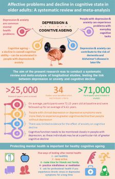
Mechanism of Rapid Antidepressant Effect of Alcohol Elucidated

by Tori Rodriguez, MA, LPC

Individuals with major depressive disorder (MDD) have double the risk of alcohol use disorders (AUDs) and vice versa, and it has previously been proposed that some people with MDD may use alcohol to self-medicate. Though alcohol can become depressant if used chronically, alcohol initially has an antidepressant effect, though the underlying mechanisms have not been identified. Findings reported in September 2016 in Nature Communications begin to elucidate the basis of this action.

Behavioral and molecular evidence of the rapid antidepressant activity of NMDA receptor (NMDAR) antagonists, which have been found to be effective within 2 hours of administration and remain so for 2 weeks, represents a significant advance in depression treatment. Antidepressant efficacy involves the induction phase and the sustained phase.

The sustained phase of rapid antidepressants requires “both new protein synthesis and an increase in protein stability… for the GABABR shift in function necessary to increase” the activity of mTORC1, a mechanistic target of rapamycin complex 1, the authors explained in their paper. Rapamycin (mTOR) is a “serine/threonine kinase essential for messenger RNA translation” and is required for the sustained impact of rapid antidepressants.

Citing previous findings that ethanol (EtOH) also blocks NMDARs in the hippocampus, scientists at the University of Texas at Austin and Wake Forest University School of Medicine in Winston-Salem, North Carolina, aimed to determine whether EtOH and NMDAR antagonists exert rapid antidepressant effects via the same synaptic pathways in rodents. They hypothesized that EtOH “has lasting antidepressant efficacy, shares the same downstream molecular signaling events as rapid antidepressants, and requires de novo protein synthesis.”

First, they found that acute exposure to EtOH led to antidepressant and anxiolytic behaviors in rodents for up to 24 hours. They then discovered that, like NMDAR antagonists, EtOH alters the expression and signaling of GABABR, increases dendritic calcium, and leads to the synthesis of new GABABRs. This synthesis requires fragile-X mental retardation protein (FMRP), an RNA-binding protein of which precise levels are needed for normal neuronal functioning.

The antidepressant effects and the changes in GABABR expression and dendritic calcium were not observed in in Fmr1-knockout (KO) mice, supporting the concept that FMRP has in important role in regulating protein synthesis after EtOH exposure, and thereby facilitating its antidepressant efficacy.

These results point to a shared molecular pathway for the antidepressant activity of EtOH and rapid antidepressants, and highlight a mechanism involved in the initial antidepressant action of alcohol. “A shift in GABABR signaling is observed with both rapid antidepressants and acute EtOH treatment, which may provide insight into the molecular basis for the high comorbidity between major depressive disorder and AUD,” the authors concluded.

http://www.psychiatryadvisor.com/addiction/rapid-antidepressant-effect-of-alcohol/article/567335/?DCMP=EMC-PA_Update_RD&cpn=psych_md%2cpsych_all&hmSubId=&NID=1710903786&dl=0&spMailingID=15723696&spUserID=MTQ4MTYyNjcyNzk2S0&spJobID=881842067&spReportId=ODgxODQyMDY3S0

New study shows that brief hyperthermia treats depression

Whole-body hyperthermia is a promising antidepressant modality that works quickly and offers prolonged benefit, according to a study recently published in the online JAMA Psychiatry.

Researchers came to that conclusion after conducting a double-blind study that randomized 30 adults with major depressive disorder to either a single session of active whole-body hyperthermia or a sham treatment that mimicked all aspects of whole-body hyperthermia except its intense heat.

The sham condition was included to strengthen the study design.

“A prior open trial found that a single session of whole-body hyperthermia reduced depressive symptoms,” researchers wrote. “However, the lack of a placebo control raises the possibility that the observed antidepressant effects resulted not from hyperthermia per se, but from nonspecific aspects of the intervention.”

Among participants randomized to sham treatment in the new study, more than 70% believed they had received whole-body hyperthermia, researchers reported, suggesting the placebo was convincing.

When researchers looked at participants’ scores on the Hamilton Depression Rating Scale throughout the 6-week period following the session, they found participants who received active whole-body hyperthermia had significantly reduced scores compared to participants who received sham treatment. Adverse events were mild.

Psych Congress Steering Committee member Charles L. Raison, MD, discussed the findings prior to their publication during a session at last year’s U.S. Psychiatric and Mental Health Congress in San Diego.

“Like ketamine, like scopolamine, and other rapid treatments for depression that are of intense interest in psychiatry, hyperthermia shows the same effect,” he said. “It doesn’t take a week or 2 to work. People feel better very, very quickly, and the effects appear to persist for an extended period of time.”

– Jolynn Tumolo

References

Janssen CW, Lowry CA, Mehl MR, et al. Whole-body hyperthermia for the treatment of major depressive disorder: a randomized clinical trial. JAMA Psychiatry. 2016 May 12. [Epub ahead of print].

Lebano L. New data support whole body hyperthermia for rapid treatment of major depression. Psych Congress Network. 2015 Sept. 10.

http://www.psychcongress.com/article/hyperthermia-provides-significant-rapid-relief-depression-study-suggests-27981

Viral and Bacterial Links to the Brain’s Decline


Herpes simplex viruses pass through the outer protein coat of a nucleus, magnified 40,000 times. Dr. Ruth Itzhak’s research published in 1997 revealed a potential link to the presence of HSV-1 (one specific variety of Herpes simplex) and the onset of Alzheimer’s in 60 percent of the cases they studied. However, she has only been able to study a low number of cases since the work has received only a cursory nod from the greater research world and little funding.

By Ed Cara

As recently as the 1970s, doctors stubbornly treated complaints of festering open sores in the stomach as a failing of diet or an inability to manage stress. Though we had long accepted the basic premise of Louis Pasteur’s germ theory—that flittering short bursts of disease and death are often caused by microscopic beings that could be stopped by sanitary food, water and specially crafted drugs—many researchers ardently resisted the idea that they could also trigger more complicated, chronic illnesses.

When it came to ulcers, no one believed that any microorganisms could endure in the acidic cauldron of our digestive system. It took the gumshoe work of Australian doctors and medical researchers Barry Marshall and Robin Warren in the 1980s to debunk that belief and discover the specific bug responsible for most chronic stomach ulcers, Helicobacter pylori. Marshall even went so far as to swallow the germ to prove the link was real and, obviously, became sick soon after. Thankfully, his self-sacrifice was eventually validated when he and Warren were awarded a Nobel Prize in 2005.

But while modern medicine has grown comfortable with the idea that even chronic physical ailments can be sparked by the living infinitesimal, there is an even bolder, more controversial proposition from a growing number of researchers. It’s the idea that certain germs, bugs and microbes can lie hidden in the body for decades, all the while slowly damaging our brains, even to the point of dementia, depression and schizophrenia.

In January 2016, a team led by Shawn Gale, an associate professor in psychology at Brigham Young University, looked at the infection history of 5,662 young to middle-aged adults alongside the results of tests intended to measure cognition. Gale’s rogues’ gallery included both parasites (the roundworm and Toxoplasma gondii ) and viruses (the hepatitis clan, cytomegalovirus, and herpes simplex virus Types 1 and 2). The team created an index of infectious disease —the more bugs a participant had been exposed to, the higher the person’s index score. It turned out that those with a higher score were more likely to have worse learning and memory skills, as well as slower information-processing speed than those with a lower score, even after controlling for other factors, like age, sex and financial status.

Aside from their shared ability to stay rooted inside us, the ways these pathogens might influence our noggins are as varied as their biology is from one another. Some, like T. gondii (often transmitted to humans via contaminated cats and infected dirt), can discreetly infest the brain and cause subtle changes to our brain chemistry, altering levels of neurotransmitters like dopamine while causing no overt signs of disease. Others, like hepatitis C, are suspected of hitching a ride onto infected white blood cells that cross the brain-blood barrier and, once inside, deplete our supply of white brain matter, the myelin-coated axons that help neurons communicate with each other and seem to actively shape how we learn. And still others, like H. pylori, could trigger a low-level but chronic inflammatory response that gradually wears down our body and mind alike.

Gale’s team found only fairly small deficits in cognition connected to infection. But other researchers, like Ruth Itzhaki, professor emeritus of molecular neurobiology at Britain’s University of Manchester, believe microbes may play an outsized role in one of the most devastating neurodegenerative disorders around: Alzheimer’s disease, which afflicted 47 million people worldwide in 2015. Last March, Itzhaki and a globe-spanning group of researchers penned an editorial in the Journal of Alzheimer’s Disease, imploring the scientific community to more seriously pursue a proposed link between Alzheimer’s and particular germs, namely herpes simplex virus Type 1 (HSV-1), Chlamydia pneumoniae and spirochetes—a diverse group of bacteria that include those responsible for syphilis and Lyme disease. The unusually direct plea, for scientists at least, was the culmination of decades of frustration.

“There’s great hostility to the microbial concept amongst certain influential people in the field, and they are the ones who usually determine whether or not one’s research grant application is successful,” says Itzhaki. “The irony is that they never provide scientific objections to the concepts—they just belittle them, so there’s nothing to rebut!”

It’s a frustration Itzhaki knows too well; in 1991, her lab published the first paper finding a clear HSV-1 link to Alzheimer’s. Since then, according to Itzhaki, over 100 published studies, from her lab and elsewhere, have been supportive of the same link. Nevertheless, Itzhaki says, the work has received only a cursory nod from the greater research world and little funding. Out of the $589 million allocated to Alzheimer’s research by the National Institutes of Health in 2015, exactly zero appeared to be spent on studying the proposed HSV-1 connection.

HSV-1 is more often known as the version of herpes that causes cold sores. Nearly all of us carry the virus from infancy; our peripheral nervous system serves as its dormant nesting ground. From there, HSV-1 can reactivate and occasionally cause mild flare-ups of disease, typically when our immune system is overwhelmed due to stress or other infections. Itzhaki’s lab, however, found that by the time we reach our golden years, the virus often migrates to the brain, where it remains capable of resurrecting itself and wreaking a new sort of havoc when opportunity presents, such as when our immune system wavers in old age.

Her team has also discovered the presence of HSV-1 in the telltale plaques—clumps of proteins in the nerve cells of the brain—used to diagnose Alzheimer’s. In mice and cell cultures infected with HSV-1, they’ve found accumulation of two proteins, beta-amyloid and tau, that form the main components of, respectively, plaques and tangles—twisted protein fibers that form inside dying cells and are another defining characteristic of Alzheimer’s. Plaques and tangles, while sometimes found in normal aging brains, have been found to overflow in the brains of deceased Alzheimer’s sufferers; neuroscientists believe these protein accumulations can cause neuron death and tissue loss. Itzhaki speculates that herpes-infected cells may either produce the proteins in an attempt to fend off HSV-1 or, because the virus itself commands them to, the proteins somehow needed to jump-start the virus’s replication.

Itzhaki, Gale and their colleagues emphasize that rather than being the sole cause of memory loss, slower reaction time or depression, viral and bacterial infections are likely just one ingredient in a soup of risk factors. But for Alzheimer’s, HSV-1 could be especially significant. Itzhaki has found that elderly people who carried both HSV-1 in the brain and the e-4 subtype of the APOE gene (responsible for creating a protein that helps transport cholesterol throughout the body) were 12 times more likely to develop Alzheimer’s than people without either.

APOE-e4, already considered a significant risk factor for Alzheimer’s and thought to make us more vulnerable to viral infection, has also been linked to a greater risk of dementia in HIV-infected patients. In a 1997 Lancet paper, Itzhaki’s group concluded that HSV-1 infection, in conjunction with APOE-e4, could account for about 60 percent of the Alzheimer’s cases they studied. Due to limited funds, however, her group was able to study only a relatively low number of cases.

“I think the proposed theory is certainly reasonable given the supporting evidence,” says Iain Campbell, a professor of molecular biology at the University of Sydney. “What is difficult to establish here is actual causality.”

It might be the case that HSV-1 and other suspects aren’t responsible for the emergence of Alzheimer’s but are simply given free rein to worsen its symptoms as the neurodegenerative disorder weakens both the immune and nervous systems. Deciphering the relationship between these latent infections and Alzheimer’s will take more dedicated research, an effort that Itzhaki feels has been stymied by the persistent lack of resources available to her and her like-minded colleagues.

As things stand, though, she believes there is enough evidence to go ahead with treatment trials; for instance, giving Alzheimer’s patients HSV-1-targeted antivirals in hopes of slowing down or stopping the progression of the disease. She and a team of clinicians are trying to obtain a grant for such a pilot clinical trial to do just that.

Exasperated as Itzhaki has been, the headwinds against her and those who share her beliefs about the brain are slowly dying down. In some cases, once-derided and obscure scientists studying how infections affect the brain are now getting some financial support. There’s Jaroslav Flegr, for example, who has for decades theorized that T. gondii could alter human behavior and even cause certain forms of schizophrenia. In the wake of increased media attention, Flegr’s volume of work on T. gondii has noticeably stepped up as well. From 2014 to 2015, he co-authored 13 papers on T. gondii, nearly twice the number he published the previous two years; the trend of increased T. gondii papers holds across all of PubMed, the largest database of published biomedical research available. “ I have no serious problem with funding of my Toxo research now,” Flegr says.

As of now, though, there have been no ulcer-related Sherlock moments to prove a link between mental dysfunction and latent infections—only indirect correlations clumping together to form a blurry snapshot of a potential crime scene. Which is why Gale and others recommend a wait-and-see approach for the public, even as they acknowledge the potentially vast implications of their research. “I wouldn’t want someone to go out tomorrow and get a whole battery of tests,” he says. “There’s still a lot we need to understand.”

http://www.newsweek.com/viral-bacterial-links-brains-decline-462194

New study may explain gene’s role in major psychiatric disorders

A new study shows the death of newborn brain cells may be linked to a genetic risk factor for five major psychiatric diseases, and at the same time shows a compound currently being developed for use in humans may have therapeutic value for these diseases by preventing the cells from dying.

In 2013, the largest genetic study of psychiatric illness to date implicated mutations in the gene called CACNA1C as a risk factor in five major forms of neuropsychiatric disease — schizophrenia, major depression, bipolar disorder, autism, and attention deficit hyperactivity disorder (ADHD). All the conditions also share the common clinical feature of high anxiety. By recognizing an overlap between several lines of research, scientists at the University of Iowa and Weill Cornell Medicine of Cornell University have now discovered a new and unexpected role for CACNA1C that may explain its association with these neuropsychiatric diseases and provide a new therapeutic target.

The new study, recently published in eNeuro, shows that loss of the CACNA1C gene from the forebrain of mice results in decreased survival of newborn neurons in the hippocampus, one of only two regions in the adult brain where new neurons are continually produced – a process known as neurogenesis. Death of these hippocampal neurons has been linked to a number of psychiatric conditions, including schizophrenia, depression, and anxiety.

“We have identified a new function for one of the most important genes in psychiatric illness,” says Andrew Pieper, MD, PhD, co-senior author of the study, professor of psychiatry at the UI Carver College of Medicine and a member of the Pappajohn Biomedical Institute at the UI. “It mediates survival of newborn neurons in the hippocampus, part of the brain that is important in learning and memory, mood and anxiety.”

Moreover, the scientists were able to restore normal neurogenesis in mice lacking the CACNA1C gene using a neuroprotective compound called P7C3-A20, which Pieper’s group discovered and which is currently under development as a potential therapy for neurodegenerative diseases. The finding suggests that the P7C3 compounds may also be of interest as potential therapies for these neuropsychiatric conditions, which affect millions of people worldwide and which often are difficult to treat.

Pieper’s co-lead author, Anjali Rajadhyaksha, associate professor of neuroscience in Pediatrics and the Feil Family Brain and Mind Research Institute at Weill Cornell Medicine and director of the Weill Cornell Autism Research Program, studies the role of the Cav1.2 calcium channel encoded by the CACNA1C gene in reward pathways affected in various neuropsychiatric disorders.

“Genetic risk factors that can disrupt the development and function of brain circuits are believed to contribute to multiple neuropsychiatric disorders. Adult newborn neurons may serve a role in fine-tuning rewarding and environmental experiences, including social cognition, which are disrupted in disorders such as schizophrenia and autism spectrum disorders,” Rajadhyaksha says. “The findings of this study provide a direct link between the CACNA1C risk gene and a key cellular deficit, providing a clue into the potential neurobiological basis of CACNA1C-linked disease symptoms.”

Several years ago, Rajadhyaksha and Pieper created genetically altered mice that are missing the CACNA1C gene in the forebrain. The team discovered that the animals have very high anxiety.

“That was an exciting finding, because all of the neuropsychiatric diseases in which this gene is implicated are associated with symptoms of anxiety,” says Pieper who also holds appointments in the UI Departments of Neurology, Radiation Oncology, Molecular Physiology and Biophysics, the Holden Comprehensive Cancer Center, and the Iowa City VA Health Care System.

By studying neurogenesis in the mice, the research team has now shown that loss of the CACNA1C gene from the forebrain decreases the survival of newborn neurons in the hippocampus – only about half as many hippocampal neurons survive in mice without the gene compared to normal mice. Loss of CACNA1C also reduces production of BDNF, an important brain growth factor that supports neurogenesis.

The findings suggest that loss of the CACNA1C gene disrupts neurogenesis in the hippocampus by lowering the production of BDNF.

Pieper had previously shown that the “P7C3-class” of neuroprotective compounds bolsters neurogenesis in the hippocampus by protecting newborn neurons from cell death. When the team gave the P7C3-A20 compound to mice lacking the CACNA1C gene, neurogenesis was restored back to normal levels. Notably, the cells were protected despite the fact that BDNF levels remained abnormally low, demonstrating that P7C3-A20 bypasses the BDNF deficit and independently rescues hippocampal neurogenesis.

Pieper indicated the next step would be to determine if the P7C3-A20 compound could also ameliorate the anxiety symptoms in the mice. If that proves to be true, it would strengthen the idea that drugs based on this compound might be helpful in treating patients with major forms of psychiatric disease.

“CACNA1C is probably the most important genetic finding in psychiatry. It probably influences a number of psychiatric disorders, most convincingly, bipolar disorder and schizophrenia,” says Jimmy Potash, MD, professor and DEO of psychiatry at the UI who was not involved in the study. “Understanding how these genetic effects are manifested in the brain is among the most exciting challenges in psychiatric neuroscience right now.”

http://www.news-medical.net/news/20160427/Study-reveals-new-function-for-CACNA1C-gene-in-psychiatric-diseases.aspx

Meditating in a tiny Iowa town to help recovery from war

By Supriya Venkatesan

At 19, I enlisted in the U.S. Army and was deployed to Iraq. I spent 15 months there — eight at the U.S. Embassy, where I supported the communications for top generals. I understand that decisions at that level are complex and layered, but for me, as an observer, some of those actions left my conscience uneasy.

To counteract my guilt, I volunteered as a medic on my sole day off at Ibn Sina Hospital, the largest combat hospital in Iraq. There I helped wounded Iraqi civilians heal or transition into the afterlife. But I still felt lost and disconnected. I was nostalgic for a young adulthood I never had. While other 20-somethings had traditional college trajectories, followed by the hallmarks of first job interviews and early career wins, I had spent six emotionally numbing years doing ruck marches, camping out on mountaintops near the demilitarized zone in South Korea and fighting someone else’s battle in Iraq.

During my deployment, a few soldiers and I were awarded a short resort stay in Kuwait. There, I had a brief but powerful experience in a meditation healing session. I wanted more. So when I returned to the United States at the end of my service, I headed to Iowa.

Forty-eight hours after being discharged from the Army, I arrived on campus at Maharishi University of Management in Fairfield, Iowa. MUM is a small liberal arts college, smack dab in the middle of the cornfields, founded by Maharishi Mahesh Yogi, the guru of transcendental meditation. I joked that I was in a quarter-life crisis, but in truth my conscience was having a crisis. Iraq left me with questions about the world and grappling with my own mortality and morality.

Readjustment was a sucker punch of culture shock. While on a camping trip for incoming students, I watched girls curl their eyelashes upon waking up and burn incense and bundles of sage to ward off negative energy. I was used to being in a similar field environment but with hundreds of guys who spit tobacco, spoke openly of their sexual escapades and played video games incessantly. Is this what it looked like to be civilian woman? Is this what spirituality looked like?

Mediation was mandatory for students on campus, and the rest of the town was composed mainly of former students or longtime followers of the maharishi. Shortly after arriving, I completed an advanced meditator course and began meditating three hours a day — a habit that is still with me five years later. Every morning, I went to a dome where students, teachers and the people of Fairfield gathered to practice meditation. In the evening, we met again for another round of meditation. During my time in Fairfield, even Oprah came to meditate in the dome.

I was incredibly lucky to have supportive mentors in the Army, but Fairfield embraced me in a maternal way. I cried for hours during post-meditation reflection. I released the trauma that is familiar to every soldier who has gone to war but is rarely discussed or even acknowledged. I let go, and I blossomed. I was emancipated of the unhealthy habits of binge-drinking and co-dependency in romantic interludes, as well as a fear that I didn’t know controlled me.

Suicide and other byproducts of post-traumatic stress disorder plague the military. In 2010, a veteran committed suicide every 65 minutes. In 2012, there were more deaths by suicide than by combat. In Iraq, one of my neighbors took his M16, put it in his mouth and shot himself. Overwhelmed with PTSD-related issues from back-to-back deployments and with no clear solution to the problem, in 2012, the Defense Department began researching meditation practices to see whether they would affect PTSD. The first study of meditation and the military population, done with Vietnam veterans in 1985, had shown 70 percent of veterans finding relief, but meditation never gained in popularity nor was it offered through veterans’ services. Even in 2010, when I learned TM, the military was alien to the concept.

But today, the results of the studies showcase immense benefits for veterans. According to the journal Military Medicine, meditation has shown a 40 percent to 55 percent reduction in symptoms of PTSD and depression among veterans. Furthermore, studies show that meditation correlates with a 42 percent reduction in insomnia and a 25 percent reduction in the stress hormone cortisol in the veteran population. To complement meditation, yoga has also been embraced as a tool for treatment by the military. With the growing acceptance of holistic approaches, psychological wounds are beginning to heal.

The four-day training course to learn TM is now available at every Veterans Affairs facility for those who have PTSD or traumatic brain injury. Even medical staff and counselors who help veterans at the VA are offered training in both TM and mindfulness meditation. Additionally, Norwich University, the oldest military college in the country, has done extensive research on TM and incoming cadets, and many military installations have integrated meditation programs into their mental health services. When I had first learned to meditate, many of my active-duty friends found it a bit too crunchy. But with the military’s recent efforts at researching meditation and funding it for all veterans, the stigma is gone, and my battle buddies see meditation as a tool for building resilience.

For me, meditation has created small but significant changes. One day, while going for a walk downtown, I stopped and patted a dog. A few minutes later, I came to a halt. I realized what I had done. While in Iraq, during a month when we were under heavy mortar attack, a bomb-sniffing K-9 had become traumatized and attacked me. This, coupled with a life-long fear of dogs, had left me guarded around the canines. I touched the scar on my elbow from where the K-9 had latched on and could no longer find the fear that had been there. Soon I was shedding all the things that held me back from living my life in an entirely unforeseen way.

For the first time in my life, I found forgiveness for those who had wronged me in the past. I literally stopped to smell the flowers on my way to work every day. And I smiled. All the freaking time. I even felt smarter. Research shows that meditation raises IQ. I’m not surprised. After graduation, I went on to complete my master’s at Columbia University.

Fairfield is also home to generations of Iowans who are born there, brought up there and die there. Many of these blue-collar Midwesterners have had animosity toward the meditators. Locals felt as if their town had been overtaken. They preferred steak to quinoa, beers at the bar to yoga and pickup trucks to carbon-reducing bicycles. And with MUM having a student body from more than 100 countries, the ethnic differences were a challenge. However, things are changing. Meditators and townspeople now fill less stereotypical roles. And with the economic boom that meditating entrepreneurs have provided the town, the differences are easier to ignore.

It was strange for me to live removed from the local Iowans. When I went shopping at the only Walmart the town had, I’d see the “Wall of Heroes” — a wall of photos of veterans from Fairfield. One day, I noticed a familiar face — a soldier from my last assignment. Fairfield and other socioeconomically depressed areas are where most military recruits come from. Here I was living among them, but not moving in step with them. Having that synchronous experience made me come back full circle. When I had first learned to meditate, my teacher had asked me what my goal was. I told her, “I want to be in the world, but not of it.” And that’s exactly what I got.

For me, this little Iowan town provided a place of respite and rejuvenation. It was easy for me to trade one lifestyle of order and discipline for another, and this provided me with nourishment and an understanding of self. Nowhere else in America can you find an entire town living and breathing the principles of Eastern mysticism. It goes way beyond taking a yoga class or going to the Burning Man festival. I continue my meditation practice and am grateful for the gifts it has provided me. But in the end, my time had come, and I had to leave. As residents would say, that was just my karma.

https://www.washingtonpost.com/posteverything/wp/2016/04/06/how-meditating-in-a-tiny-iowa-town-helped-me-recover-from-war/

Virtual Reality Therapy Shows Promise Against Depression

An immersive virtual reality therapy could help people with depression to be less critical and more compassionate towards themselves, reducing depressive symptoms, finds a new study from UCL (University College London) and ICREA-University of Barcelona.

The therapy, previously tested by healthy volunteers, was used by 15 depression patients aged 23-61. Nine reported reduced depressive symptoms a month after the therapy, of whom four experienced a clinically significant drop in depression severity. The study is published in the British Journal of Psychiatry Open and was funded by the Medical Research Council.

Patients in the study wore a virtual reality headset to see from the perspective of a life-size ‘avatar’ or virtual body. Seeing this virtual body in a mirror moving in the same way as their own body typically produces the illusion that this is their own body. This is called ’embodiment’.

While embodied in an adult avatar, participants were trained to express compassion towards a distressed virtual child. As they talked to the child it appeared to gradually stop crying and respond positively to the compassion. After a few minutes the patients were embodied in the virtual child and saw the adult avatar deliver their own compassionate words and gestures to them. This brief 8-minute scenario was repeated three times at weekly intervals, and patients were followed up a month later.

“People who struggle with anxiety and depression can be excessively self-critical when things go wrong in their lives,” explains study lead Professor Chris Brewin (UCL Clinical, Educational & Health Psychology). “In this study, by comforting the child and then hearing their own words back, patients are indirectly giving themselves compassion. The aim was to teach patients to be more compassionate towards themselves and less self-critical, and we saw promising results. A month after the study, several patients described how their experience had changed their response to real-life situations in which they would previously have been self-critical.”

The study offers a promising proof-of-concept, but as a small trial without a control group it cannot show whether the intervention is responsible for the clinical improvement in patients.

“We now hope to develop the technique further to conduct a larger controlled trial, so that we can confidently determine any clinical benefit,” says co-author Professor Mel Slater (ICREA-University of Barcelona and UCL Computer Science). “If a substantial benefit is seen, then this therapy could have huge potential. The recent marketing of low-cost home virtual reality systems means that methods such as this could potentially be part of every home and be used on a widespread basis.”

Publication: Embodying self-compassion within virtual reality and its effects on patients with depression. Falconer, CJ et al. British Journal of Psychiatry Open (February, 2016)

Risk of suicide increases 3X after a concussion

New research published in the Canadian Medical Association Journal shows that even mild concussions sustained in ordinary community settings might be more detrimental than anyone anticipated; the long-term risk of suicide increases threefold in adults if they have experienced even one concussion. That risk increases by a third if the concussion is sustained on a weekend instead of a weekday—suggesting recreational concussions are riskier long-term than those sustained on the job.

“The typical patient I see is a middle-aged adult, not an elite athlete,” says Donald Redelmeier, a senior scientist at the University of Toronto and one of the study’s lead authors. “And the usual circumstances for acquiring a concussion are not while playing football; it is when driving in traffic and getting into a crash, when missing a step and falling down a staircase, when getting overly ambitious about home repairs—the everyday activities of life.”

Redelmeier and his team wanted to examine the risks of the concussions acquired under those circumstances. They identified nearly a quarter of a million adults in Ontario who were diagnosed with a mild concussion over a timespan of 20 years—severe cases that resulted in hospital admission were excluded from the study—and tracked them for subsequent mortality due to suicide. It turned out that more than 660 suicides occurred among these patients, equivalent to 31 deaths per 100,000 patients annually—three times the population norm. On average, suicide occurred almost six years after the concussion. This risk was found to be independent of demographics or previous psychiatric conditions, and it increased with additional concussions.

For weekend concussions, the later suicide risk increased to four times the norm. Redelmeier and his fellow researchers had wondered whether the risk would differ between occupational and recreational concussions. They did not have information about how the concussions happened, so they used day of the week as a proxy. Although they do not know why weekend risk is indeed higher, they suspect it may be because on weekends medical staff may not be as available or accessible or people may not seek immediate care.

Although the underlying causes of the connection between concussion and suicide are not yet known, Redelmeier says that there were at least three potential explanations. A concussion may be a marker but not necessarily a mechanism of subsequent troubles—or, in other words, people who sustain concussions may already have baseline life imbalances that increase their risks for depression and suicide. “But we also looked at the subgroup of patients who had no past psychiatric history, no past problems, and we still found a significant increase in risk. So I don’t think that’s the entire story,” he notes. One of the more likely explanations, he says, is that concussion causes brain injury such as inflammation (as has been found in some studies) from which the patient may never fully recover. Indeed, a study conducted in 2014 found that sustaining a head injury leads to a greater risk of mental illness later in life. The other possibility is that some patients may not give themselves enough time to get better before returning to an ordinary schedule, leading to strain, frustration and disappointment—which, in turn, may result in depression and ultimately even suicide.

Lea Alhilali, a physician and researcher at the Barrow Neurological Institute who did not participate in this study, uses diffusion tensor imaging (an MRI technique) to measure the integrity of white matter in the brain. Her team has found similarities between white matter degeneration patterns in patients with concussion-related depression and noninjured patients with major depressive disorder—particularly in the nucleus accumbens, or the “reward center” of the brain. “It can be difficult to tease out what’s related to an injury and what’s related to the circumstances surrounding the trauma,” Alhilali says. “There could be PTSD, loss of job, orthopedic injuries that can all influence depression. But I do believe there’s probably an organic brain injury.”

Alhilali points to recent studies on chronic traumatic encephalopathy (CTE), a progressive degenerative brain disease associated with repeated head traumas. Often linked to dementia, depression, loss of impulse control and suicide, CTE was recently diagnosed in 87 of 91 deceased NFL players. Why, then, she says, should we not suspect that concussion causes other brain damage as well?

This new study may only represent the tip of the iceberg. “We’re only looking at the most extreme outcomes, at taking your own life,” Redelmeier says. “But for every person who dies from suicide, there are many others who attempt suicide, and hundreds more who think about it and thousands more who suffer from depression.”

More research needs to be done; this study was unable to take into account the exact circumstances under which the concussions were sustained. Redelmeier’s research examined only the records of adults who sought medical attention, it did not include more severe head injuries that required hospitalization or extensive emergency care. To that extent, his findings may have underestimated the magnitude of the absolute risks at hand.

Yet many people are not aware of these risks.

Redelmeier is adamant that people should take concussions seriously. “We need to do more research about prevention and recovery,” he says. “But let me at least articulate three things to do: One, give yourself permission to get some rest. Two, when you start to feel better, don’t try to come back with a vengeance. And three, even after you’re feeling better, after you’ve rested properly, don’t forget about it entirely. If you had an allergic reaction to penicillin 15 years ago, you’d want to mention that to your doctor and have it as a permanent part of your medical record. So, too, if you’ve had a concussion 15 years ago.”

http://www.scientificamerican.com/article/a-single-concussion-may-triple-the-long-term-risk-of-suicide1/

Why do older white men have higher risk of suicide?

Older men of European descent (white men) have significantly higher suicide rates than any other demographic group in the United States, including older women across ethnicities and older men of African, Latino, or Indigenous decent, according to research published in Men and Masculinities.

In her latest addition to suicide research, Silvia Sara Canetto, PhD, professor in the Department of Psychology at Colorado State University, has found that older white men have higher suicide rates yet fewer burdens associated with aging. They are less likely to experience widowhood, have better physical health and fewer disabilities than older women, and have more economic resources than older women across ethnicities and ethnic minority older men.

Rather than being due to physical aging adversities, therefore, increased suicide rates among older white men in the United States may be because they are less psychologically equipped to deal with the normal challenges of aging; likely because of their privilege until late adulthood, Dr Canetto asserted.
Another important factor in white men’s vulnerability to suicide once they reach late life may be dominant cultural scripts of masculinity, aging, and suicide, Dr Canetto said. A particularly damaging cultural script may be the belief that suicide is a masculine response to “the indignities of aging.” This idea implies that suicide is justified or even glorified among men.

To illustrate these cultural scripts, Dr Canetto examined two famous suicide cases and their accompanying media coverage. The founder of Kodak, George Eastman, died of suicide at age 77. His biographer said that Eastman was “unprepared and unwilling to face the indignities of old age.”

American journalist and author Hunter S. Thompson died of suicide in 2005 at age 67, and was described by friends as having triumphed over “the indignities of aging.” Both of these suicides were covered in the press through scripts of conventional “white” masculinity, Dr Canetto stated. “The dominant story was that their suicide was a rational, courageous, powerful choice,” she said in a statement.

Canetto’s research challenges the idea that high suicide rates are inevitable among older white men. Canetto notes that older men are not the most suicide-prone group everywhere in the world; in China, for example, women at reproductive age are the demographic with the highest rate of suicide. This is additional evidence that suicide in older white men is culturally determined and thus preventable.

Dr Canetto’s research shows that cultural scripts may offer a new way of understanding and preventing suicide. The “indignities of aging” suicide script and the belief that suicide is a masculine, powerful response to aging can and should be challenged, Dr Canetto said.

Canetto SS. Suicide: Why Are Older Men So Vulnerable? Men Masc. 2015; doi:10.1177/1097184X15613832.

The Power of Music in Alleviating Dementia Symptoms

by Tori Rodriguez, MA, LPC

As the search continues for effective drug treatments for dementia, patients and caregivers may find some measure of relief from a common, non-pharmaceutical source. Researchers have found that music-related memory appears to be exempt from the extent of memory impairment generally associated with dementia, and several studies report promising results for several different types of musical experiences across a variety of settings and formats.

“We can say that perception of music can be intact, even when explicit judgments and overt recognition have been lost,” Manuela Kerer, PhD, told Psychiatry Advisor. “We are convinced that there is a specialized memory system for music, which is distinct from other domains, like verbal or visual memory, and may be very resilient against Alzheimer’s disease.”

Kerer is a full-time musical composer with a doctoral degree in psychology who co-authored a study on the topic while working at the University of Innsbruck in Austria. She and her colleagues investigated explicit memory for music among ten patients with early-state Alzheimer’s disease (AD) and ten patients with mild cognitive impairment (MCI), and compared their performance to that of 23 healthy participants. Not surprisingly, the patient group demonstrated worse performance on tasks involving verbal memory, but they did significantly better than controls on the music-perceptional tasks of detecting distorted tunes and judging timbre.

“The temporal brain structures necessary for verbal musical memory were mildly affected in our clinical patients, therefore attention might have shifted to the discrimination tasks which led to better results in this area,” she said. “Our results enhance the notion of an explicit memory for music that can be distinguished from other types of explicit memory — that means that memory for music could be spared in this patient group.”

Other findings suggest that music might even improve certain aspects of memory among people with dementia. In a randomized controlled trial published in last month in the Journal of Alzheimer’s Disease, music coaching interventions improved multiple outcomes for both patients with dementia and their caregivers. The researchers divided 89 pairs of patients with dementia and their caregivers into three groups: two groups were assigned to caregiver-led interventions that involved either singing or listening to music, while a third group received standard care. Before and after the 10-week intervention, and six months after the intervention, participants were assessed on measures of mood, quality of life and neuropsychological functioning.

Results showed that the singing intervention improved working memory among patients with mild dementia and helped to preserve executive function and orientation among younger patients, and it also improved the well-being of caregivers. The listening intervention was found to have a positive impact on general cognition, working memory and quality of life, particularly among patients in institutional care with moderate dementia not caused by AD. Both interventions led to reductions in depression.

The findings suggest that “music has the power to improve mood and stimulate cognitive functions in dementia, most likely by engaging limbic and medial prefrontal brain regions, which are often preserved in the early stages of the illness,” study co-author Teppo Särkämö, PhD, a researcher at the University of Helsinki, Finland, told Psychiatry Advisor. “The results indicate that when used regularly, caregiver-implemented musical activities can be an important and easily applicable way to maintain the emotional and cognitive well-being of persons with dementia and also to reduce the psychological burden of family caregivers.”

Singing has also been shown to increase learning and retention of new verbal material in patients with AD, according to research published this year in the Journal of Clinical & Experimental Neuropsychology, and findings published in 2013 show that listening to familiar music improves the verbal narration of autobiographical memories in such patients. Another study found that a music intervention delivered in a group format reduced depression and delayed the deterioration of cognitive functions, especially short-term recall, in patients with mild and moderate dementia. Group-based music therapy appears to also decrease agitation among patients in all stages of dementia, as described in a systematic review published in 2014 in Nursing Times.

n addition to the effects of singing and listening to music on patients who already have dementia, playing a musical instrument may also offer some protection against the condition, according to a population-based twin study reported in 2014 in the International Journal of Alzheimer’s Disease. Researchers at the University of Southern California found that older adults who played an instrument were 64% less likely than their non-musician twin to develop dementia or cognitive impairment.

“Playing an instrument is a unique activity in that it requires a wide array of brain regions and cognitive functions to work together simultaneously, throughout both the right and left hemispheres,” co-author Alison Balbag, PhD, told Psychiatry Advisor. While the study did not examine causal mechanisms, “playing an instrument may be a very effective and efficient way to engage the brain, possibly granting older musicians better maintained cognitive reserve and possibly providing compensatory abilities to mitigate age-related cognitive declines.”

She notes that clinicians might consider suggesting that patients incorporate music-making into their lives as a preventive activity, or encouraging them to keep it up if they already play an instrument.
Further research, particularly neuroimaging studies, is needed to elucidate the mechanisms behind the effects of music on dementia, but in the meantime it could be a helpful supplement to patients’ treatment plans. “Music has considerable potential and it should be introduced much more in rehabilitation and neuropsychological assessment,” Kerer said.

http://www.psychiatryadvisor.com/alzheimers-disease-and-dementia/neurocognitive-neurodegenerative-memory-musical-alzheimers/article/452120/3/

References

Kerer M, Marksteiner J, Hinterhuber H, et al. Explicit (semantic) memory for music in patients with mild cognitive impairment and early-stage Alzheimer’s disease. Experimental Aging Research; 2013; 39(5):536-64.

Särkämö T, Laitinen S, Numminen A, et al. Clinical and Demographic Factors Associated with the Cognitive and Emotional Efficacy of Regular Musical Activities in Dementia. Journal of Alzheimer’s Disease; 2015; published online ahead of print.

Palisson J, Roussel-Baclet C, Maillet D, et al. Music enhances verbal episodic memory in Alzheimer’s disease. Journal of Clinical & Experimental Neuropsychology; 2015; 37(5):503-17.

El Haj M, Sylvain Clément, Luciano Fasotti, Philippe Allain. Effects of music on autobiographical verbal narration in Alzheimer’s disease. Journal of Neurolinguistics; 2013; 26(6): 691–700.

Chu H, Yang CY, Lin Y, et al. The impact of group music therapy on depression and cognition in elderly persons with dementia: a randomized controlled study. Biological Research for Nursing; 2014; 16(2):209-17.

Craig J. Music therapy to reduce agitation in dementia. Nursing Times; 2014; 110(32-33):12-5.
Balbag MA, Pedersen NL, Gatz M. Playing a Musical Instrument as a Protective Factor against Dementia and Cognitive Impairment: A Population-Based Twin Study. International Journal of Alzheimer’s Disease; 2014; 2014: 836748.

CPAP therapy demonstrated to reduce depression in adults with obstructive sleep apnea

A new study shows that depressive symptoms are extremely common in people who have obstructive sleep apnea, and these symptoms improve significantly when sleep apnea is treated with continuous positive airway pressure therapy.

Results show that nearly 73 percent of sleep apnea patients (213 of 293 patients) had clinically significant depressive symptoms at baseline, with a similar symptom prevalence between men and women. These symptoms increased progressively and independently with sleep apnea severity.

However, clinically significant depressive symptoms remained in only 4 percent of the sleep apnea patients who adhered to CPAP therapy for 3 months (9 of 228 patients). Of the 41 treatment adherent patients who reported baseline feelings of self-harm or that they would be “better dead,” none reported persisting suicidal thoughts at the 3-month follow-up.

“Effective treatment of obstructive sleep apnea resulted in substantial improvement in depressive symptoms, including suicidal ideation,” said senior author David R. Hillman, MD, clinical professor at the University of Western Australia and sleep physician at the Sir Charles Gairdner Hospital in Perth. “The findings highlight the potential for sleep apnea, a notoriously underdiagnosed condition, to be misdiagnosed as depression.”

Study results are published in the September issue of the Journal of Clinical Sleep Medicine.

The American Academy of Sleep Medicine reports that obstructive sleep apnea (OSA) is a common sleep disease afflicting at least 25 million adults in the U.S. Untreated sleep apnea increases the risk of other chronic health problems including heart disease, high blood pressure, Type 2 diabetes, stroke and depression.

The study group comprised 426 new patients referred to a hospital sleep center for evaluation of suspected sleep apnea, including 243 males and 183 females. Participants had a mean age of 52 years. Depressive symptoms were assessed using the validated Patient Health Questionnaire (PHQ-9), and the presence of obstructive sleep apnea was determined objectively using overnight, in-lab polysomnography. Of the 293 patients who were diagnosed with sleep apnea and prescribed CPAP therapy, 228 were treatment adherent, which was defined as using CPAP therapy for an average of 5 hours or more per night for 3 months.

According to the authors, the results emphasize the importance of screening people with depressive symptoms for obstructive sleep apnea. These patients should be asked about common sleep apnea symptoms including habitual snoring, witnessed breathing pauses, disrupted sleep, and excessive daytime sleepiness.

http://www.eurekalert.org/pub_releases/2015-09/aaos-ctr092215.php