Phantom Eye Patients See and Feel with Missing Eyeballs

by Elizabeth Preston

Amputees often feel eerie sensations from their missing limbs. These “phantom limb” feelings can include pain, itching, tingling, or even a sense of trying to pick something up. Patients who lose an eye may have similar symptoms—with the addition of actual phantoms.

Phantom eye syndrome (PES) had been studied in the past, but University of Liverpool psychologist Laura Hope-Stone and her colleagues recently conducted the largest study of PES specifically in patients who’d lost an eye to cancer.

The researchers sent surveys to 239 patients who’d been treated for uveal melanoma at the Liverpool Ocular Oncology Centre. All of these patients had had one eye surgically removed. Some of their surgeries were only 4 months in the past; others had taken place almost 4 and a half years earlier. Three-quarters of the patients returned the surveys, sharing details about how they were doing in their new monocular lives.

Sixty percent of respondents said they had symptoms of phantom eye syndrome. These symptoms included pain, visual sensations, or the impression of actually seeing with the missing eye.

Patients with visual symptoms most often saw simple shapes and colors. But some people reported more distinct images, “for example, resembling wallpaper, a kaleidoscope, or fireworks, or even specific scenes and people,” the authors write.

Then there were the ghosts.

Some people said they had seen strangers haunting their fields of vision, as in these survey responses:

A survey isn’t a perfect way to measure how common PES is overall. But Hope-Stone says there were enough survey responses to produce helpful data for doctors who treat patients with eye cancer.

“We can now tell whether certain kinds of patients are more likely to have phantom symptoms,” she says. For example, “PES is more common in younger patients, and having pain in the non-existent eye is more likely in patients who are anxious and depressed, although we don’t know why.”

About a fifth of PES patients, understandably, said they were disturbed by their symptoms. A similar number found them “pleasurable,” Hope-Stone says.

Doctors aren’t sure exactly why phantom eye syndrome occurs. Since different patients have different symptoms, Hope-Stone says, “I suspect that…there may be a range of causes.”

For that matter, phantom limbs are still mysterious to doctors too. “Human perception is a complex process,” Hope-Stone explains. Even when our sensory organs are gone—the vision receptors in our eyes, the pain and touch receptors in our hands—the nerves and brain areas that used to talk to those organs keep working just fine. “Interactions between [these systems] may contribute to phantom sensations,” she says, although “the exact mechanisms are unclear.”

Even if they don’t know why it happens, doctors can warn their patients about the kinds of symptoms they’re likely to experience—and the ghosts they might see.

http://blogs.discovermagazine.com/inkfish/2015/06/05/phantom-eye-patients-see-and-feel-with-missing-eyeballs/#.VtM-OfkrIgv

People who exercise at middle age might have bigger brains later on

Poor physical fitness in middle age might be associated with a smaller brain size later on, according to a study published in an online issue of Neurology.

Brains shrink as people age, and the atrophy is related to cognitive decline and increased risk for dementia, a researcher said, and exercise reduces that deterioration and cognitive decline.

In this study, more than 1,500 people at an average age of 40 and without dementia or heart disease took a treadmill test. Twenty years later, they took another test, along with MRI brain scans. The study found those who didn’t perform as well on the treadmill test — a sign of poor fitness — had smaller brains 20 years later.

Among those who performed lower, people who hadn’t developed heart problems and weren’t using medication for blood pressure had the equivalent of one year of accelerated brain aging. Those who had developed heart problems or were using medication had the equivalent of two years of accelerated brain aging.

Their exercise capacity was measured using the length of time participants could exercise on the treadmill before their heart rate reached a certain level. Researchers measured heart rate and blood pressure responses to an early stage on the treadmill test, which provides a good picture for a person’s fitness level, according to the study author Nicole Spartano, a postdoctoral fellow at the Boston University School of Medicine.

Physical fitness is evolving as a significant factor related to cognitive health in older age. A study published in May 2015 found that higher levels of physical fitness in middle-aged adults were associated with larger brain volumes five years later.

This study shows that for people with heart disease, fitness might be particularly important for prevention of brain aging, Spartano said.

“We found that poor physical fitness in midlife was linked to more rapid brain aging two decades later,” she said. “This message may be especially important for people with heart disease or at risk for heart disease, in which we found an even stronger relationship between fitness and brain aging.”

The researchers also found that people with higher blood pressure and heart rate during exercise were more likely to have smaller brain sizes 20 years later. People with poor physical fitness usually have higher blood pressure and heart rate responses to low levels of exercise compared to people who exercise more, Spartano said

“From other studies, we know that exercise training programs that improve fitness may increase blood flow and oxygen to the brain over the short term,” Spartano said. “Over the course of a lifetime, improved blood flow may have an impact on brain aging and prevent cognitive decline in older age.”

The study suggests promotion of physical fitness during middle age is an important step toward ensuring healthy brain aging.

“The broad message,” Spartano said, “is that health and lifestyle choices that you make throughout your life may have consequences many years later.”

http://www.cnn.com/2016/02/15/health/poor-fitness-smaller-brain/index.html

Region of the brain that responds specifically to music identified.

M.I.T. researchers Nancy Kanwisher, Josh H. McDermott and Sam Norman-Haignere have uncovered specific parts of the brain that are activated primarily by music — and not, say, human speech or ambient sound.

In fact, according to the findings they published in the journal Neuron, the circuits that “light up” to different kinds of sound are located in completely different parts of the auditory cortex.

n unpacking this groundbreaking study, M.I.T. News explains that by utilizing a new method working with functional magnetic resonance imaging (fMRI), the researchers were able to identify six different neural population response patterns in 10 human subjects who were each played 165 sound clips. In summary, “one population responded most to music, another to speech, and the other four to different acoustic properties such as pitch and frequency.”

Dr. Horman-Haignere, the lead author of the findings, told the New York Times that, “the sound of a solo drummer, whistling, pop songs, rap, almost everything that has a musical quality to it, melodic or rhythmic” would activate the part of the auditory cortex called the sulcus, or major crevice.

Josef Rauschecker, director of the Laboratory of Integrative Neuroscience and Cognition at Georgetown University, praised the study, noting that “the idea that the brain gives specialized treatment to music recognition, that it regards music as fundamental a category as speech, is very exciting to me.

“There are theories that music is older than speech or language,” he added. “Some even argue that speech evolved from music.”

Though it’s still unclear what particular features of music are lighting up that part of the brain, the study proves something that we suspected all along: though we may not know how to describe what good music is, our bodies certainly know it when they hear it.

http://www.billboard.com/articles/news/6873880/music-brain-effect-scientists-mit-study

Scientific evidence that bad habits are wired into your brain

by Ben Taub

Bad habits are never easy to kick, and the reason could be down to more than just flimsy will power. According to a new study into the neurological mechanisms behind habit formation, such problematic modes of behavior can actually cause changes in the way the brain is wired, to the point where our little (or not so little) imperfections become written into our mental machinery.

This process is mediated via the dorsolateral striatum, a brain region that processes sensory-motor signals in order to stimulate the striatal projection neurons (SPNs) of another part of the brain called the basal ganglia, which has been associated with controlling habitual behavior. The SPNs in the basal ganglia are arranged in two pathways, called the direct and indirect pathways.

Previous research has indicated that the SPNs of the direct pathway stimulate action, while those of the indirect pathway inhibit action. As such, the direct and indirect pathways are sometimes described as the “go” and “stop” pathways, respectively.

To test how these mechanisms become disrupted by the formation of habits, a team of researchers from Duke University conducted an experiment in which mice were trained to press a lever in order to receive a sugary pellet. When the supply of these treats was later stopped, some mice continued to press the lever – indicating they had formed a habit – while others did not. The researchers then used a dye to observe the firing of neurons in the basal ganglia of the mice, noting the differences between those who had formed a habit and those who had not.

Describing their findings in the journal Neuron, the study authors reveal that firing rates increased for both the direct and indirect pathways in habit-forming mice, and that the order in which these neurons fired had become disrupted. More specifically, they found that the SNPs of the “go” pathway tended to fire earlier than those of the “stop” pathway, which would appear to explain why the mice habitually performed a particular action.

Furthermore, they noted that this disparity between direct and indirect SNPs was apparent throughout the basal ganglia, rather than just in those neurons involved in the lever-pressing task. Because of this, they suggest that the formation of a single habit causes “broad modifications” to the neural firing pathways of the brain, which may then make a person more vulnerable to developing other habits. In other words, it may be possible to develop a “habit-forming brain.”

To conclude their study, the researchers attempted to erase the habit that some of the mice had developed. To do so, they began rewarding mice with treats if they stopped pressing the lever, and then once again traced the neural patterns in those who overcame their habit.

This was found to result in a reduction in direct SNP firing, but did not affect indirect SNP firing. Therefore, the study authors conclude that while the formation of a habit distorts both pathways, the erasure of this behavior only affects one of the two pathways.

Whether or not this research can be used to develop new treatments for those with harmful habits such as addictions remains to be seen. For instance, while these findings raise the possibility of erasing habits by stimulating certain neurons, such as through a technique known as transcranial magnetic stimulation, the side effects of any such intervention could ultimately outweigh the benefits, since the neurons of the basal ganglia are involved in a wide range of processes beyond those associated with habit formation.

http://www.iflscience.com/brain/bad-habits-may-be-wired-your-brain

Risk of suicide increases 3X after a concussion

New research published in the Canadian Medical Association Journal shows that even mild concussions sustained in ordinary community settings might be more detrimental than anyone anticipated; the long-term risk of suicide increases threefold in adults if they have experienced even one concussion. That risk increases by a third if the concussion is sustained on a weekend instead of a weekday—suggesting recreational concussions are riskier long-term than those sustained on the job.

“The typical patient I see is a middle-aged adult, not an elite athlete,” says Donald Redelmeier, a senior scientist at the University of Toronto and one of the study’s lead authors. “And the usual circumstances for acquiring a concussion are not while playing football; it is when driving in traffic and getting into a crash, when missing a step and falling down a staircase, when getting overly ambitious about home repairs—the everyday activities of life.”

Redelmeier and his team wanted to examine the risks of the concussions acquired under those circumstances. They identified nearly a quarter of a million adults in Ontario who were diagnosed with a mild concussion over a timespan of 20 years—severe cases that resulted in hospital admission were excluded from the study—and tracked them for subsequent mortality due to suicide. It turned out that more than 660 suicides occurred among these patients, equivalent to 31 deaths per 100,000 patients annually—three times the population norm. On average, suicide occurred almost six years after the concussion. This risk was found to be independent of demographics or previous psychiatric conditions, and it increased with additional concussions.

For weekend concussions, the later suicide risk increased to four times the norm. Redelmeier and his fellow researchers had wondered whether the risk would differ between occupational and recreational concussions. They did not have information about how the concussions happened, so they used day of the week as a proxy. Although they do not know why weekend risk is indeed higher, they suspect it may be because on weekends medical staff may not be as available or accessible or people may not seek immediate care.

Although the underlying causes of the connection between concussion and suicide are not yet known, Redelmeier says that there were at least three potential explanations. A concussion may be a marker but not necessarily a mechanism of subsequent troubles—or, in other words, people who sustain concussions may already have baseline life imbalances that increase their risks for depression and suicide. “But we also looked at the subgroup of patients who had no past psychiatric history, no past problems, and we still found a significant increase in risk. So I don’t think that’s the entire story,” he notes. One of the more likely explanations, he says, is that concussion causes brain injury such as inflammation (as has been found in some studies) from which the patient may never fully recover. Indeed, a study conducted in 2014 found that sustaining a head injury leads to a greater risk of mental illness later in life. The other possibility is that some patients may not give themselves enough time to get better before returning to an ordinary schedule, leading to strain, frustration and disappointment—which, in turn, may result in depression and ultimately even suicide.

Lea Alhilali, a physician and researcher at the Barrow Neurological Institute who did not participate in this study, uses diffusion tensor imaging (an MRI technique) to measure the integrity of white matter in the brain. Her team has found similarities between white matter degeneration patterns in patients with concussion-related depression and noninjured patients with major depressive disorder—particularly in the nucleus accumbens, or the “reward center” of the brain. “It can be difficult to tease out what’s related to an injury and what’s related to the circumstances surrounding the trauma,” Alhilali says. “There could be PTSD, loss of job, orthopedic injuries that can all influence depression. But I do believe there’s probably an organic brain injury.”

Alhilali points to recent studies on chronic traumatic encephalopathy (CTE), a progressive degenerative brain disease associated with repeated head traumas. Often linked to dementia, depression, loss of impulse control and suicide, CTE was recently diagnosed in 87 of 91 deceased NFL players. Why, then, she says, should we not suspect that concussion causes other brain damage as well?

This new study may only represent the tip of the iceberg. “We’re only looking at the most extreme outcomes, at taking your own life,” Redelmeier says. “But for every person who dies from suicide, there are many others who attempt suicide, and hundreds more who think about it and thousands more who suffer from depression.”

More research needs to be done; this study was unable to take into account the exact circumstances under which the concussions were sustained. Redelmeier’s research examined only the records of adults who sought medical attention, it did not include more severe head injuries that required hospitalization or extensive emergency care. To that extent, his findings may have underestimated the magnitude of the absolute risks at hand.

Yet many people are not aware of these risks.

Redelmeier is adamant that people should take concussions seriously. “We need to do more research about prevention and recovery,” he says. “But let me at least articulate three things to do: One, give yourself permission to get some rest. Two, when you start to feel better, don’t try to come back with a vengeance. And three, even after you’re feeling better, after you’ve rested properly, don’t forget about it entirely. If you had an allergic reaction to penicillin 15 years ago, you’d want to mention that to your doctor and have it as a permanent part of your medical record. So, too, if you’ve had a concussion 15 years ago.”

http://www.scientificamerican.com/article/a-single-concussion-may-triple-the-long-term-risk-of-suicide1/

New research links subgroups of schizophrenia to specific visualized brain anomalies

An international team of researchers has linked specific symptoms of schizophrenia with various anatomical characteristics in the brain, according to research published in NeuroImage.

By analyzing the brain’s anatomy with magnetic resonance imaging (MRI), researchers from the University of Granada, Washington University in St. Louis, and the University of South Florida have demonstrated the existence of distinctive subgroups among patients with schizophrenia who suffer from different symptoms.

These findings could herald a significant step forward in diagnosing and treating schizophrenia.

To perform the study, the researchers conducted the MRI technique “diffusion tensor imaging” on 36 healthy participants and 47 schizophrenic participants.

The researchers found that tests on schizophrenic participants revealed various abnormalities in parts of the corpus callosum, a bundle of neural fibers that connects the left and right cerebral hemispheres and is essential for effective interhemispheric communication.

Different anomalies in the corpus callosum were associated with different symptoms in the schizophrenic participants. An anomaly in one part of the brain structure was associated with strange and disorganized behavior; another anomaly was associated with disorganized thought and speech, as well as negative symptoms such as a lack of emotion; and other anomalies were associated with hallucinations.

In 2014, this same research group proved that schizophrenia is not a single illness. The team demonstrated the existence of 8 genetically distinct disorders, each with its own symptoms. Igor Zwir, PhD, and Javier Arnedo from the University of Granada’s Department of Computer Technology and Artificial Intelligence found that different sets of genes were strongly linked with different clinical symptoms.

“The current study provides further evidence that schizophrenia is a heterogeneous group of disorders, as opposed to a single illness, as was previously thought to be case,” Dr Zwir said in a statement.

While current treatments for schizophrenia tend to be generic regardless of the symptoms exhibited by each patient, the researchers believe that in the future, analyzing how specific gene networks are linked to various brain features and specific symptoms will help develop treatments that are adapted to each patient’s individual disorder.

To conduct the analysis of the gene groups and brain scans, the researchers developed a new, complex analysis of the relationships between different types of data and recommendations regarding new data. The system is similar to that used by companies such as Netflix to determine what movies they want to broadcast.

“To conduct the research, we did not begin by studying individuals who had certain schizophrenic symptoms in order to determine whether they had the corresponding brain anomalies,” said Dr Zwir in a statement. “Instead, we first analyzed the data, and that’s how we discovered these patterns. This type of information, combined with data on the genetics of schizophrenia, will someday be of vital importance in helping doctors treat the disorders in a more precise and effective way.”

Reference
Arnedo J, Mamah D, Baranger DA, et al. Decomposition of brain diffusion imaging data uncovers latent schizophrenias with distinct patterns of white matter anisotropy. NeuroImage. 2015; doi:10.1016/j.neuroimage.2015.06.083.

http://www.psychiatryadvisor.com/schizophrenia-and-psychoses/types-subgroups-schizophrenia-linked-various-different-brain-anomalies-corpus-callosum/article/470226/?DCMP=EMC-PA_Update_rd&cpn=psych_md&hmSubId=&hmEmail=5JIkN8Id_eWz7RlW__D9F5p_RUD7HzdI0&NID=&dl=0&spMailingID=13630678&spUserID=MTQ4MTYyNjcyNzk2S0&spJobID=720090900&spReportId=NzIwMDkwOTAwS0

DARPA program aims to develop an implantable neural interface capable of connecting with one million neurons

A new DARPA program aims to develop an implantable neural interface able to provide unprecedented signal resolution and data-transfer bandwidth between the human brain and the digital world. The interface would serve as a translator, converting between the electrochemical language used by neurons in the brain and the ones and zeros that constitute the language of information technology. The goal is to achieve this communications link in a biocompatible device no larger than one cubic centimeter in size, roughly the volume of two nickels stacked back to back.

The program, Neural Engineering System Design (NESD), stands to dramatically enhance research capabilities in neurotechnology and provide a foundation for new therapies.

“Today’s best brain-computer interface systems are like two supercomputers trying to talk to each other using an old 300-baud modem,” said Phillip Alvelda, the NESD program manager. “Imagine what will become possible when we upgrade our tools to really open the channel between the human brain and modern electronics.”

Among the program’s potential applications are devices that could compensate for deficits in sight or hearing by feeding digital auditory or visual information into the brain at a resolution and experiential quality far higher than is possible with current technology.

Neural interfaces currently approved for human use squeeze a tremendous amount of information through just 100 channels, with each channel aggregating signals from tens of thousands of neurons at a time. The result is noisy and imprecise. In contrast, the NESD program aims to develop systems that can communicate clearly and individually with any of up to one million neurons in a given region of the brain.

Achieving the program’s ambitious goals and ensuring that the envisioned devices will have the potential to be practical outside of a research setting will require integrated breakthroughs across numerous disciplines including neuroscience, synthetic biology, low-power electronics, photonics, medical device packaging and manufacturing, systems engineering, and clinical testing. In addition to the program’s hardware challenges, NESD researchers will be required to develop advanced mathematical and neuro-computation techniques to first transcode high-definition sensory information between electronic and cortical neuron representations and then compress and represent those data with minimal loss of fidelity and functionality.

To accelerate that integrative process, the NESD program aims to recruit a diverse roster of leading industry stakeholders willing to offer state-of-the-art prototyping and manufacturing services and intellectual property to NESD researchers on a pre-competitive basis. In later phases of the program, these partners could help transition the resulting technologies into research and commercial application spaces.

To familiarize potential participants with the technical objectives of NESD, DARPA will host a Proposers Day meeting that runs Tuesday and Wednesday, February 2-3, 2016, in Arlington, Va. The Special Notice announcing the Proposers Day meeting is available at https://www.fbo.gov/spg/ODA/DARPA/CMO/DARPA-SN-16-16/listing.html. More details about the Industry Group that will support NESD is available at https://www.fbo.gov/spg/ODA/DARPA/CMO/DARPA-SN-16-17/listing.html. A Broad Agency Announcement describing the specific capabilities sought will be forthcoming on http://www.fbo.gov.

NESD is part of a broader portfolio of programs within DARPA that support President Obama’s brain initiative. For more information about DARPA’s work in that domain, please visit: http://www.darpa.mil/program/our-research/darpa-and-the-brain-initiative.

http://www.darpa.mil/news-events/2015-01-19

Thanks to Kebmodee for bringing this to the It’s Interesting community.

Direct brain to brain communication

By Jerry Adler
SMITHSONIAN MAGAZINE

Telepathy, 2015: At the Center for Sensorimotor Neural Engineering of the University of Washington, a young woman dons an electroencephalogram cap, studded with electrodes that can read the minute fluctuations of voltage across her brain. She is playing a game, answering questions by turning her gaze to one of two strobe lights labeled “yes” and “no.” The “yes” light is flashing at 13 times a second, the “no” at 12, and the difference is too small for her to perceive, but sufficient for a computer to detect in the firing of neurons in her visual cortex. If the computer determines she is looking at the “yes” light, it sends a signal to a room in another building, where another woman is sitting with a magnetic coil positioned behind her head. A “yes” signal activates the magnet, causing a brief disturbance in the second subject’s visual field, a virtual flash (a “phosphene”) that she describes as akin to the appearance of heat lightning on the horizon. In this way, the first woman’s answers are conveyed to another person across the campus, going “Star Trek” one better: exchanging information between two minds that aren’t even in the same place.

For nearly all of human history, only the five natural senses were known to serve as a way into the brain, and language and gesture as the channels out. Now researchers are breaching those boundaries of the mind, moving information in and out and across space and time, manipulating it and potentially enhancing it. This experiment and others have been a “demonstration to get the conversation started,” says researcher Rajesh Rao, who conducted it along with his colleague Andrea Stocco. The conversation, which will likely dominate neuroscience for much of this century, holds the promise of new technology that will dramatically affect how we treat dementia, stroke and spinal cord injuries. But it will also be about the ethics of powerful new tools to enhance thinking, and, ultimately, the very nature of consciousness and identity.

That new study grew out of Rao’s work in “brain-computer interfaces,” which process neural impulses into signals that can control external devices. Using an EEG to control a robot that can navigate a room and pick up objects—which Rao and his colleagues demonstrated as far back as 2008—may be commonplace someday for quadriplegics.

In what Rao says was the first instance of a message sent directly from one human brain to another, he enlisted Stocco to help play a basic “Space Invaders”-type game. As one person watched the attack on a screen and communicated by using only thought the best moment to fire, the other got a magnetic impulse that caused his hand, without conscious effort, to press a button on a keyboard. After some practice, Rao says, they got quite good at it.

“That’s nice,” I said, when he described the procedure to me. “Can you get him to play the piano?”

Rao sighed. “Not with anything we’re using now.”

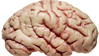
For all that science has studied and mapped the brain in recent decades, the mind remains a black box. A famous 1974 essay by the philosopher Thomas Nagel asked, “What Is It Like to Be a Bat?” and concluded that we will never know; another consciousness—another person’s, let alone a member of another species—can never be comprehended or accessed. For Rao and a few others to open that door a tiny crack, then, is a notable achievement, even if the work has mostly underscored how big a challenge it is, both conceptually and technologically.

The computing power and the programming are up to the challenge; the problem is the interface between brain and computer, and especially the one that goes in the direction from computer to brain. How do you deliver a signal to the right group of nerve cells among the estimated 86 billion in a human brain? The most efficient approach is an implanted transceiver that can be hard-wired to stimulate small regions of the brain, even down to a single neuron. Such devices are already in use for “deep brain stimulation,” a technique for treating patients with Parkinson’s and other disorders with electrical impulses. But it’s one thing to perform brain surgery for an incurable disease, and something else to do it as part of an experiment whose benefits are speculative at best.

So Rao used a technique that does not involve opening the skull, a fluctuating magnetic field to induce a tiny electric current in a region of the brain. It appears to be safe—his first volunteer was his collaborator, Stocco—but it is a crude mechanism. The smallest area that can be stimulated in this way, Rao says, is not quite half an inch across. This limits its application to gross motor movements, such as hitting a button, or simple yes-or-no communication.

Another way to transmit information, called focused ultrasound, appears to be capable of stimulating a region of the brain as small as a grain of rice. While the medical applications for ultrasound, such as imaging and tissue ablation, use high frequencies, from 800 kilohertz up to the megahertz range, a team led by Harvard radiologist Seung-Schik Yoo found that a frequency of 350 kilohertz works well, and apparently safely, to send a signal to the brain of a rat. The signal originated with a human volunteer outfitted with an EEG, which sampled his brainwaves; when he focused on a specific pattern of lights on a computer screen, a computer sent an ultrasound signal to the rat, which moved his tail in response. Yoo says the rat showed no ill effects, but the safety of focused ultrasound on the human brain is unproven. Part of the problem is that, unlike magnetic stimulation, the mechanism by which ultrasound waves—a form of mechanical energy—creates an electric potential isn’t fully understood. One possibility is that it operates indirectly by “popping” open the vesicles, or sacs, within the cells of the brain, flooding them with neurotransmitters, like delivering a shot of dopamine to exactly the right area. Alternatively, the ultrasound could induce cavitation—bubbling—in the cell membrane, changing its electrical properties. Yoo suspects that the brain contains receptors for mechanical stimulation, including ultrasound, which have been largely overlooked by neuroscientists. Such receptors would account for the phenomenon of “seeing stars,” or flashes of light, from a blow to the head, for instance. If focused ultrasound is proven safe, and becomes a feasible approach to a computer-brain interface, it would open up a wide range of unexplored—in fact, barely imagined—possibilities.

Direct verbal communication between individuals—a more sophisticated version of Rao’s experiment, with two connected people exchanging explicit statements just by thinking them—is the most obvious application, but it’s not clear that a species possessing language needs a more technologically advanced way to say “I’m running late,” or even “I love you.” John Trimper, an Emory University doctoral candidate in psychology, who has written about the ethical implications of brain-to-brain interfaces, speculates that the technology, “especially through wireless transmissions, could eventually allow soldiers or police—or criminals—to communicate silently and covertly during operations.” That would be in the distant future. So far, the most content-rich message sent brain-to-brain between humans traveled from a subject in India to one in Strasbourg, France. The first message, laboriously encoded and decoded into binary symbols by a Barcelona-based group, was “hola.” With a more sophisticated interface one can imagine, say, a paralyzed stroke victim communicating to a caregiver—or his dog. Still, if what he’s saying is, “Bring me the newspaper,” there are, or will be soon, speech synthesizers—and robots—that can do that. But what if the person is Stephen Hawking, the great physicist afflicted by ALS, who communicates by using a cheek muscle to type the first letters of a word? The world could surely benefit from a direct channel to his mind.

Maybe we’re still thinking too small. Maybe an analog to natural language isn’t the killer app for a brain-to-brain interface. Instead, it must be something more global, more ambitious—information, skills, even raw sensory input. What if medical students could download a technique directly from the brain of the world’s best surgeon, or if musicians could directly access the memory of a great pianist? “Is there only one way of learning a skill?” Rao muses. “Can there be a shortcut, and is that cheating?” It doesn’t even have to involve another human brain on the other end. It could be an animal—what would it be like to experience the world through smell, like a dog—or by echolocation, like a bat? Or it could be a search engine. “It’s cheating on an exam if you use your smartphone to look things up on the Internet,” Rao says, “but what if you’re already connected to the Internet through your brain? Increasingly the measure of success in society is how quickly we access, digest and use the information that’s out there, not how much you can cram into your own memory. Now we do it with our fingers. But is there anything inherently wrong about doing it just by thinking?”

Or, it could be your own brain, uploaded at some providential moment and digitally preserved for future access. “Let’s say years later you have a stroke,” says Stocco, whose own mother had a stroke in her 50s and never walked again. “Now, you go to rehab and it’s like learning to walk all over again. Suppose you could just download that ability into your brain. It wouldn’t work perfectly, most likely, but it would be a big head start on regaining that ability.”

Miguel Nicolelis, a creative Duke neuroscientist and a mesmerizing lecturer on the TED Talks circuit, knows the value of a good demonstration. For the 2014 World Cup, Nicolelis—a Brazilian-born soccer aficionado—worked with others to build a robotic exoskeleton controlled by EEG impulses, enabling a young paraplegic man to deliver the ceremonial first kick. Much of his work now is on brain-to-brain communication, especially in the highly esoteric techniques of linking minds to work together on a problem. The minds aren’t human ones, so he can use electrode implants, with all the advantages that conveys.

One of his most striking experiments involved a pair of lab rats, learning together and moving in synchrony as they communicated via brain signals. The rats were trained in an enclosure with two levers and a light above each. The left- or right-hand light would flash, and the rats learned to press the corresponding lever to receive a reward. Then they were separated, and each fitted with electrodes to the motor cortex, connected via computers that sampled brain impulses from one rat (the “encoder”), and sent a signal to a second (the “decoder”). The “encoder” rat would see one light flash—say, the left one—and push the left-hand lever for his reward; in the other box, both lights would flash, so the “decoder” wouldn’t know which lever to push—but on receiving a signal from the first rat, he would go to the left as well.

Nicolelis added a clever twist to this demonstration. When the decoder rat made the correct choice, he was rewarded, and the encoder got a second reward as well. This served to reinforce and strengthen the (unconscious) neural processes that were being sampled in his brain. As a result, both rats became more accurate and faster in their responses—“a pair of interconnected brains…transferring information and collaborating in real time.” In another study, he wired up three monkeys to control a virtual arm; each could move it in one dimension, and as they watched a screen they learned to work together to manipulate it to the correct location. He says he can imagine using this technology to help a stroke victim regain certain abilities by networking his brain with that of a healthy volunteer, gradually adjusting the proportions of input until the patient’s brain is doing all the work. And he believes this principle could be extended indefinitely, to enlist millions of brains to work together in a “biological computer” that tackled questions that could not be posed, or answered, in binary form. You could ask this network of brains for the meaning of life—you might not get a good answer, but unlike a digital computer, “it” would at least understand the question. At the same time, Nicolelis criticizes efforts to emulate the mind in a digital computer, no matter how powerful, saying they’re “bogus, and a waste of billions of dollars.” The brain works by different principles, modeling the world by analogy. To convey this, he proposes a new concept he calls “Gödelian information,” after the mathematician Kurt Gödel; it’s an analog representation of reality that cannot be reduced to bytes, and can never be captured by a map of the connections between neurons (“Upload Your Mind,” see below). “A computer doesn’t generate knowledge, doesn’t perform introspection,” he says. “The content of a rat, monkey or human brain is much richer than we could ever simulate by binary processes.”

The cutting edge of this research involves actual brain prostheses. At the University of Southern California, Theodore Berger is developing a microchip-based prosthesis for the hippocampus, the part of the mammal­ian brain that processes short-term impressions into long-term memories. He taps into the neurons on the input side, runs the signal through a program that mimics the transformations the hippocampus normally performs, and sends it back into the brain. Others have used Berger’s technique to send the memory of a learned behavior from one rat to another; the second rat then learned the task in much less time than usual. To be sure, this work has only been done in rats, but because degeneration of the hippocampus is one of the hallmarks of dementia in human beings, the potential of this research is said to be enormous.

Given the sweeping claims for the future potential of brain-to-brain communication, it’s useful to list some of the things that are not being claimed. There is, first, no implication that humans possess any form of natural (or supernatural) telepathy; the voltages flickering inside your skull just aren’t strong enough to be read by another brain without electronic enhancement. Nor can signals (with any technology we possess, or envision) be transmitted or received surreptitiously, or at a distance. The workings of your mind are secure, unless you give someone else the key by submitting to an implant or an EEG. It is, however, not too soon to start considering the ethical implications of future developments, such as the ability to implant thoughts in other people or control their behavior (prisoners, for example) using devices designed for those purposes. “The technology is outpacing the ethical discourse at this time,” Emory’s Trimper says, “and that’s where things get dicey.” Consider that much of the brain traffic in these experiments—and certainly anything like Nicolelis’ vision of hundreds or thousands of brains working together—involves communicating over the Internet. If you’re worried now about someone hacking your credit card information, how would you feel about sending the contents of your mind into the cloud?There’s another track, though, on which brain-to-brain communication is being studied. Uri Hasson, a Princeton neuroscientist, uses functional magnetic resonance imaging to research how one brain influences another, how they are coupled in an intricate dance of cues and feedback loops. He is focusing on a communication technique that he considers far superior to EEGs used with transcranial magnetic stimulation, is noninvasive and safe and requires no Internet connection. It is, of course, language.

Read more: http://www.smithsonianmag.com/innovation/why-brain-brain-communication-no-longer-unthinkable-180954948/#y1xADWfAk1VkKIJc.99

Uploading Our Minds into Digital Space


Human cortical neurons in the brain. (David Scharf/Corbis)

By Jerry Adler
Smithsonian Magazine

Ken Hayworth, a neuroscientist, wants to be around in 100 years but recognizes that, at 43, he’s not likely to make it on his own. Nor does he expect to get there preserved in alcohol or a freezer; despite the claims made by advocates of cryonics, he says, the ability to revivify a frozen body “isn’t really on the horizon.” So Hayworth is hoping for what he considers the next best thing. He wishes to upload his mind—his memories, skills and personality—to a computer that can be programmed to emulate the processes of his brain, making him, or a simulacrum, effectively immortal (as long as someone keeps the power on).

Hayworth’s dream, which he is pursuing as president of the Brain Preservation Foundation, is one version of the “technological singularity.” It envisions a future of “substrate-independent minds,” in which human and machine consciousness will merge, transcending biological limits of time, space and memory. “This new substrate won’t be dependent on an oxygen atmosphere,” says Randal Koene, who works on the same problem at his organization, Carboncopies.org. “It can go on a journey of 1,000 years, it can process more information at a higher speed, it can see in the X-ray spectrum if we build it that way.” Whether Hayworth or Koene will live to see this is an open question. Their most optimistic scenarios call for at least 50 years, and uncounted billions of dollars, to implement their goal. Meanwhile, Hayworth hopes to achieve the ability to preserve an entire human brain at death—through chemicals, cryonics or both—to keep the structure intact with enough detail that it can, at some future time, be scanned into a database and emulated on a computer.

That approach presumes, of course, that all of the subtleties of a human mind and memory are contained in its anatomical structure—conventional wisdom among neuroscientists, but it’s still a hypothesis. There are electrochemical processes at work. Are they captured by a static map of cells and synapses? We won’t know, advocates argue, until we try to do it.

The initiatives require a big bet on the future of technology. A 3-D map of all the cells and synapses in a nervous system is called a “connectome,” and so far researchers have produced exactly one, for a roundworm called Caenorhabditis elegans, with 302 neurons and about 7,000 connections among them. A human brain, according to one reasonable estimate, has about 86 billion neurons and 100 trillion synapses. And then there’s the electrochemical activity on top of that. In 2013, announcing a federal initiative to produce a complete model of the human brain, Francis Collins, head of the National Institutes of Health, said it could generate “yottabytes” of data—a million million million megabytes. To scan an entire human brain at the scale Hayworth thinks is necessary—effectively slicing it into virtual cubes ten nanometers on a side—would require, with today’s technology, “a million electron microscopes running in parallel for ten years.” Mainstream researchers are divided between those who regard Hayworth’s quest as impossible in practice, and those, like Miguel Nicolelis of Duke University, who consider it impossible in theory. “The brain,” he says, “is not computable.”

And what does it mean for a mind to exist outside a brain? One immediately thinks of the disembodied HAL in 2001: A Space Odyssey. But Koene sees no reason that, if computers continue to grow smaller and more powerful, an uploaded mind couldn’t have a body—a virtual one, or a robotic one. Will it sleep? Experience hunger, pain, desire? In the absence of hormones and chemical neurotransmitters, will it feel emotion? It will be you, in a sense, but will you be it?

These questions don’t trouble Hayworth. To him, the brain is the most sophisticated computer on earth, but only that, and he figures his mind could also live in one made of transistors instead. He hopes to become the first human being to live entirely in cyberspace, to send his virtual self into the far future.

Read more: http://www.smithsonianmag.com/innovation/quest-upload-mind-into-digital-space-180954946/#OBRGToqVzeqftrBt.99

Scientists claim to have localized the brain region of Christmas Spirit

It being a widespread phenomenon, researchers were interested to determine where in the human body holiday people find their penchant for “merriment, gifts, delightful smells, and copious amounts of good food.” They recruited 26 participants to undergo functional magnetic resonance imaging (fMRI), given this technology has long been used to locate emotional and functional centers in the human brain; feelings such as joy, sorrow, and disgust.

Participants watched a series of images through video goggles as they were being scanned. It was a continual series of 84 images, where each one was displayed for two seconds each and were organized in a way that after six consecutive images of all things Christmas, there were then six everyday images with similar form and features. Afterward, participants answered questionnaires about their Christmas traditions (if any), feelings associated with Christmas, and ethnicity.

Based on their answers, 10 were put into the “Christmas group,” 10 were put into the “non-Christmas group,” and six ended up being excluded for either too strong a spirit or non-positive associations with Christmas. The Christmas group was comprised entirely of ethnic Danes, while the non-Christmas group consisted of people from Pakistan, India, Iraq, and Turkey.

The scans showed an increase of brain activity in the primary visual cortex of both groups when the images were Christmas-themed compared to everyday images. The Christmas group, however, also experienced increased activity in the somatosensory cortex. When comparing brain activation maps of both groups, researchers found five areas with more neural activity among the Christmas group responding to Christmas images than those in the non-Christmas group.

After the primary visual and somatosensory cortex, the left primary motor and premotor cortex, right inferior/superior parietal lobe, and bilateral primary somatosensory cortex were also activated. These parts of the brain have been associated with spirituality, somatic sense, and recognizing facial emotional.

“There is a cerebral response when people view Christmas images, and there are differences in this response between people who celebrate Christmas compared with those with no Christmas traditions,” researchers wrote. “Cerebral perfusion was similar between the two groups, despite the Christmas group’s yearly yuletide feast.”

Researchers propose the Christmas spirit is a functional neurological network, which they realize their colleagues may not agree with. They anticipate the argument that “studies such as the present one overemphasize the importance of localized brain activity and that attempts to localize complex emotions in the brain contribute little to the understanding of these emotions.” But, they added, “with the good spirit of the holiday they reject these negative perspectives.”

“We generally believe that fMRI is an outstanding technology for exploring the brain, but that any fMRI experiment is only as good as its hypothesis, design, and interpretation,” they explained. “While celebrating the current results at a subsequent Christmas party, we discussed some limitations of the study.”

They continued: “For instance, the study design doesn’t distinguish whether the observed activation is Christmas specific or the result of any combination of joyful, festive, or nostalgic emotions in general. The paired Christmas/non-Christmas pictures might have been systematically different in a way that we were not aware of — for example, the ‘Christmas pictures’ containing more red color. Maybe the groups were different in other ways apart from the obvious cultural difference.”

But even they can acknowledge further research into this topic is necessary to identify the factors affecting one’s response to Christmas. Understanding how this spirit works as a neurological network could “be an important first step in transcultural neuroscience and the associations humans have with their festive traditions.”

Source: Haddock BT et al. Evidence of a Christmas spirit network in the brain: functional MRI study. BMJ Open. 2015.

http://www.medicaldaily.com/lack-christmas-spirit-may-be-traced-back-brain-bah-humbug-365468