Depression speeds up brain ageing

Psychologists at the University of Sussex have found a link between depression and an acceleration of the rate at which the brain ages. Although scientists have previously reported that people with depression or anxiety have an increased risk of dementia in later life, this is the first study that provides comprehensive evidence for the effect of depression on decline in overall cognitive function (also referred to as cognitive state), in a general population.

For the study, published today, Thursday 24 May 2018, in the journal Psychological Medicine, researchers conducted a robust systematic review of 34 longitudinal studies, with the focus on the link between depression or anxiety and decline in cognitive function over time. Evidence from more than 71,000 participants was combined and reviewed. Including people who presented with symptoms of depression as well as those that were diagnosed as clinically depressed, the study looked at the rate of decline of overall cognitive state – encompassing memory loss, executive function (such as decision making) and information processing speed – in older adults.

Importantly, any studies of participants who were diagnosed with dementia at the start of study were excluded from the analysis. This was done in order to assess more broadly the impact of depression on cognitive ageing in the general population. The study found that people with depression experienced a greater decline in cognitive state in older adulthood than those without depression. As there is a long pre-clinical period of several decades before dementia may be diagnosed, the findings are important for early interventions as currently there is no cure for the disease.

Lead authors of the paper, Dr Darya Gaysina and Amber John from the EDGE (Environment, Development, Genetics and Epigenetics in Psychology and Psychiatry) Lab at the University of Sussex, are calling for greater awareness of the importance of supporting mental health to protect brain health in later life.

Dr Gaysina, a Lecturer in Psychology and EDGE Lab Lead, comments: “This study is of great importance – our populations are ageing at a rapid rate and the number of people living with decreasing cognitive abilities and dementia is expected to grow substantially over the next thirty years.

“Our findings should give the government even more reason to take mental health issues seriously and to ensure that health provisions are properly resourced. We need to protect the mental wellbeing of our older adults and to provide robust support services to those experiencing depression and anxiety in order to safeguard brain function in later life.”

Researcher Amber John, who carried out this research for her PhD at the University of Sussex adds: “Depression is a common mental health problem – each year, at least 1 in 5 people in the UK experience symptoms. But people living with depression shouldn’t despair – it’s not inevitable that you will see a greater decline in cognitive abilities and taking preventative measures such as exercising, practicing mindfulness and undertaking recommended therapeutic treatments, such as Cognitive Behaviour Therapy, have all been shown to be helpful in supporting wellbeing, which in turn may help to protect cognitive health in older age.”

The research paper, ‘Affective problems and decline in cognitive state in older adults’ will be available at: https:// doi.org/10.1017/S0033291718001137 from Thursday 24 May 2018.

http://www.sussex.ac.uk/broadcast/read/44977

In Japan, a Buddhist Funeral Service for Robot Dogs


An electronics repair company gives a compassionate farewell to mechanical pets, with a traditional ceremony held in a historic temple.

By James Burch
A traveler happening upon a funeral for robot dogs might be taken aback.

Is this a performance art statement about modern life? Is it a hoax? A practical joke?

But this is actually a religious ceremony, and the emotions expressed by the human participants are genuine.

A dog-shaped robot—as opposed to say, a dish on wheels with a built-in vacuum cleaner—represented a focus on entertainment and companionship. When Sony released the AIBO (short for “artificial intelligence robot”) in 1999, 3,000 units—the greater share of the first run—were sold to the Japanese market. At an initial cost of $3,000 in today’s money, those sold out in 20 minutes.

But AIBOs never became more than a niche product, and in 2006 Sony canceled production. In seven years, they’d sold 150,000 of the robots.

Some AIBO owners had already become deeply attached to their pet robots, though. And here is where the story takes an unexpected turn.

AIBOs aren’t like a remote-control car. They were designed to move in complex, fluid ways, with trainability and a simulated mischievous streak. (Meet Sophia, the robot that almost seems human.)

Over time, they would come to “know” their human companions, who grew attached to them as if they were real dogs. (Learn how playing games helped build the modern world.)

The AIBOs’ programs included both doggish behaviors, like tail-wagging, and humanlike actions, such as dancing, and—in later models—speech.

So when Sony announced in 2014 that they would no longer support updates to the aging robots, some AIBO owners heard a much more somber message: Their pet robot dogs would die. The community of devoted owners began sharing tips on providing care for their pets in the absence of official support.

Nobuyuki Norimatsu didn’t intend to create a cyberhospital. According to Nippon.com, the former Sony employee, who founded the repair company A-Fun in a Chiba Prefecture, a Tokyo suburb, simply felt a duty to stand by the company’s products. (Watch sunlight create a heart inside a Chiba Prefecture cave.)

And then came a request to repair an AIBO. Nippon.com reports that, at first, no one knew exactly what to do, but months of trial and error saw the robodog back on its feet. Soon, A-Fun had a steady demand for AIBO repairs—which could only be made by cannibalizing parts from other, defunct AIBOs.

Hiroshi Funabashi, A-Fun’s repairs supervisor, observes that the company’s clients describe their pets’ complaints in such terms as “aching joints.” Funabashi realized that they were not seeing a piece of electronic equipment, but a family member.

And Norimatsu came to regard the broken AIBOs his company received as “organ donors.” Out of respect for the owners’ emotional connection to the “deceased” devices, Norimatsu and his colleagues decided to hold funerals.

A-Fun approached Bungen Oi, head priest of Kōfuku-ji, a Buddhist temple in Chiba Prefecture’s city of Isumi. Oi agreed to take on the duty of honoring the sacrifice of donor AIBOs before their disassembly. In 2015, the centuries-old temple held its first robot funeral for 17 decommissioned AIBOs. Just as with the repairs, demand for funeral ceremonies quickly grew.

The most recent service, in April 2018, brought the total number of dearly departed AIBOs to about 800. Tags attached to the donor bodies record the dogs’ and owners’ names.

Services include chanting and the burning of incense, as they would for the human departed. A-Fun employees attend the closed ceremonies, serving as surrogates for the “families” of the pets, and pliers are placed before the robodogs in place of traditional offerings like fruit. Robots even recite Buddhist sutras, or scriptures. (Meet a master of Japanese Tea Ceremony.)

According to Head Priest Oi, honoring inanimate objects is consistent with Buddhist thought. Nippon.com quotes the priest: “Even though AIBO is a machine and doesn’t have feelings, it acts as a mirror for human emotions.” Speaking with videographer Kei Oumawatari, Oi cites a saying, “Everything has Buddha-nature.”

AIBOs and similar robots are especially popular among the elderly, and limited research hints that robots could potentially act like therapy animals—though attachment to machines could also be a symptom of loneliness, an increasing concern in Japan. (READ: Will a robot be your friend or steal your job?)

Sony has now introduced a new line of more advanced AIBOs, and although they are apparently not technologically compatible with their predecessors, it would seem they stand a good chance of finding similar popularity with those who can appreciate the soul of a machine.

Though AIBO funerals are closed to the public, travelers in Japan can at other times visit the Isumi’s historic Kōfuku-ji, one of several temples in the region including work by the master wood carver IHACHI. Isumi tourist info (Click on “Select Language” in the upper right for English.)

To learn about other personal robots, such as Paro, a therapeutic seal-bot, visit the permanent exhibit “Create your future” at Miraikan, the National Museum of Emerging Science and Innovation in Tokyo.

https://www.nationalgeographic.com/travel/destinations/asia/japan/in-japan–a-buddhist-funeral-service-for-robot-dogs/

Thanks to Kebmodee for bringing this to the It’s Interesting community.

How ancestors of living birds survived asteroid strike

The ancestors of modern birds may have survived the asteroid strike that wiped out the rest of their kin by living on the forest floor.

The new theory, based on studying fossilised plants and ornithological data, helps explain how birds came to dominate the planet.

The asteroid impact 66 million years ago laid waste to the world’s forests.

Ground-dwelling bird ancestors managed to survive, eventually taking to the trees when the flora recovered.

“It seems clear that being a relatively small-bodied bird capable of surviving in a tree-less world would have conferred a major survival advantage in the aftermath of the asteroid strike,” said Dr Daniel Field of the Milner Centre for Evolution at the University of Bath.

We already know that the early ancestors of modern birds were probably capable of flight, and relatively small in size.

Scientists have now pieced together their ecology to better understand how these partridge-like bird ancestors managed to avoid destruction in a particularly bleak moment in the Earth’s history.

“Teasing these stories from the rock record is a challenge when the action took place over 66 million years ago, over a relatively short period of time,” said Dr Field, who led a team of UK, US and Swedish researchers.

The plant fossil record shows that the asteroid caused global deforestation and extinction of most flowering plants, destroying the habitats of tree-dwelling animals.

Birds didn’t move back into the trees again until the forests recovered thousands of years later.

“The recovery of canopy-forming trees such as palms and pines happened much later, which coincides with the evolution and explosion of diversity of tree-dwelling birds,” said Dr Antoine Bercovici from Smithsonian Institution.

The researchers found that once the forests had recovered, birds began to adapt to living in trees, acquiring shorter legs than their ground-dwelling ancestors and various specialisations for perching on branches.

They eventually diversified into ostriches and their relatives, chickens and their relatives, and ducks and their relatives.

“Perhaps the best modern analogue for one of the surviving birds lineages are modern tinamous – this is a modern group of flying relatives of ostriches: they are relatively small bodied, and live on the ground,” said Dr Field.

Today’s “amazing living bird diversity can be traced to these ancient survivors”, he added.

The research is published in the journal Current Biology.

http://www.bbc.com/news/science-environment-44226534

Ultrasound Fires Up the Auditory Cortex—Even Though Animals Can’t Hear It


Ultrasound activates auditory pathways in the rodent brain (red arrows) regardless of where in the brain the ultrasound-generating transducer is placed.

By Abby Olena

Activating or suppressing neuronal activity with ultrasound has shown promise both in the lab and the clinic, based on the ability to focus noninvasive, high-frequency sound waves on specific brain areas. But in mice and guinea pigs, it appears that the technique has effects that scientists didn’t expect. In two studies published today (May 24) in Neuron, researchers demonstrate that ultrasound activates the brains of rodents by stimulating an auditory response—not, as researchers had presumed, only the specific neurons where the ultrasound is focused.

“These papers are a very good warning to folks who are trying to use ultrasound as a tool to manipulate brain activity,” says Raag Airan, a neuroradiologist and researcher at Stanford University Medical Center who did not participate in either study, but coauthored an accompanying commentary. “In doing these experiments going forward [the hearing component] is something that every single experimenter is going to have to think about and control,” he adds.

Over the past decade, researchers have used ultrasound to elicit electrical responses from cells in culture and motor and sensory responses from the brains of rodents and primates. Clinicians have also used so-called ultrasonic neuromodulation to treat movement disorders. But the mechanism by which high frequency sound waves work to exert their influence is not well understood.

The University of Minnesota’s Hubert Lim studies ways to restore hearing, but many of the strategies that his group uses are invasive, such as cochlear implants, which require surgery to insert a device inside the ear. He says that he and his colleagues were excited by the prospect of using noninvasive and precise ultrasound to activate the parts of the brain responsible for hearing.

Lim’s team started by stimulating the brains of guinea pigs with audible noise or with pulsed ultrasound directly over the auditory cortex. They were surprised to observe similar neuronal responses to the two different stimuli because ultrasound is outside the spectrum that the guinea pigs—and humans—can hear. The researchers also found that the rodents’ neurons showed comparable electrical activity in the auditory cortex regardless of where in the brain the researchers directed the ultrasound. This raised the question: are the animals’ brains responding directly to the ultrasound or to responses of the auditory system?

When the authors cut the guinea pigs’ auditory nerves or removed their cochlear fluid, the guinea pigs stopped responding to the ultrasound and to audible noise. Lim’s team concluded that what must be happening is ultrasound moves through brain tissue and vibrates the cochlear fluid. This vibration then triggers auditory signaling and indirectly activates the auditory cortex and other brain regions, rather than ultrasound having a direct effect on the activity of the neurons.

“I am actually very hopeful that ultrasound can be a powerful tool that can not only modulate but also treat different neurologic and psychiatric disorders, and that it can achieve a noninvasive yet localized activation,” says Lim. “But what we’re trying to show in this paper is that there are many confounding effects that are actually happening with ultrasound, and we have to remove those effects to really see how it’s activating the brain.”

A coauthor on the companion study, Mikhail Shapiro of Caltech, says that previous work showing that it is possible to apply ultrasound to the brains of mice and rats to elicit electrical activity and movement in their limbs left him and his colleagues curious about how it works. To determine where and when neural activation happens, they applied ultrasonic pulses to the brains of transgenic mice that have neurons that light up when stimulated. As with guinea pigs, ultrasound is inaudible to mice.

“To our surprise, we found that the main activation pattern that we were seeing was not in the region where we were applying the ultrasound directly, but actually in the auditory areas of the brain, those responsible for processing information about sound,” Shapiro tells The Scientist.

Consistent with the findings of Lim and colleagues, Shapiro and his coauthors determined that the mouse brains lit up across the cortex, starting from the auditory cortex. And as in the guinea pigs, the mouse neurons responded similarly to ultrasound and audible sounds. The researchers also showed that both ultrasound and audible noise elicited motor movements that decreased when they used chemicals to deafen the mice.

“We’re not trying to imply that [the effects of ultrasound observed in previous studies are] due to this auditory side effect,” says Shapiro. “We’re very optimistic that now that we know that it’s there, we will be able to design ways to get around it and still be able to use this technology scientifically.”

Shy Shoham, a neuroscientist and biomedical engineer at New York University Langone Medical Center who did not participate in the studies, tells The Scientist that these papers highlight how careful researchers must be in the future when using ultrasound to modify neuronal function. “In the field of neural stimulation in general, we should always be very concerned about off-target effects,” he says. We must “delineate what is real and what isn’t.”

“The big take home point here is that we need to take care of the auditory effects,” says Kim Butts Pauly, who studies ultrasound neuromodulation at Stanford University Medical Center and who coauthored the accompanying commentary with Airan. “There’s been very compelling data from other studies that ultrasound can stimulate the brain and change recordings from the brain that are completely separate from any auditory effects. As we get rid of the auditory effects, then the more subtle effects may become apparent.”

H. Guo et al., “Ultrasound produces extensive brain activation via a cochlear pathway,” Neuron, doi:10.1016/j.neuron.2018.04.036, 2018.

T. Sato et al., “Ultrasonic neuromodulation causes widespread cortical activation via an indirect auditory mechanism,” Neuron, doi:10.1016/j.neuron.2018.05.009, 2018.

https://www.the-scientist.com/?articles.view/articleNo/54652/title/Ultrasound-Fires-Up-the-Auditory-Cortex-Even-Though-Animals-Can-t-Hear-It/

Semir Osmanagić claims he has found the world’s oldest pyramids in Bosnia, and shown that they generate Nikola Tesla’s predicted torison fields of standing energy.

By JON AUSTIN

Scientist Semir Osmanagić claims a series of triangular-shaped hills in his native Bosnia, are artificial pyramids that are bigger and older than those in Egypt.

Despite mainstream archaeologists saying they are just natural rock formations, Mr Osmanagic has made another bold claim that he has found Nikola Tesla’s so-called “torison fields of standing energy” at the Bosnian Pyramids site, which means we could now “communicate with aliens”.

Mr Telsa was a Serbian-American inventor, physicist, and futurist, who contributed to the design of the AC electricity supply system in 1888.

His ideas became more left-field and experimental towards the end of the 1800s, and he devised the theory of “standing waves” of energy coming from Earth that meant electricity could be transmitted wirelessly over long distances.

Mr Osmanagić has claimed the alleged discovery at one of the “34,000 year old” pyramids he calls the Pyramid of the Sun “changes the history of planet” and could lead to intergalactic communication.

He wrote: “The discovery of Tesla’s standing waves at the top of the Bosnian Pyramid of the Sun— which are believed to travel faster than the speed of light, while not losing strength as they pass through cosmic bodies—prove the existence of something referred to as a cosmic web or cosmic internet which allow for a immediate intergalactic communication throughout the universe.

“Recorded energetic phenomena above the Pyramid of the Sun at Visoko seek a different definition of a pyramid compared to conventional, dogmatic explanations.

“The pyramids are energy boosters that send and receive information through the Sun.”

Tesla devised a theory of standing waves saying they travel faster than light, meaning they could “move through other cosmic bodies without wasting energy.”

Mr Osmanagić claims on the surface of and underneath the Bosnian Pyramid of the Sun, archaeological digs have found quartz crystals. The crystal is present in the underground tunnels as well, a mineral he says receives then amplifies energy.

He claims there are seven levels of tunnels inside the pyramid and that this amplifies the intensity of the energy.

Osmanagić also supports the ancient aliens theory that advanced beings came to Earth thousands of years ago to help build the pyramids.

He added: “Life originated thanks to an intervention on our planet, species on Earth change in the long term through experiments where evolution plays a minor role, and homo sapiens is the result of genetic engineering.

“And, of course, we are not the first nor the most advanced civilisation in the history of the planet.”

Boston University’s archaeological professor, Curtis Runnels, has been one of many to attempt to put the Bosnian Pyramid claims to bed.

He said: “Early prehistoric cultures, including village farmers of the Neolithic period [back to 9,000 years ago], and before them Stone Age hunters and gatherers, did not have populations large enough or social structures organised in ways that would have permitted the creation of pyramids on a large scale.

“Pyramidal shapes offer the least resistance to such forces, and are common forms in nature.”

https://www.express.co.uk/news/weird/773789/Bosnian-Pyramid-Nikola-Tesla-standing-waves-aliens

Early life trauma in men associated with changes in sperm that are later transmitted to their children.

Exposure to early life trauma can lead to poor physical and mental health in some individuals, which can be passed on to their children. Studies in mice show that at least some of the effects of stress can be transmitted to offspring via environmentally-induced changes in sperm miRNA levels.

A new epigenetics study raises the possibility that the same is true in humans. It shows for the first time that the levels of the same two sperm miRNAs change in both men and mice exposed to early life stress. In mice, the negative effects of stress are transmitted to offspring. The study is published On May 23rd in Translational Psychiatry.

“The study raises the possibility that some of the vulnerability of children is due to Lamarckian type inheritance derived from their parents’ experiences,” said Larry Feig, Ph.D., professor of Developmental, Molecular and Chemical Biology at Tufts University School of Medicine and member of the Cell, Molecular and Developmental Biology and Neuroscience programs at the Sackler School of Graduate Biomedical Sciences at Tufts.

The human part of the study utilized the Adverse Childhood Experiences (ACE) questionnaire as an indicator of men’s early life trauma. The ACE Study questionnaire includes 10 yes or no questions about one’s experiences until the age of 18, including physical, verbal, or sexual abuse, and physical or emotional neglect. Other questions relate to one’s family members. Four or more yes answers put one at significantly increased risk for future mental and physical health problems. According to a ChildTrends research brief published in 2014, a remarkably high percentage (~10 percent) of the population report scores at or above this cutoff.

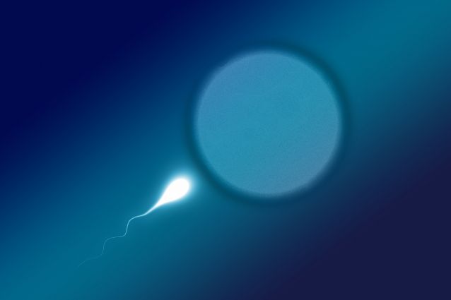
miRNAs constitute a newly appreciated type of gene regulator, where each miRNA controls a distinct set of genes. Until recently, sperm from fathers were thought to contribute only DNA to the mother’s egg upon fertilization, but new data in mice indicate that sperm also contribute miRNAs that influence the next generation. Sperm miRNA expression in humans is known to be affected by environmental factors, such as smoking and obesity, but no human study to date has documented the effects of stress.

The new study found that among 28 Caucasian male volunteers, the expression of two highly related sperm miRNAs, miR-449 and miR-34, were inversely proportional to the men’s ACE scores. Men with the most extensive early abuse (highest ACE scores) had as much as a 300-fold reduction in the two sperm miRNAs compared to men with the least abuse.

The idea that these changes can affect the next generation is supported by additional findings in the study, e.g.:

the same sperm miRNA changes that take place in men with high ACE scores also occur in mice exposed to early life social instability stress, which Feig’s lab has shown previously leads to anxiety and sociability defects in female offspring of stressed males for at least three generations;
these two sets of miRNAs are known to work together in mice to allow proper development of the brain and sperm;
in humans, miR-34c has been implicated in promoting early embryo development;
the mouse studies showed that the decline in these sperm miRNA levels is transmitted to the next generation; and
when these embryos mature, these miRNAs are also reduced in the sperm of their male offspring who pass on stress behaviors to their female offspring.
“This is the first study to show that stress is associated with altered levels of sperm miRNAs in humans. We are currently setting up a new, larger study in men, and additional experiments in mice that could yield further support for the idea that changes in these sperm miRNAs do, in fact, contribute to an elevation of stress-related disorders across generations,” said David Dickson, an M.D./Ph.D. student at Tufts and first author of the study.

“Looking to the future, we may be able to figure out a way to restore the low miRNA levels found in men exposed to extreme trauma, because epigenetic changes, such as stress-induced decreases in sperm miRNA expression, are reversible, unlike genetic changes that alter the DNA sequence,” Dickson added.

For example, obesity has been shown to alter specific sperm miRNA levels in men, while bariatric surgery and subsequent weight loss can reverse the changes. In addition, Isabelle Mansuy’s lab has reversed some of the negative effects of stress in mice across generations by exposing mice to an “enriched environment” that involves extensive social interactions, exercise and opportunities to explore their surroundings.

Feig pointed out that in addition to focusing on the potential transgenerational effects of stress, there is a growing appreciation that physicians should collect information on childhood trauma for the sake of the patients who are experiencing this early trauma.

This is because “childhood abuse, trauma and dysfunction adds to the risk of future physical and psychiatric maladies, and significant exposure to abusive and/or dysfunctional families is remarkably common. Moreover, sensitivity to PTSD has been shown to correlate with ACE score, implying the ACE questionnaire could be used as a screening tool to identify people who should take extra precaution to avoid potentially traumatic experiences,” he said.

“However, some people may not answer the ACE survey accurately due to inaccurate recall or because of the sensitive nature of many of the questions, particularly in settings that do not allow anonymity and/or where their answers could affect their future. Thus, discovery of unbiased markers for early trauma, like specific sperm miRNA content, could complement ACE surveys in some clinical settings to bolster preventative medicine,” he concluded.

The authors note that the relatively small sample size limits their ability to more deeply explore the association between ACE scores and miRNA expression. In addition, a longitudinal study with information on behavioral and psychological factors throughout adulthood, with repeated measurements of sperm miRNA content, could allow for further exploration on the effect of cumulative exposure to childhood trauma on miRNA.

Additional authors are Jessica Paulus, Sc.D., Tufts Medical Center as well as Tufts University School of Medicine and the Sackler School; Virginia Mensah, M.D., formerly in Feig’s lab with Women & Infants Hospital and the Warren Alpert Medical School at Brown University and now with the Reproductive Science Center of New Jersey; Janis Lem, Ph.D., Tufts Medical Center; Lorena Saavedra-Rodriguez, Ph.D., formerly a postdoctoral fellow in Feig’s laboratory at Tufts and now with a biopharmaceutical company; and Adrienne Gentry, D.O. and Kelly Pagidas, M.D., University of Louisville School of Medicine.

This study was supported by awards from the National Institute of Mental Health of the National Institutes of Health (R01MH107536), as well as the Tufts Center for Neuroscience Research (National Institute of Neurological Disorders and Stroke of the NIH, P30NS047243). The content is solely the responsibility of the authors and does not necessarily represent the official views of the National Institutes of Health or other funders.

Dickson, D.A., Paulus, J.K., Mensah, V., Lem, J., Saavedra-Rodriguez, L., Gentry, A., Pagidas, K., and Feig, L. A. (2018). Reduced levels of miRNAs 449 and 34 in sperm of mice and men exposed to early life stress. Translational Psychiatry. https://doi.org/10.1038/s41398-018-0146-2

https://now.tufts.edu/news-releases/early-life-trauma-men-associated-reduced-levels-sperm-micrornas

Pluto May Have Formed from 1 Billion Comets


This view of Pluto’s Sputnik Planitia nitrogen-ice plain was captured by NASA’s New Horizons spacecraft during its flyby of the dwarf planet in July 2015.

At its heart, Pluto may be a gigantic comet.

Researchers have come up with a new theory about the dwarf planet’s origins after taking a close look at Sputnik Planitia, the vast nitrogen-ice glacier that constitutes the left lobe of Pluto’s famous “heart” feature.

“We found an intriguing consistency between the estimated amount of nitrogen inside the glacier and the amount that would be expected if Pluto was formed by the agglomeration of roughly a billion comets or other Kuiper Belt objects similar in chemical composition to 67P, the comet explored by Rosetta,” Chris Glein, a scientist at the Southwest Research Institute (SwRI) in San Antonio, said in a statement.

The European Space Agency’s Rosetta mission orbited Comet 67P/Churyumov-Gerasimenko from 2014 through 2016. The orbiting mothership also dropped a lander named Philae onto the icy body, pulling off the first-ever soft touchdown on a comet’s surface. (The Kuiper Belt is the ring of frigid objects beyond Neptune’s orbit; Pluto is the belt’s largest resident.)

Glein and his SwRI colleague Hunter Waite devised the new Pluto-formation scenario after analyzing data from Rosetta and NASA’s New Horizons mission, which flew by Pluto in July 2015.

The scientists also made some inferences about the dwarf planet’s evolution in their new study, which was published online Wednesday (May 23) in the journal Icarus.

“Our research suggests that Pluto’s initial chemical makeup, inherited from cometary building blocks, was chemically modified by liquid water, perhaps even in a subsurface ocean,” Glein said.

Glein and Waite aren’t claiming to have nailed down Pluto’s origin definitively; a “solar model,” in which the dwarf planet coalesced from cold ices with a chemical composition closer to that of the sun, also remains in play, the duo said.

“This research builds upon the fantastic successes of the New Horizons and Rosetta missions to expand our understanding of the origin and evolution of Pluto,” Glein said.

“Using chemistry as a detective’s tool, we are able to trace certain features we see on Pluto today to formation processes from long ago,” he added. “This leads to a new appreciation of the richness of Pluto’s ‘life story,’ which we are only starting to grasp.”

Rosetta’s mission ended in September 2016, when the probe’s handlers steered it to an intentional crash-landing on 67P’s surface. New Horizons’ work, however, is far from done. The NASA spacecraft is speeding toward a flyby of a small Kuiper Belt object known officially as 2014 MU69 (and unofficially as Ultima Thule). This close encounter, which will occur on Jan. 1, 2019, about 1 billion miles (1.6 billion kilometers) beyond Pluto’s orbit, is the centerpiece of New Horizons’ extended mission.

https://www.space.com/40687-pluto-formation-1-billion-comets.html

A Hangover Pill? Tests on drunk mice show promise

“Civilization begins with distillation,” said William Faulkner, a writer and drinker. Although our thirst for alcohol dates back to the Stone Age, nobody has figured out a good way to deal with the ensuing hangover after getting drunk.

As a chemical engineering professor and wine enthusiast, I felt I needed to find a solution. As frivolous as this project may sound, it has serious implications. Between 8 and 10 percent of emergency room visits in America are due to acute alcohol poisoning. Alcohol is the leading risk factor for premature deaths and disability among people aged 15-49 and its abuse leads to serious health problems, including cardiovascular and liver cancer. Despite these sobering facts, current treatments for alcohol overdose largely rely on the body’s own enzymes to break down this drug.

I decided to design an antidote that could help people enjoy wine or cocktails or beer without a hangover, and at the same time create a lifesaving therapy to treat intoxication and overdose victims in the ER. I chose to create capsules filled with natural enzymes usually found in liver cells to help the body process the alcohol faster.

Together with professor Cheng Ji, an expert in liver diseases from Keck School of Medicine at the University of Southern California, and my graduate student Duo Xu, we developed an antidote and tested it in mice.

Inspired by the body’s approach for breaking down alcohol, we chose three natural enzymes that convert alcohol into harmless molecules that are then excreted. That might sound simple, because these enzymes were not new, but the tricky part was to figure out a safe, effective way to deliver them to the liver.

To protect the enzymes, we wrapped each of them in a shell, using a material the U.S. Food and Drug Administration had already approved for pills. We then injected these nanocapsules into the veins of drunk mice where they hurtled through the circulatory system, eventually arriving in the liver where they entered the cells and served as mini–reactors to digest alcohol.

We showed that in inebriated mice (which fall asleep much faster than drunk humans), the treatment decreased the blood alcohol level by 45 percent in just four hours compared to mice that didn’t receive any. Meanwhile, the blood concentration of acetaldehyde – a highly toxic compound that is carcinogenic, causes headaches and vomiting, makes people blush after drinking, and is produced during the normal alcohol metabolism – remained extremely low. The animals given the drug woke from their alcohol-induced slumber faster than their untreated counterparts – something all college students would appreciate.

The ability to efficiently break down alcohol quickly should help patients wake up earlier and prevent alcohol poisoning. It should also protect their liver from alcohol–associated stress and damage.

We are currently completing tests to ensure that our nanocapsules are safe and don’t trigger unexpected or dangerous side effects. If our treatments prove effective in animals, we could begin human clinical trials in as early as one year.

This sort of antidote won’t stop people from going too far when consuming alcohol, but it could help them recover quicker.

https://www.technologynetworks.com/neuroscience/articles/a-hangover-pill-tests-on-drunk-mice-show-promise-302970?utm_campaign=NEWSLETTER_TN_Neuroscience_2017&utm_source=hs_email&utm_medium=email&utm_content=63148685&_hsenc=p2ANqtz-_9-CBGC564lH1Jr5Fxrauf8vQZ42sDx9gSSQj_dPJTj3gm3QDvY74R4WiynR1vM5L7tdtTLBIV40iEWBKcEB7JzwFUnQ&_hsmi=63148685

Shipwreck laden with billions in treasure discovered off Colombia


An estimated $17 billion was lost when the San José sank in 1708.


The sinking of the San José as depicted in a 1772 landscape by British painter Samuel Scott. (Photo: Samuel Scott)


The decorative carvings on these cannons allowed researchers to confirm the wreck as the remains of the San José. (Photo: REMUS image, Woods Hole Oceanographic Institution)


Do these centuries-old tea cups sit upon treasure worth more than $17 billion? (Photo: REMUS image, Woods Hole Oceanographic Institution)

The wreck of the San José, a long-lost treasure galleon of the Spanish Navy, has finally been located off the coast of Colombia.

The Woods Hole Oceanographic Institution (WHOI), with permission from the Colombian government, made the announcement of the discovery earlier this week, adding that the wreck’s final resting spot has been a closely guarded secret for almost three years.

“We’ve been holding this under wraps out of respect for the Colombian government,” Rob Munier, WHOI’s vice president for marine facilities and operations, told the AP.

Part of the Spanish treasure fleet, the San José was a 64-gun, three-masted galleon built in 1698 and tasked with shipping vast quantities of gold, silver, emeralds and jewelry from the South American colonies to Spain. On June 8, 1708, the vessel was sailing with the treasure fleet when it was ambushed by a British squadron off Colombia. During the battle that ensued, the San José’s powder magazines detonated, destroying the ship and taking an estimated $17 billion in previous metals and gems to the bottom of the sea.

Of the 600 people aboard, only 11 survived the sinking.

According to the WHOI, the final resting spot of the San José was finally discovered more than 300 years later at an undisclosed site off the coast of Cartagena, Colombia, on Nov. 27, 2015. An international team of scientists and engineers located the wreck at a depth of some 2,000 feet using side sonar and an autonomous underwater vehicle called REMUS 6000. The vehicle, which also mapped the wreck of the Titanic in 2010, was able to provide positive identification of the San José by locating and photographing the legendary vessel’s distinctive cannons.

“The wreck was partially sediment-covered, but with the camera images from the lower altitude missions, we were able to see new details in the wreckage and the resolution was good enough to make out the decorative carving on the cannons,” WHOI engineer and expedition leader Mike Purcell said in a statement. “MAC’s (Maritime Archaeology Consultants) lead marine archaeologist, Roger Dooley, interpreted the images and confirmed that the San José had finally been found.”

While the WHOI or any of the other participating agencies did not reveal whether any of the gold, silver, or emeralds the San José was carrying was detected, the images captured by REMUS do show several cannons, tea cups, and ceramic jugs littering the wreck site.

Should Colombia successfully excavate the site, the WHOI says the government is planning on building a “a museum and world-class conservation laboratory” to preserve and publicly display the wreck’s contents.

“We are pleased to have played a part in settling one of the great shipwreck mysteries for the benefit of the Colombian people and maritime history buffs worldwide,” WHOI Vice President for Marine Facilities and Operations Rob Munier said in the same statement. “We look forward to our continued involvement to answer the basic oceanographic research questions associated with the find.”

https://www.mnn.com/lifestyle/arts-culture/blogs/shipwreck-laden-billions-treasure-discovered-colombia