Light Enables Long-Term Memory Maintenance in Fruit Flies


S. Inami et al., “Environmental light is required for maintenance of long-term memory in Drosophila,” J Neurosci, 40:1427–39, 2020.

by Diana Kwon

As Earth rotates around its axis, the organisms that inhabit its surface are exposed to daily cycles of darkness and light. In animals, light has a powerful influence on sleep, hormone release, and metabolism. Work by Takaomi Sakai, a neuroscientist at Tokyo Metropolitan University, and his team suggests that light may also be crucial for forming and maintaining long-term memories.

The puzzle of how memories persist in the brain has long been of interest to Sakai. Researchers had previously demonstrated, in both rodents and flies, that the production of new proteins is necessary for maintaining long-term memories, but Sakai wondered how this process persisted over several days given cells’ molecular turnover. Maybe, he thought, an environmental stimulus, such as the light-dark cycles, periodically triggered protein production to enable memory formation and storage.

Sakai and his colleagues conducted a series of experiments to see how constant darkness would affect the ability of Drosophila melanogaster to form long-term memories. Male flies exposed to light after interacting with an unreceptive female showed reduced courtship behaviors toward new female mates several days later, indicating they had remembered the initial rejection. Flies kept in constant darkness, however, continued their attempts to copulate.

The team then probed the molecular mechanisms of these behaviors and discovered a pathway by which light activates cAMP response element-binding protein (CREB)—a transcription factor previously identified as important for forming long-term memories—within certain neurons found in the mushroom bodies, the memory center in fly brains.

“The fact that light is essential for long-term memory maintenance is fundamentally interesting,” says Seth Tomchick, a neuroscientist at the Scripps Research Institute in Florida who wasn’t involved in the study. However, he adds, “more work will be necessary” to fully characterize the molecular mechanisms underlying these effects.

https://www.the-scientist.com/the-literature/lasting-memories-67441?utm_campaign=TS_DAILY%20NEWSLETTER_2020&utm_source=hs_email&utm_medium=email&utm_content=87927085&_hsenc=p2ANqtz-_7gIn7Nu8ghtWiBtiy6oqTctJuYb31bx6bzhbcV3gVpx0-YoIVNtAhnXXNJT0GC496PAntAiSvYpxLdVAnvITlfOG96g&_hsmi=87927085

Spain’s oldest woman (113 years old) speaks about surviving COVID


Branyas lives in Olot, a city in Catalonia.

By Jack Guy and Al Goodman

A 113-year-old woman, thought to be the oldest in Spain, has said she feels fine after surviving a brush with coronavirus.

Video footage of Maria Branyas, who was born on March 4 1907, shows the super-centenarian speaking to the director of the care home where she lives in Olot, Catalonia.

“In terms of my health I am fine, with the same minor annoyances that anyone can have,” said Branyas in the video. It was recorded Monday, a spokeswoman for the care home told CNN.

Branyas recovered after a mild case of Covid-19. Her battle started shortly after her family visited her on March 4 to celebrate her 113th birthday, the spokeswoman said.

The family has not been able to visit in person since then. Branyas has lived for 18 years in her own private room at the Santa Maria del Tura nursing home, which is run by the Institute of the Order of San Jose of Gerona, affiliated with the Roman Catholic Church, the spokeswoman said.

Branyas was born in San Francisco in the United States, where her father worked as a journalist, reports the AFP news agency.

Over the course of her long life she has survived two world wars as well as the 1918 flu pandemic, which killed more than 50 million people around the world.

Although Branyas recovered from coronavirus, two residents of the same home died of it. The situation at the care home has since improved, said the spokeswoman.

Spain’s state of emergency, in effect since March 14, has strict confinement measures that remain in place. But with the infection and death rates now declining, the government has lifted some lockdown measures in certain parts of the country, on what it says will be a gradual reopening of activity.

But the initial lifting of these restrictions did not apply to Olot, where Branyas lives.

https://www.cnn.com/2020/05/13/europe/spain-oldest-woman-coronavirus-survivor-scli-intl/index.html

Virtual reality experiments show that cannabis increases propensity for false memories

by Amy Schleunes

When Lilian Kloft stumbled across a 2015 study showing a connection between cannabis use and susceptibility to false memories, she found herself wondering about the legal implications of the results. The study had discovered that heavy users of cannabis were more likely than controls to form false memories—recollections of events that never occurred, for example, or warped memories of events that did—even when they were not at the moment “high.”

This kind of false remembering can pose difficulties for people gathering reliable testimony in the event of a crime, says Kloft, a PhD student in psychopharmacology and forensic psychology at Maastricht University in the Netherlands. Consequently, the growing acceptance of cannabis worldwide raises questions not only about how the drug affects memory, but also about how law enforcement officials should conduct interviews with suspects, victims, and witnesses who may be under the influence or regular users of the drug.

In order to further investigate the connection between cannabis and false memory formation, Kloft and collaborators recruited 64 volunteers for a series of experiments. Participants, who were occasional cannabis users, were given a vaporizer containing either cannabis or a hemp placebo and then told to inhale deeply and hold their breath for 10 seconds. After that, the researchers tested them in three different tasks designed to induce false memories.

In the first task, the team asked the volunteers to memorize lists of words, and then to pick out those words from test lists that also included dummy words. As expected, both the sober and the intoxicated participants falsely remembered some of the dummy words. But while the sober participants mostly falsely remembered words that were strongly associated with words on the original lists, the intoxicated participants also selected less-related and completely unrelated terms.

In the next two tasks, the researchers wanted to see if they could induce false memories by providing misinformation to the participants. Hoping to imbue these tests with more real-world relevance than a list of words, Kloft and colleagues designed two immersive virtual reality scenarios involving common crimes. In the first, the “eyewitness scenario,” participants observed a fight on a train platform, after which a virtual co-witness recounted the incident but with several errors, including falsely recalling a police dog that wasn’t part of the altercation. In the “perpetrator scenario,” participants entered a crowded bar and were instructed to commit a crime themselves—to steal a purse.

The researchers observed a range of effects associated with cannabis as the intoxicated subjects interacted in these virtual environments. Some participants laughed and talked to the virtual characters in the scenarios, Kloft reports, while others became paranoid and required assistance in stealing the purse. “One person even ran away so quickly that they ripped out the whole VR setup and it fell to the ground,” she says. When researchers interviewed the participants afterward using a combination of leading and non-leading questions, those who were intoxicated showed higher rates of false memory for both the eyewitness and perpetrator scenarios compared with controls.

To look for longer-term effects of cannabis, the experimenters called the subjects back a week later and tested them again on the word lists, this time with a few different dummy words thrown in. They also re-interviewed the subjects about the VR scenarios using a combination of old and new questions. As before, they found lower memory accuracy in the word-association test in those who had been intoxicated compared with sober participants. There were no statistically significant differences between the groups for the virtual reality scenarios, a result that Kloft says may indicate memory decay over time in all participants.

Cognitive neuropsychopharmacologist Manoj Doss, a postdoc at Johns Hopkins University who was not involved in the study, has used word association and other tasks in his own research to demonstrate that tetrahydrocannabinol (THC), the main psychoactive ingredient in cannabis, increases false memories when participants attempt to retrieve information they’d previously learned. Doss says that the study by Kloft and collaborators is novel not only because it employs virtual reality, but because it shows that both the real-world scenarios and the word association task can induce false memories.

For the tests administered after one week, however, Doss notes that it’s difficult to determine if the researchers were observing actual false memories, because participants might remember both the accurate and the dummy information they encountered in the original experiment. In the follow-up test, “people might say yes to the things they’re not supposed to just because they saw them in that first test,” says Doss. He suggests that increasing the number of items tested, as well as separately analyzing the new and previously used word tests and interview questions, could reveal a higher incidence of false memories in the delayed test for the participants who took cannabis.

Giovanni Marsicano, a neuroscientist at the University of Bordeaux in France who did not participate in the research, says that the new results match up with findings he’s made in mice: animals that receive injections of THC are more likely than controls to associate unrelated stimuli—itself a sort of false memory. His work has also shown that a cannabinoid receptor known as CB1 that is highly abundant in the hippocampus and prefrontal cortex probably plays a key role in the formation of these incidental associations. One of this receptor’s main jobs is to decrease the release of neurotransmitters. Marsicano hypothesizes that when the CB1 receptor is activated, neural signaling is inhibited in such a way that the brain is less able to separate correct from incorrect information.

Roger Pertwee, a pharmacologist at the University of Aberdeen in the UK who was not involved in the research, says that the Dutch team’s results aren’t surprising given what’s known about how cannabinoids affect memory. Unlike endogenous cannabinoids, which tend to selectively activate some CB1 receptors and not others, compounds in cannabis activate all CB1 receptors at once; this indiscriminate activation may also somehow contribute to the formation of false memories, explains Pertwee, who works with GW Pharmaceuticals, a company that makes prescription medicines derived from cannabis.

In the future, Kloft says she’s interested in looking at how people regard the memories they form when high in order to find out whether they “trust” those memories. “Are they confident in them, and is there any strategy they pursue to correct for their probably impaired memory?”

Study coauthor Elizabeth Loftus, a cognitive psychologist and human memory expert at the University of California, Irvine, says that the team’s study should prompt people to think about best practices when it comes to intoxicated witnesses. “The law recognizes that there are vulnerable witnesses who need extra special care and attention when you’re interviewing them: young children, people with mental disabilities, sometimes the elderly are included in that category,” Loftus says. “Might not [people who are high] be another example of . . . vulnerable witnesses where you’ve got to be extra careful?”

https://www.the-scientist.com/notebook/cannabis-increases-propensity-for-false-memories-67473?utm_campaign=TS_DAILY%20NEWSLETTER_2020&utm_source=hs_email&utm_medium=email&utm_content=87631437&_hsenc=p2ANqtz–3qIL8Nng2JbtXCj4SM5wciCtEP1dVQlCZ5bcSGcfOZ4lZ6v_Hyruet-yvSuzp2a67Xy5el2TdFX8Tpyb8oU7OBsMjdg&_hsmi=87631437

Even 400,000 years ago, the loss of animal species took a toll on humans


A mammoth is depicted on the walls of the Rouffignac caves in France.

By CHRISTIAN COTRONEO

Research published recently in the journal Time and Mind, suggests even our ancient ancestors missed a species they hunted when it disappeared or migrated elsewhere.

That’s because their relationship with animals was much more nuanced than a simple sustenance-based dynamic. Animals were not only hunted, but revered.

“The disappearance of a species that supported human existence for millennia triggered not only technological and social changes but also had profound emotional and psychological effects,” the authors note in the study.

To reach that conclusion, Tel Aviv University researchers looked at hunter-gatherer societies at various points in human history — from as far back as 400,000 years ago to the present — and noted the complex “multidimensional connection” between humans and animals. In all, 10 case studies suggested that bond was existential, physical, spiritual, and emotional

“There has been much discussion of the impact of people on the disappearance of animal species, mostly through hunting,” the study’s lead author Eyal Halfon explains in a press release. “But we flipped the issue to discover how the disappearance of animals — either through extinction or migration — has affected people.”

An animal’s sudden absence, researchers noted, resonates deeply — both emotionally and psychologically — among people who relied on those animals for food. The researchers suspect understanding that impact could help brace us for the dramatic environmental changes happening today.

“We found that humans reacted to the loss of the animal they hunted — a significant partner in deep, varied and fundamental ways,” Halfon notes in the release.

“Many hunter-gatherer populations were based on one type of animal that provided many necessities such as food, clothing, tools and fuel,” he adds. “For example, until 400,000 years ago prehistoric humans in Israel hunted elephants. Up to 40,000 years ago, residents of Northern Siberia hunted the woolly mammoth. When these animals disappeared from those areas, this had major ramifications for humans, who needed to respond and adapt to a new situation. Some had to completely change their way of life to survive.”

A Siberian community, for example, adapted to the disappearance of wooly mammoths by migrating east — and becoming the first known settlers in Alaska and northern Canada. In central Israel, researchers noted, the change from elephants to deer as a hunting source brought physical changes to the humans who lived there. They had to develop agility and social connections, rather than the brute strength required to take down elephants.

But an animal’s disappearance from an environment also created powerful emotional ripples.

“Humans felt deeply connected to the animals they hunted, considering them partners in nature, and appreciating them for the livelihood and sustenance they provided,” Halfon explains. “We believe they never forgot these animals — even long after they disappeared from the landscape.”

Indeed, researchers cite engravings of mammoths and seals from the Late Paleolithic period in Europe as compelling examples of that emotional connection. Both species were likely long gone from that region by the time the engravings were made.

“These depictions reflect a simple human emotion we all know very well: longing,” Halfon notes. “Early humans remembered the animals that disappeared and perpetuated them, just like a poet who writes a song about his beloved who left him.”

Those feelings may even involve a sense of guilt — and maybe even a lesson for a society that lost an animal species.

“Indigenous hunter-gatherer societies have been very careful to maintain clear rules about hunting. As a result, when an animal disappears, they ask: ‘Did we behave properly? Is it angry and punishing us? What can we do to convince it to come back?'” explains study co-author Ran Barkai. “Such a reaction has been exhibited by modern-day hunter-gatherer societies as well.”

https://www.mnn.com/earth-matters/animals/stories/loss-animal-species-human-toll-prehistoric-study?utm_source=Weekly+Newsletter&utm_campaign=7bdab7543a-RSS_EMAIL_CAMPAIGN_WED0506_2020&utm_medium=email&utm_term=0_fcbff2e256-7bdab7543a-40844241

Dr. Richard Friedman, Who Debunked Homosexuality Myth, Dies at 79


Dr. Richard C. Friedman in an undated photo. “Straight people had the same personality issues, and they got away with murder,” his wife said, “but gay people were stigmatized, and he didn’t think that was right.” In an important book, he challenged the widely held Freudian notion that same-sex attraction was curable, finding it instead rooted in biology.

By Kim Severson

In the 1980s, when marriage and adopting children seemed impossible dreams for gay men, the psychoanalyst Richard C. Friedman became their champion.

His 1988 book, “Male Homosexuality: A Contemporary Psychoanalytic Perspective,” showed that sexual orientation was largely biological and presented a case that helped undermine the belief held by most Freudian analysts at the time that homosexuality was a pathology that could somehow be cured.

“I felt an ethical obligation to find the reasons for anti-homosexual prejudice,” he once told an interviewer. His wife, Susan Matorin, a clinical social worker at the Weill Medical College of Cornell, put it more plainly: “Straight people had the same personality issues, and they got away with murder, but gay people were stigmatized, and he didn’t think that was right.”

Dr. Friedman’s motivation wasn’t political. “He very much felt like you followed the science, and it didn’t matter what the political backdrop was,” his son, Jeremiah, a screenwriter in Los Angeles, said in a phone interview.

Although the American Psychiatric Association, the dominant mental health organization in the United States, changed its diagnostic manual in 1973 and stopped classifying homosexuality as an illness, psychoanalysts continued to describe homosexuality as a perversion, and many believed it could be cured.

Dr. Friedman, using studies of identical twins and theories of developmental psychology, made a scholarly rather than ideological case that biology rather than upbringing played a significant role in sexual orientation.

It was a direct challenge to popular Freudian theories and thrust him into the center of debates among the more established heavyweights of psychoanalysis. It led to a model in which analyst and patient simply assumed that homosexuality was intrinsic, said Jack Drescher, a professor of psychiatry at Columbia University who knew Dr. Friedman and would later offer his own critiques of Dr. Friedman’s theory as new approaches to working with gay and lesbian patients emerged.

“Given that he was a younger colleague, it was brave of him to take older experts on,” Professor Drescher said. But it was in keeping with who he was. “He had an edge and wasn’t afraid of anybody,” he said.

Dr. Friedman died on March 31 at his home in Manhattan. Though the specific cause was not clear, Ms. Matorin said, he had for years been grappling with a number of health problems, including cardiac and metabolic conditions. He was 79.

Richard C. Friedman was born on Jan. 20, 1941, in the Bronx, the oldest of three sons of William Friedman and Henrietta Fuerstein. His father was a food inspector for the city; his mother a teacher.

His parents instilled in their sons a deep love of learning — all three would go on to become doctors — and of music, insisting on violin and piano lessons. Dr. Friedman would help pay for medical school by playing the accordion at events, and he remained an excellent pianist.


Dr. Friedman’s 1988 book showed that sexual orientation was largely biological and presented a case that helped undermine the belief held by most Freudian analysts that homosexuality was a pathology that could be cured.

At the time, a child could still get beaten on the streets of the Bronx for being, like Richard, Jewish, and his family was deeply affected by genocide in Europe during World War II.

While he was at the Bronx High School of Science, he received a National Merit Scholarship and used it to attend Bard College in Annandale-on-Hudson, N.Y., graduating in 1961. Five years later he graduated from the University of Rochester School of Medicine and Dentistry and became a psychiatric resident at the New York State Psychiatric Institute and the Columbia Presbyterian Medical Center, both in Manhattan.

Although he was best known for his work on human sexuality, Dr. Friedman was equally proud of a study he did at the medical center that showed that medical interns performed poorly when they were sleep-deprived. The work helped change how medical schools trained up-and-coming doctors.

After enlisting in the United States Army Medical Corps, he became chief of inpatient psychiatry at William Beaumont Army Medical Center in El Paso, Texas, where he treated traumatized young men returning from the Vietnam War. It was there, his son said, that his suspicion of ingrained authority deepened.

Dr. Friedman would go on to become a clinical professor of psychiatry at Weill Cornell Medical College and a faculty member at Columbia University. He published more books and numerous articles on human sexuality, working with Dr. Jennifer Downey, a Manhattan psychiatrist and Columbia professor. He was also the longtime editor of the journal Psychodynamic Psychiatry.

Intellectually restless, Dr. Friedman was a civic-minded student of history who was well-versed in Shakespeare, a devoted reader of biographies and a fan of opera, not to mention the New York Knicks.

He was also a methodical man with distinct tastes, his family said. He always carried a copy of the United States Constitution, and without fail he would slip on gaberdine pants, an oxford shirt, a tie and a blue blazer when he went to his office on Manhattan’s Upper West Side. Saturdays were more casual. He left off the tie.

In addition to his wife and son, he is survived by two daughters from a previous marriage, Heidi Friedman and Carla Greene; two brothers, Daniel and Joseph; and two grandchildren.

Although his critics found him to be unyielding in his views, coming off as if he thought he was the smartest person in the room (and often he was), he had a thriving private practice and devoted patients.

One was the author Andrew Solomon, whose book “The Noonday Demon: An Atlas of Depression” won the National Book Award for nonfiction in 2001. He was Dr. Friedman’s patient for 25 years. Without him, Mr. Solomon said, he might never have understood that as a gay man he could be married and have a family, or that he was capable of professional accomplishment.

“What was most striking was just his confidence and clarity,” Mr. Solomon said.

The presence of receptors for SARS-CoV-2 on a wide variety of human cells supports the idea that COVID-19 is not just a respiratory disease but an illness across multiple organs.


Human cell types within corresponding organs that express the genes for both ACE2 and CTSL (green dot) or both ACE2 and TMPRSS2 (orange dot).

by Chris Baraniuk

When the SARS-CoV-2 virus enters the human body, it breaks into cells with the help of two proteins that it finds there, ACE2 and TMPRSS2. While there has been much discussion of viral infection in gut and lung cells, researchers have dug into massive gene expression datasets to show that other potential target cells also producing ACE2 and TMPRSS2 are scattered throughout the body—including in the heart, bladder, pancreas, kidney, and nose. There are even some in the eye and brain.

The results, published in a preprint on bioRxiv April 21, show that such cells are strikingly abundant. Many are epithelial cells, which line the outer surface of organs. The new findings add to an emerging picture of SARS-CoV-2 as a virus that can target cells in many places in the human body, rather than being focused on a particular organ or part of the respiratory tract.

Cardiologist Frank Ruschitzka at the University Hospital of Zürich and colleagues separately published a letter in The Lancet April 17 in which they described how virus particles had been found in the vascular endothelium, a thin layer of cells lining blood vessels in various organs of the body, for instance.

“This is not just a virus pneumonia,” Ruschitzka, who was not involved in the latest study, tells The Scientist, referring to COVID-19. “This is a disease like we have never seen before—it is not an influenza, it hits the vessels all over, it hits the heart as well.”

To uncover the locations of cells bearing ACE2 and TMPRSS2, the preprint researchers turned to the Human Cell Atlas, a project that has allowed scientists to pool together data on human cells since 2016.

By scouring single-cell sequencing records of around 1.2 million individual cells from human tissue samples, the team was able to find out which of those cells produce both ACE2 and TMPRSS2, and note their locations in the body. The analysis used 16 unpublished datasets of lung and airway cells and 91 published datasets spanning a range of human organs.

Coauthor Christoph Muus, a graduate student at Harvard University and the Broad Institute, explains that while the data show cells in many locations in the body produce SARS-CoV-2 receptors, it’s not certain that the virus can infect all of those tissues.

“Expressing the receptor is a necessary condition but not necessarily a sufficient condition,” he says. For example, potential target cells were found in the testes, but scientists still don’t know if SARS-CoV-2 infects and replicates in that part of the body.

Jeremy Kamil, a virologist at Louisiana State University Health Shreveport, says the preprint provides important details about the human body that may help scientists understand how SARS-CoV-2 infects hosts. By finding viral protein fragments in tissue samples from patients who died because of COVID-19, scientists might be able to firm up which organs are genuine sites of infection, he adds.

“I’d say this paper gives people a roadmap at where you might want to look in the body to understand where this virus is going,” he says.

One limitation of the work is that relatively little metadata about the people who donated tissue samples were available for the various datasets, though information about age and gender were included in many. The researchers don’t know, for example, whether there was an ethnicity bias in the data, whether patients had pre-existing conditions, or whether they were taking any medications. All of these things could affect gene expression in particular cells.

Smoking status was available for a subset of the data, and the team used this to show that smoking is correlated with a greater expression of the ACE2 gene in the upper airway, but lower expression in certain lung cells. Further research is needed to understand whether this affects smokers’ susceptibility to COVID-19. Data from China suggest that smokers are 14 times more likely to develop a severe form of the disease.

Some researchers from the same group using similar data have also recently published papers in Cell and Nature. In those cases, the researchers focused on certain groups of cells. The study reported in Nature examined cells potentially involved in viral transmission and found that nasal epithelial cells, in particular, were associated with expression of ACE2 and TMPRSS2. The authors report that the virus might exploit cells that secrete fluids in the nasal passage, which might help it spread from one person to another in droplets released, say, when someone sneezes.

The Cell study, meanwhile, also found ACE2 and TMPRSS2 transcripts in nasal, gut, and lung cells but the researchers also found that the protein interferon activated ACE2 expression in vitro. The human body uses interferon to fight infections, so it is not clear whether the protein is of overall benefit or detriment to COVID-19 patients.

The use of so many different data sources backs up the validity of the preprint authors’ findings, says Marta Gaglia, a molecular biologist at Tufts University. She agrees with the researchers that discovering ACE2- and TMPRSS2-producing cells in various places around the body does not prove the virus can always infect such cells.

“I think the reality is that most of the problems come from the lung,” she adds. Plus, while doctors treating COVID-19 patients may detect problems in multiple organs, those issues might not necessarily be caused directly by SARS-CoV-2 infection, says Gaglia. A problematic immune system response, for instance, could damage certain tissues in the body as an indirect consequence of viral infection.

https://www.the-scientist.com/news-opinion/receptors-for-sars-cov-2-present-in-wide-variety-of-human-cells-67496?utm_campaign=TS_DAILY%20NEWSLETTER_2020&utm_source=hs_email&utm_medium=email&utm_content=87213170&_hsenc=p2ANqtz-_vGzY0JSZbqON-CbrWnU2wp22vNPAa-zcPDPoSZR69MA0qXhi3ukYIXekJJKZ_A_GfMi8lV1cuO5y2DnnkhV-rdYFrPQ&_hsmi=87213170

New Medical Device to Detect Precancerous Lesions in the Esophagus Wins 2020 Edison Award

A medical device based on technology developed by three faculty members from Case Western Reserve University and University Hospitals Cleveland Medical Center (UH) has won a prestigious 2020 Edison Best New Product Award.

EsoCheck, a device designed to help detect precancerous changes in the esophagus, was named a “Silver” winner of the 2020 Edison Best New Product Awards in the “Medical/Dental – Testing Solutions” subcategory.

Esophageal adenocarcinomas have increased more than five-fold in recent years and are a highly lethal cancer, with less than 20% 5-year survival. These cancers arise from a precursor lesion of Barrett’s esophagus (BE), which is an abnormal cell type that arises in the lower esophagus.

EsoCheck is a swallowable balloon-based device that, in a simple five-minute outpatient exam, can collect cells from the lower region of the esophagus to help determine if Barrett’s disease is present. Unlike endoscopy, the current method for examining the esophagus, EsoCheck does not require a patient to undergo sedation, lose a day of work or need a companion for transportation.

The EsoCheck device works together with EsoGuard, a companion molecular assay that tests the DNA from the cells retrieved by EsoCheck for the presence of genetic changes indicative of the presence or absence of Barrett’s disease.

Lucid Diagnostics, a subsidiary of New York-based PAVmed Inc., licensed the EsoCheck and EsoGuard technology through the Case Western Reserve University Technology Transfer Office in 2018.

The EsoCheck device and EsoGuard DNA test were co-invented by Amitabh Chak, MD, (Professor of Medicine at the Case Western Reserve School of Medicine and gastroenterologist at the University Hospitals Digestive Health Institute); Sanford Markowitz, MD, PhD, (Ingalls Professor of Cancer Genetics and Medicine at the School of Medicine and an oncologist at University Hospitals Seidman Cancer Center); and Joseph Willis, MD,(Professor of Pathology at the School of Medicine and Pathology Vice-Chair for translational research at UH).

The technology was developed as part of the Case Comprehensive Cancer Center’s GI SPORE (Gastrointestinal Specialized Program of Research Excellence) and BETRNet (Barrett’s Esophagus Translational Research Network) programs led by Markowitz and Chak, and was first tested in humans in a clinical trial led by Chak at University Hospitals.

Further support for the clinical assay development was derived from a National Cancer Institute award led by Willis. The development was also supported by the Case-Coulter partnership and the State of Ohio Third Frontier Technology Validation Start-up Fund.

Last fall, the new EsoCheck method for examining the esophagus received clearance from the U.S. Food and Drug Administration for clinical use, and, this February, the companion EsoGuard DNA test for Barrett’s detection received breakthrough designation from the FDA.

Since 1987, the Edison Awards, named after Thomas Alva Edison, have recognized some of the most innovative products and business leaders in the world. They’re among the most prestigious accolades, honoring excellence in new product and service development, marketing, design and innovation.

About University Hospitals / Cleveland, Ohio

Founded in 1866, University Hospitals serves the needs of patients through an integrated network of 18 hospitals, more than 50 health centers and outpatient facilities, and 200 physician offices in 16 counties throughout northern Ohio. The system’s flagship academic medical center, University Hospitals Cleveland Medical Center, located in Cleveland’s University Circle, is affiliated with Case Western Reserve University School of Medicine. The main campus also includes University Hospitals Rainbow Babies & Children’s Hospital, ranked among the top children’s hospitals in the nation; University Hospitals MacDonald Women’s Hospital, Ohio’s only hospital for women; University Hospitals Harrington Heart & Vascular Institute, a high-volume national referral center for complex cardiovascular procedures; and University Hospitals Seidman Cancer Center, part of the NCI-designated Case Comprehensive Cancer Center. UH is home to some of the most prestigious clinical and research programs in the nation, including cancer, pediatrics, women’s health, orthopedics, radiology, neuroscience, cardiology and cardiovascular surgery, digestive health, transplantation and urology. UH Cleveland Medical Center is perennially among the highest performers in national ranking surveys, including “America’s Best Hospitals” from U.S. News & World Report. UH is also home to Harrington Discovery Institute at University Hospitals – part of The Harrington Project for Discovery & Development. UH is one of the largest employers in Northeast Ohio with 28,000 physicians and employees. Advancing the Science of Health and the Art of Compassion is UH’s vision for benefitting its patients into the future, and the organization’s unwavering mission is To Heal. To Teach. To Discover. Follow UH on LinkedIn, Facebook @UniversityHospitals and Twitter @UHhospitals. For more information, visit UHhospitals.org.

https://finance.yahoo.com/news/medical-device-developed-cwru-uh-123000489.html

This Could Be the First Record of Someone Getting Killed by a Meteorite


A drawing of a meteorite falling in Ukraine in 1866.

By Ryan F. Mandelbaum

A team of scientists think they’ve found the oldest evidence of a meteorite striking and killing a person, according to a new report published in the journal journal Meteoritics & Planetary Science.

Given the hype around space rocks hitting Earth, there are surprisingly few records of meteorites striking people, much less killing anyone. But scientists at the Ego University and Trakya University in Turkey and the SETI Institute in the United States found a 1888 record from General Directorate of State Archives of the Presidency of the Republic of Turkey that contains three manuscripts that seem to recount a death-by-meteorite event.

The first manuscript, written on September 13, 1888, details a fireball occurring the month before in the evening, over a village whose exact location the scientists couldn’t determine. Smoke and fire accompanied the flash, and meteorites rained from the sky for 10 minutes. One man died and another was injured and paralyzed as a result of the event. A second manuscript contained a request forwarded to Sultan Abdul Hamid II asking what should be done about the event. A third also recounts the events and mentions that a man named Ahmed Munir Pasha sent a letter with “a stone piece” to the Grand Vizier.

Basically, on August 22, 1888, a meteor exploded over a village in Turkey, killing one man and paralyzing another. On September 13, a local legislator reported the event; the central government heard about it on October 8; and the sultan heard about it on October 9, according to the translations in the new paper, titled “Earliest evidence of a death and injury by a meteorite.”

Translating these documents came with its challenges—Ottoman Turkish is difficult to read, the scientists explained. The researchers noted that there are still a lot more records awaiting digitization, and they don’t have any physical evidence of the 1888 impact. Regardless, this would be the earliest known record definitively stating that a meteorite killed someone.

Meteorite deaths are exceedingly rare. Most recently, a bus driver in India named V. Kamaraj died in an apparent meteorite strike in Natrampalli back in 2016, though scientific experts, including at NASA, refuted the claim. The National Resource Council estimates that 91 people should die in meteorite-related accidents every year, but there aren’t records of these deaths. Injury-by-meteorite is perhaps more common—over 1,600 people were injured when a meteor fell over Chelyabinsk in Russia in 2013, and famously, Ann Hewlett Hodges was hit and slightly hurt by a meteorite in Alabama in 1954.

Earth is big enough that the odds of dying from a meteorite impact are exceedingly slim.

https://gizmodo.com/this-could-be-the-first-record-of-someone-getting-kille-1843045054

How exercise and diet affect coronavirus risk

by MARY JO DILONARDO

How you move and how you eat could have an impact on how your body responds when faced with the coronavirus. Like so many other health complications, diet and exercise seem to affect the body’s ability to fight COVID-19 — the disease caused by the coronavirus — and its complications.

Exercise and COVID-19 complications

Regular exercise may help reduce the risk of acute respiratory distress syndrome (ARDS), a dangerous and potentially fatal condition caused by COVID-19, according to new research.

ARDS results when fluid builds up in in the tiny air sacs in the lungs, according to the Mayo Clinic. When this happens, lungs aren’t able to fill completely because of the fluid. That means less oxygen reaches the bloodstream, so organs don’t have enough oxygen to function.

Zhen Yan of the University of Virginia School of Medicine says medical research findings “strongly support” the possibility that exercise can prevent or at least reduce the severity of ARDS. Between 3% to 17% of all people with COVID-19 develop ARDS, according to the Centers for Disease Control and Prevention (CDC). An estimated 20% to 42% of hospitalized COVID-19 patients will develop ARDS. The range for patients admitted to intensive care is 67% to 85%.

According to earlier research, ARDS has a mortality rate as high as 45% for severe cases.

“All you hear now is either social distancing or ventilator, as if all we can do is either avoid exposure or rely on a ventilator to survive if we get infected,” Yan said in a statement. “The flip side of the story is that approximately 80% of confirmed COVID-19 patients have mild symptoms with no need of respiratory support. The question is, ‘Why?’ Our findings about an endogenous antioxidant enzyme provide important clues and have intrigued us to develop a novel therapeutic for ARDS caused by COVID-19.”

Yan, the director of the Center for Skeletal Muscle Research at UVA’s Robert M. Berne Cardiovascular Research Center, reviewed medical research of an antioxidant known as extracellular superoxide dismutase (EcSOD). The antioxidant protects tissues from damage and increases healing. It’s naturally made by muscles, but production is increased during cardiovascular exercise. The results of the findings were published in Redox Biology.
According to Yan’s analysis, even just one workout session can increase production of the antioxidant. So, he’s encouraging people to find a way to exercise while making sure to maintain social distancing.

“We cannot live in isolation forever,” he said. “Regular exercise has far more health benefits than we know. The protection against this severe respiratory disease condition is just one of the many examples.”

How diet impacts coronavirus risk

In addition to exercise, diet plays a key role in how our bodies respond to the coronavirus. We know that underlying conditions are what make so many people susceptible to COVID-19. Those with obesity, Type 2 diabetes, heart disease or high blood pressure are at the highest risk. Many of these conditions are impacted by diet.

But it’s not just making a few smart food choices once in a while. It’s a complete lifestyle change that can be affected by everything from where and how you live to culture, resources and habits.

“Healthy living is very difficult for Americans facing relentless advertising for processed and unhealthy foods, addictive (salt and sugar) ultra-processed food, entrenched and culturally-reinforced taste preferences, limited access to healthy foods for many Americans, public policy that subsidizes disease-promoting foods, sedentary behavior, and a health care and medical education system that still largely emphasizes sick care over prevention,” writes Casey Means, M.D., a practicing physician with a clinical focus on nutrition, nutrigenomics and disease prevention, and Grady Means, a writer and former corporate strategy consultant, in The Hill.

Poor diet is “now the leading cause of poor health in the U.S.,” Dr. Dariush Mozaffarian, dean of the Freidman School of Nutrition Science and Policy at Tufts University, told Jane E. Brody of The New York Times. Fewer than one American adult in five is metabolically healthy, he said.

“Only 12 percent of Americans are without high blood pressure, high cholesterol, diabetes or pre-diabetes,” he said. “The statistics are horrifying, but unlike COVID they happened gradually enough that people just shrugged their shoulders. However, beyond age, these are the biggest risk factors for illness and death from COVID-19.”

Metabolic syndrome is a cluster of conditions including high blood pressure, high blood glucose, poor cholesterol, high triglycerides and excess abdominal fat, according to the American Heart Association. Metabolic health and the immune system influence each other. When the former is lacking, infections can increase.

Many people are turning to unhealthy comfort foods during this crisis. Others are limited in what they can find because of empty store shelves. But the biggest problem is those who live in food deserts and poor communities that never had access to healthy foods in the first place.

“The COVID pandemic has cast a glaring light on longstanding costly and life-threatening inequities in American society. Those living in economically challenged communities, and especially people of color, are bearing the heaviest burden of COVID-19 infections. But while diet-related disorders increase vulnerability to the virus, limited national attention has been paid to lack of access to nutritionally wholesome foods that can sustain metabolic health and support a vigorous immune system,” Brody writes.

“Clearly, when this pandemic subsides, a lot more attention to the American diet will be needed to ward off future medical, economic and social calamities from whatever pathogen next comes down the pike.”

https://www.mnn.com/health/fitness-well-being/stories/how-exercise-and-diet-affect-coronavirus-risk-and-complications?utm_source=Weekly+Newsletter&utm_campaign=7e2aecbd8c-RSS_EMAIL_CAMPAIGN_MON0427_2020&utm_medium=email&utm_term=0_fcbff2e256-7e2aecbd8c-40844241

‘Zoom fatigue’ is taxing the brain. Here’s why that happens.


The unprecedented explosion of video calling in response to the pandemic has launched an unofficial social experiment.

BY JULIA SKLAR

JODI EICHLER-LEVINE FINISHED teaching a class over Zoom on April 15, and she immediately fell asleep in the guest bedroom doubling as her office. The religion studies professor at Lehigh University in Pennsylvania says that while teaching is always exhausting, she has never “conked out” like that before.

Until recently, Eichler-Levine was leading live classes full of people whose emotions she could easily gauge, even as they navigated difficult topics—such as slavery and the Holocaust—that demand a high level of conversational nuance and empathy. Now, like countless people around the world, the COVID-19 pandemic has thrust her life into a virtual space. In addition to teaching remotely, she’s been attending a weekly department happy hour, an arts-and-crafts night with friends, and a Passover seder—all over the videoconferencing app Zoom. The experience is taking a toll.

“It’s almost like you’re emoting more because you’re just a little box on a screen,” Eichler-Levine says. “I’m just so tired.”

So many people are reporting similar experiences that it’s earned its own slang term, Zoom fatigue, though this exhaustion also applies if you’re using Google Hangouts, Skype, FaceTime, or any other video-calling interface. The unprecedented explosion of their use in response to the pandemic has launched an unofficial social experiment, showing at a population scale what’s always been true: virtual interactions can be extremely hard on the brain.

“There’s a lot of research that shows we actually really struggle with this,” says Andrew Franklin, an assistant professor of cyberpsychology at Virginia’s Norfolk State University. He thinks people may be surprised at how difficult they’re finding video calls given that the medium seems neatly confined to a small screen and presents few obvious distractions.

Zoom gloom

Humans communicate even when they’re quiet. During an in-person conversation, the brain focuses partly on the words being spoken, but it also derives additional meaning from dozens of non-verbal cues, such as whether someone is facing you or slightly turned away, if they’re fidgeting while you talk, or if they inhale quickly in preparation to interrupt.

These cues help paint a holistic picture of what is being conveyed and what’s expected in response from the listener. Since humans evolved as social animals, perceiving these cues comes naturally to most of us, takes little conscious effort to parse, and can lay the groundwork for emotional intimacy.

However, a typical video call impairs these ingrained abilities, and requires sustained and intense attention to words instead. If a person is framed only from the shoulders up, the possibility of viewing hand gestures or other body language is eliminated. If the video quality is poor, any hope of gleaning something from minute facial expressions is dashed.

“For somebody who’s really dependent on those non-verbal cues, it can be a big drain not to have them,” Franklin says. Prolonged eye contact has become the strongest facial cue readily available, and it can feel threatening or overly intimate if held too long.

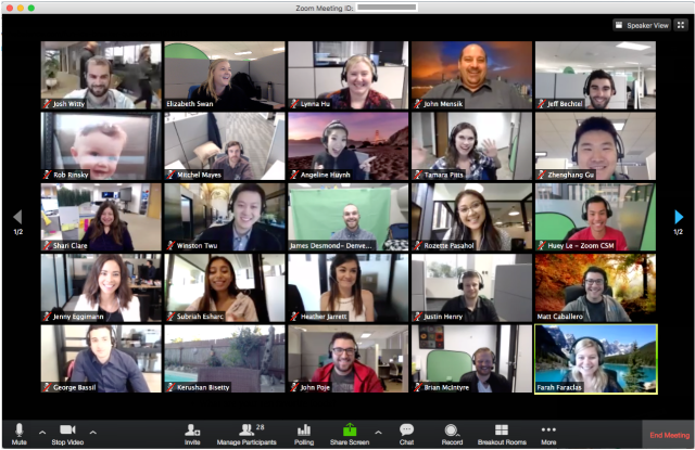
Multi-person screens magnify this exhausting problem. Gallery view—where all meeting participants appear Brady Bunch-style—challenges the brain’s central vision, forcing it to decode so many people at once that no one comes through meaningfully, not even the speaker.

“We’re engaged in numerous activities, but never fully devoting ourselves to focus on anything in particular,” says Franklin. Psychologists call this continuous partial attention, and it applies as much to virtual environments as it does to real ones. Think of how hard it would be to cook and read at the same time. That’s the kind of multi-tasking your brain is trying, and often failing, to navigate in a group video chat.

This leads to problems in which group video chats become less collaborative and more like siloed panels, in which only two people at a time talk while the rest listen. Because each participant is using one audio stream and is aware of all the other voices, parallel conversations are impossible. If you view a single speaker at a time, you can’t recognize how non-active participants are behaving—something you would normally pick up with peripheral vision.

For some people, the prolonged split in attention creates a perplexing sense of being drained while having accomplished nothing. The brain becomes overwhelmed by unfamiliar excess stimuli while being hyper-focused on searching for non-verbal cues that it can’t find.

That’s why a traditional phone call may be less taxing on the brain, Franklin says, because it delivers on a small promise: to convey only a voice.

Zoom boon

By contrast, the sudden shift to video calls has been a boon for people who have neurological difficulty with in-person exchanges, such as those with autism who can become overwhelmed by multiple people talking.

John Upton, an editor at the New Jersey-based news outlet Climate Central, recently found out he is autistic. Late last year, he was struggling with the mental load of attending packed conferences, engaging during in-person meetings, and navigating the small-talk that’s common in work places. He says these experiences caused “an ambiguous tension, a form of anxiety.”

As a result, he suffered a bout of autistic burnout and struggled to process complicated information—which he says is normally his strength—leading to feelings of helplessness and futility. To combat the issue, he began transitioning to working mostly from home and stacking all in-person meetings on Thursdays, to get them out of the way.

Now that the pandemic has pushed his coworkers to be remote as well, he has observed their video calls lead to fewer people talking and less filler conversation at the beginning and end of each meeting. Upton says his sense of tension and anxiety has been reduced to the point of being negligible.

This outcome is supported by research, says the University of Québec Outaouais’s Claude Normand, who studies how people with developmental and intellectual disabilities socialize online. People with autism tend to have difficulty understanding when it’s their turn to speak in live conversations, she notes. That’s why the frequent lag between speakers on video calls may actually help some autistic people. “When you’re Zooming online, it’s clear whose turn it is to talk,” Normand says.

However, other people on the autism spectrum may still struggle with video chatting, as it can exacerbate sensory triggers such as loud noise and bright lights, she adds.

On the whole, video chatting has allowed human connections to flourish in ways that would have been impossible just a few years ago. These tools enable us to maintain long-distance relationships, connect workrooms remotely, and even now, in spite of the mental exhaustion they can generate, foster some sense of togetherness during a pandemic.

It’s even possible Zoom fatigue will abate once people learn to navigate the mental tangle video chatting can cause. If you’re feeling self-conscious or overstimulated, Normand recommends you turn off your camera. Save your energy for when you absolutely want to perceive the few non-verbal cues that do come through, such as during the taxing chats with people you don’t know very well, or for when you want the warm fuzzies you get from seeing someone you love. Or if it’s a work meeting that can be done by phone, try walking at the same time.

“Walking meetings are known to improve creativity, and probably reduce stress as well,” Normand says.

https://www.nationalgeographic.com/science/2020/04/coronavirus-zoom-fatigue-is-taxing-the-brain-here-is-why-that-happens/