For years, scientists have known that mitochondria—the power source of cells—play a role in brain disorders such as depression, bipolar disorder, anxiety and stress responses. But recently scientists at the University of Maryland School of Medicine (UMSOM) have identified significant mitochondrial changes in brain cells that take place in cocaine addiction, and they have been able to block them.
In mice exposed repeatedly to cocaine, UMSOM researchers were able to identify an increase in a molecule that plays a role in mitochondria division (or fission) in a reward region of the brain. Researchers were able to block this change by using a special chemical, Mdivi-1. The researchers also blocked responses to cocaine by genetically manipulating the fission molecule within the mitochondria of brain cells, according to research published in Neuron.
“We are actually showing a new role for mitochondria in cocaine-induced behavior, and it’s important for us to further investigate that role,” said Mary Kay Lobo, PhD, Associate Professor of Anatomy and Neurobiology.
The researchers initially studied the mitochondria in cocaine-exposed mice and determined that mitochondria fission increased in the major reward region of the brain. To confirm this same change in humans, researchers were able to identify similar changes in the mitochondrial fission molecule in tissue collected from post mortem individuals who were cocaine dependents.
Dr. Lobo said that this latest research could help UMSOM researchers better understand changes in brain cells and mitochondria from other addictive disorders. “We are interested to see if there are mitochondrial changes when animals are taking opiates. That is definitely a future direction for the lab,” she said.
“This research is another great example of our ground-breaking work at the University of Maryland School of Medicine to better understand the biology behind drug addiction,” said E. Albert Reece, MD, PhD, MBA, University Executive Vice President of Medical Affairs and the John Z. and Akiko K. Bowers Distinguished Professor and Dean at the University of Maryland School of Medicine.
Germany’s Green Belt is one of Europe’s most unique open spaces: a once heavily militarized stretch of the Iron Curtain that’s now a natural wonderland filled with a variety of threatened animal species.
by Matt Hickman
Although the Berlin Wall came crashing down on Nov. 9, 1989, there’s another important milestone for a reunified Germany that was ushered in this month. As of Feb. 5, 2018, the heavily fortified concrete barrier that divided the German capital beginning in 1961 has now been down longer than it was up: 28 years, two months and 27 days.
That being said, it’s sometimes easy to forget that the physical and ideological divide between East and West wasn’t just limited to a famous 90-some-mile wall in Berlin.
Predating the Berlin Wall by 16 years and located nearly 100 miles east, the Inner German Border was the true physical manifestation of the Iron Curtain: a 870-mile frontier that ran the entire length of the divided country from the Baltic Sea in the north to the former Czechoslovakia in the south. On one side of this 650-foot-wide strip of land stood the Federal Republic of Germany (FRG) and on the other — just beyond an extensive network of dog runs, minefields, concrete watchtowers, bunkers, booby traps and forbidding electrified barbed wire fences — stood the German Democratic Republic (GDR), a communist dictatorship that remained firmly in the grasp of the Soviet Union until the dissolution of the Eastern Bloc.
Remnants of the “Death Strip” that once severed Germany still exist — so called because hundreds of East Germans perished while attempting to flee the GDR for less totalitarian pastures. Many of the old watchtowers, fortifications and short stretches of fence have been preserved. Here, history, no matter how painful, hasn’t been paved over and replaced with shopping malls and tract housing. And as such, the scars of a divided Germany remain. But what unusual and beautiful scars they are.
Almost the entirety of the Inner German Border has been reclaimed by Mother Nature as part of a sprawling wildlife reserve and outdoor recreation area known as Das Grüne Band — the Green Belt. Encompassing large swaths of undisturbed countryside and farmland in addition to the border zone, in some ways the Green Belt — often described as a “living monument to reunification” and a “memory landscape” — remains a no man’s land given that a wide variety of plants and animals, many rare and endangered, positively rule.
Germany’s Green Belt isn’t entirely continuous. However, most of this exclusion zone-turned-wildlife haven remains in a near-natural state.
From ‘death zone into a lifeline’
Rich in biodiversity and largely unhampered by 21st century human development, the Green Belt is a project of German environmental group Bund Naturschutz (BUND) that dates back to 1989. However, work had begun on the non-fortified western side of the border zone much earlier after conservationists noticed that this woeful place was also a wildlife magnet. “The division of Germany was a travesty that robbed people of their freedom, but a positive side effect was the way the sealed border allowed nature to flourish,” Eckhard Selz, a park ranger hailing from the former East Germany, explained to the Guardian in 2009.
In a 2017 NBC News profile, conservationist Kai Frobel, considered by many to be the father of the Green Belt, explained that “nature essentially has been given a 40-year holiday” in the erstwhile border area, which itself has been transformed from a “death zone into a lifeline.”
“When we grew up in this area, we all thought that this monster of a border line had been built for eternity,” 58-year-old Frobel says of his teenage years spent as a budding conservationist hailing from Colburg, a Bavarian town located on the western side of the border but largely surrounded by the GDR. “No one, really no one, believed in German reunification at the time.”
When the Iron Curtain collapsed, Frobel and his fellow conservationists, including many from the former East Germany, rushed to protect and preserve the border zone. The worry was that the largely untouched area would give way to roads, housing and massive commercial farming operations — a “brown belt,” if you will. Vital wildlife habitats just recently discovered would be lost.
With governmental backing, the Green Belt became the first German nature conservation project to involve parties from both sides of a nation that had just been fused back together. Decades later, an impressive 87 percent of the Green Belt, which passes through nine of Germany’s 16 states, remains in an undeveloped or near-natural state. While there are some gaps in this unusually elongated wildlife refuge, BUND is continually working to restore them and prevent other sections from giving way to development.
“You will find no other place in Germany with the richness of habitats and species that the Green Belt provides,” Frobel tells NBC News.
A Cold War era concrete watchtower still stands along the eastern section of what was once the notorious Inner German Border.
The one upside of a nation-dividing no man’s land
In October of last year, Frobel, along with Inge Sielman and Hubert Weiger, were awarded the German government’s top environmental prize for their tireless work preserving and protecting the old Inner German Border and environs. (The trio received a combined 245,00 euros or roughly $284,300.)
As Deutsche Welle explains, the Green Belt’s dual function as a historical site and wildlife refuge is more vital today than ever. Many animals, forced to seek out new habitats due to encroaching development in outlying areas of the German countryside, are flocking to the protected area in record numbers.
“The Green Belt is now home to countless natural wonders that have been crowded out in other areas,” German President Frank-Walter Steinmeir explained at October’s Germany Environmental Prize ceremony, held in the city of Brunswick.
Tranquil, sobering and biologically diverse, the Green Belt is popular amongst hikers, cyclists, birders and history buffs alike.
In total, conservationists believe the Green Belt to be home to upwards of 1,200 plant and animal species that are endangered or near-extinct in Germany, including the lady’s slipper orchid, the Eurasian otter, wildcats and the European tree frog. The Green Belt also hosts a large number of rare and threatened birds such as the black stork.
“We discovered that over 90 percent of the bird species that were rare or highly endangered in Bavaria — such as the whinchat, the corn bunting and the European nightjar — could be found in the Green Belt. It became a final retreat for many species, and it still is today,” Frobel tells Deutsche Welle.
One less rare species found in growing abundance throughout the Green Zone are tourists. Germany has long touted the region as a sustainable “soft” tourism hotspot, particularly in recent years. Laced with hiking trails and dotted with nature viewing areas along with a fair number of memorials, museums, quaint villages and a handful of crumbling leftovers from the Cold War era, the Green Zone passes through already tourism-friendly nature regions including the Franconian and Thuringian forests, the Harz Mountains and the verdant floodplain of the river Elbe.
In addition to local conservation groups, a number of local tourism authorities are working alongside BUND to promote the natural splendors of the once inaccessible border region. “Numerous cycling and hiking trails along the Green Belt connect special points of experience and information,” reads the Green Belt tourism page. “You can see cranes and northern geese from observation ramparts, conquer castles and palaces, descend into diminutive mining pits, climb border towers, dart along old border trails in the dark, or be inspired by works of art.”
With informative signs guiding the way and pointing out important sights, the Green Belt is described as a ‘memory landscape.
A model for something much bigger
Of course, Germany wasn’t the only country cracked by the Iron Curtain.
For nearly four decades, the entire European continent was split between East and West with little movement between the two sides. And much like the heralded conservation area that’s flourishing in a once-divided Deutschland, the European Green Belt Initiative aims to protect biodiversity along the line of former Iron Curtain but on a much more ambitious scale.
Stretching from the Barents Sea on the Russian/Norwegian border and along the Baltic coast before cutting through the heart of Central Europe and terminating at the Adriatic and Black seas, the 7,500-mile European Green Belt links 24 individual countries through a winding necklace of national parks, nature preserves and other protected areas.
As in Germany, many of these European border regions were largely restricted/avoided during their existence. And so, wildlife moved in and flourished in relative solitude.
“Unwittingly, the once-divided Europe encouraged the conservation and development of valuable habitats. The border area served as a retreat for many endangered species,” explains the European Green Belt website.
Founded in 2003 and very much modeled on the work of BUND in Germany, the European Green Belt Initiative is a burgeoning grassroots movement comprised of around 150 governmental and non-governmental conservation organizations hailing from a diverse number of countries.
And in addition to inspiring a band of protected wilderness that bisects the European continent, the many successes of Germany’s Green Belt have also inspired South Korean officials to reach out to Frobel and his colleagues and discuss ways that the Korean Demilitarized Zone could some day (emphasis on some day) be transformed into a protected wildlife area.
“Conservationists are already preparing a so-called Green Belt Korea, and are in close consultation with us,” Frobel told Deutsche Welle in a 2017 interview with Deutsch Welle. He points out that the Korean Demilitarized Zone, home to “a well-preserved biodiverse habitat,” is the “only region in the world that can be compared with Germany before 1989.”
“They are using Germany’s Green Belt as its model for when reunification comes — even though the situation doesn’t look too good at the moment,” says Frobel.
In one of the first successes of its kind, researchers from Case Western Reserve University School of Medicine and six other institutions have inhibited the spreading of cancer cells from one part of the body to another. In doing so, they relied on a new model of how cancer metastasizes that emphasizes epigenetics, which examines how genes are turned on and off.
In a study published in Nature Medicine, the investigators—including scientists from the National Cancer Institute and Cleveland Clinic—used innovative epigenetic-centered techniques to halt the spread of bone cancer (osteosarcoma) cells to the lungs in mice.
The large majority of deaths associated with osteosarcoma are due to the spreading of the cancer to the lungs, a process known as metastasis. Most human osteosarcoma cases occur in children and young adults between the ages of 10 and 30, with teens the most frequently affected age group. Clinical outcomes for patients with osteosarcoma have not improved for more than 30 years, and there are currently no approved targeted anti-metastatic therapies for the disease in wide clinical use.
“More than 90 percent of all cancer deaths are the result of tumor metastasis, not primary-site tumors,” said the study’s senior author, Peter Scacheri, professor of genetics and genome sciences at Case Western Reserve University School of Medicine and member of the Case Comprehensive Cancer Center. “While many of the genes responsible for metastasis have been identified, the mechanisms that control these genes are not well defined. Our findings demonstrate that altered gene-enhancer activity is fundamental to a cancer cell’s ability to metastasize.”
Gene enhancers are short segments of DNA that, when bound by specialized proteins called transcription factors, function like switches to activate genes. This process is critical for normal development, as when a single fertilized egg develops into the many different cell types that comprise the body.
There are tens of thousands of gene enhancers in each cell, far more than the number of genes; they will be in different “on” and “off” positions in, for example, eye and heart cells (or gradients thereof, like a dimmer switch’s effects on the brightness-level of a light). These distinctive “on and off” profiles lend cells their unique characteristics, even though they all have the same DNA.
But faulty enhancer regulation appears to contribute to tumor-formation and subsequent spreading of cancer cells. In addition, different cancers are distinguishable by different enhancer patterns.
In this new study, the authors show that the on-off switches of cancer cells that have metastasized are in different positions than in the cells of the source tumor.
“Metastasis results from a complex set of traits acquired by tumor cells, distinct from those necessary for tumors to form in the first place,” said the study’s lead author, James J. Morrow, a medical student in the Medical Scientist Training Program at Case Western Reserve University School of Medicine. “Unfortunately, searching for gene mutations that drive metastasis has not substantially improved outcomes for patients with metastatic disease. Five-year survival rates for cancer patients with regional or localized disease have significantly improved for many types of cancer. But with few exceptions, outcomes for patients with metastatic cancer have remained stagnant.
“It is well established that primary tumor formation is driven by a combination of genetic and epigenetic events,” he continued. “So based on the knowledge that enhancers drive both normal cell development and tumor-formation, we hypothesized that they may play a similar role in the transition of cancer cells from one developmentally distinct tissue to another during metastatic progression.”
Through epigenomic profiling experiments, the Case Western Reserve-led researchers consistently identified certain bunched clusters of enhancers—known as metastatic variant enhancer loci (Met-VELs)—near cancer genes in lung metastases of patients with osteosarcoma, indicating that they were central to the metastatic process. Using experimental mouse models, the researchers then showed that growth of osteosarcoma cells in the lung can be mitigated by using BET inhibitors (anti-cancer drugs currently in clinical trials), which broadly interrupt the function of Met-VELs in driving gene expression.
Second, they demonstrated that the metastatic capacity of osteosarcoma cells can be diminished by blocking expression of individual genes regulated by Met-VELs or the transcription factors driving that regulation. They verified that a particular Met-VEL-linked gene, Tissue Factor (F3), is essential for metastatic colonization. Specifically, interrupting the signaling and pro-coagulant (blood clotting) functions of F3 with antibodies inhibiting these functions was sufficient to prevent metastasis. Additionally, they showed that deleting a single Met-VEL regulating F3 expression via the TALEN gene-editing process achieved a similar effect.
“Our experiments show that removing a single enhancer of the F3 gene in tumor cells virtually eliminates their ability to metastasize in mice,” said Scacheri. “Collectively, our findings establish that enhancer elements endow tumor cells with metastatic capacity and that targeted inhibition of genes associated with enhancer alterations, or deleting altered enhancers themselves is sufficient to block metastatic colonization and proliferation. While our work focused on lung metastasis in osteosarcoma, the findings have implications for other types of metastatic cancer as well.”
The Case Western Reserve University School of Medicine focus on epigenetics in the new study represents a break with the prevailing model for metastasis, which largely explores mutations in the genes—not if or how certain genes are turned on or off. And the preponderance of current cancer research takes place on the early stages of disease, such as how tumors are formed and on what distinguishes cancer cells from normal cells, and not on metastasis. Additionally, most cancer medications and treatments today were developed to kill primary tumors, not cancer cells that have spread elsewhere.
Scientists at The Scripps Research Institute (TSRI) have achieved a major milestone toward designing a safe and effective vaccine to both treat heroin addiction and block lethal overdose of the drug. Their research, published today in the journal Molecular Pharmaceutics, shows how a new anti-heroin formulation that is safe in animal models remains stable at room temperature for at least 30 days. As a result, the vaccine is close to being ready for human testing.
“The heroin vaccine is one step closer to clinical evaluation,” says Candy S. Hwang, PhD, first author of the study and a research associate at TSRI.
According to the National Institute on Drug Abuse, 15,446 Americans died from heroin overdose between 2000 and 2016, and the mortality rates are increasing. Heroin abuse has been further fueled by a rise in prescription opioid abuse—studies show that opioid pain reliever users are 40 times more likely to abuse heroin.
The first formulation of the heroin vaccine was developed in 2013 by a team led by Kim D. Janda, PhD, the Ely R. Callaway Jr. Professor of Chemistry and member of the Skaggs Institute for Chemical Biology at TSRI. It has been shown to be effective—and safe—in both mouse and non-human primate models.
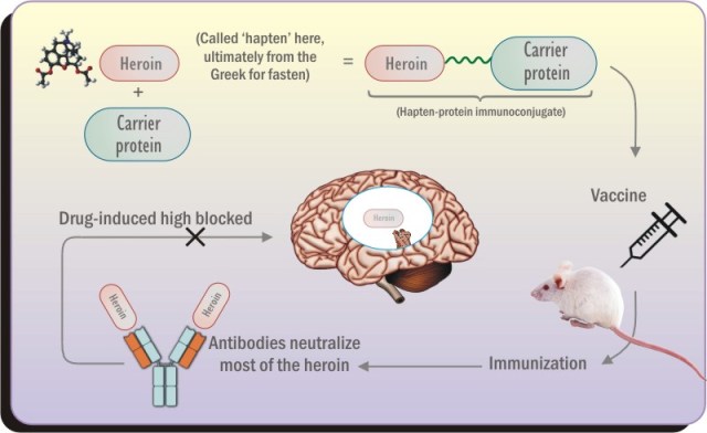
The vaccine works by training the immune system antibodies to recognize and bind to heroin molecules, blocking the drug from reaching the brain to cause a “high.” Researchers believe that blocking the high of heroin will help eliminate the motivation for many recovering addicts to relapse into drug use.
The heroin molecule does not naturally prompt an antibody response, so researchers attach it to a carrier protein that alerts the immune system to start making antibodies. Scientists also add an ingredient called an adjuvant to the vaccine, which boosts the immune response and makes the vaccine more effective.
Hwang says, “Our goal was to prepare a vaccine that could be advanced to clinical trials. As such, we were looking for the best combination of ‘hapten’ (the heroin molecule), carrier protein and adjuvant to keep the vaccine both stable for transport and storage but still efficacious.”
For the new study, the researchers investigated how 20 different carrier protein/adjuvant combinations worked, including shelf stability based on temperature and storage time and whether the formulation was a liquid or powder.
Their experiments in rodent models showed that the best vaccine formulation contained a carrier protein called tetanus toxoid (TT) and adjuvants called alum and CpG ODN. The discovery that alum worked best as an adjuvant was especially significant since alum is one of the few adjuvants used in vaccines already approved by the U.S. Food and Drug Administration. The researchers also found that there was no difference in how well it worked between the liquid and powder versions of this formulation.
Hwang notes that the best vaccine formulation showed protection against lethal doses of heroin. This is particularly important as many heroin addicts have succumb to overdose and death during their attempts to quit the drug.
With this new study, the researchers have shown that the vaccine is safe and effective in animal models, stable under clinical conditions and reliant on an already-approved adjuvant. The next step is to find a producer to make the vaccine on a large scale.
“We believe that a heroin vaccine would be tremendously beneficial for people who have a heroin substance use disorder but have found difficulty in trying to quit,” says Hwang.
In addition to Hwang and Janda, authors of the study, “Enhancing Efficacy and Stability of an Anti-Heroin Vaccine: Examination of Antinociception, Opioid Binding Profile, and Lethality,” were Paul T. Bremer, Cody J. Wenthur, Beverly Ellis and Bin Zhou of The Scripps Research Institute; and Sam On Ho, SuMing Chiang and Gary Fujii of Molecular Express, Inc.
The study was supported by the National Institutes of Health (grants UH3DA041146, F32AI126628, F32DA043323, R42DA040422 and R44AI094770).
An Amazon molly, Poecilia formosa, an asexual fish species native to Texas that is entirely female.
By Shana Hutchin
Highlights
The Amazon molly has flourished by defying nature’s odds to reproduce asexually, cloning themselves by duping the male fish of another species to waste their germplasm
Females steal the entire genome of their host males, keep it for one generation and then throw it out again
The existence of Amazon mollies back anywhere from 100,000 to 200,000 years ago to a sexual reproduction event involving two different species of fish
Faculty Fellow Dr. Manfred Schartl led the international team that recently sequenced the first Amazon molly fish genome
No species is immune from the suffering of unrequited love, but scientists expect to learn volumes about the biological basis of sex from the newly sequenced genome of an all-female, asexual Texas native – the Amazon molly fish – that has thrived as a master of male manipulation over millennia.
The fresh waters along the Texas-Mexico border serve as home to this evolutionary anomaly – a fish that has flourished by defying nature’s odds to reproduce asexually through a natural form known as parthenogenesis in which growth and development of embryos occurs without fertilization, resulting only in daughters that are true clones of their mothers.
Texas A&M University Hagler Institute for Advanced Study (HIAS) Faculty Fellow Dr. Manfred Schartl led the international team that recently sequenced the first Amazon molly genome and the genomes of the original parental species that created this unique fish in an effort to better understand how its reproduction deviates from the male-female sexual norm and why the Amazon molly as a species has fared so well in the process.
The evolution of sex
The findings from their National Institutes of Health-funded research are published online today (Feb. 12) in the Nature research journal Nature Ecology & Evolution.
“The existence of two sexes, male and female, is one of the oldest and most widespread phenomena in biology,” says Schartl, a world leader in cellular and molecular biology of Xiphophorus model systems including platyfish and swordtails. “Studies on the exceptional case of asexuality helps us to better understand the biological meaning and evolution of sex.”
Animals that reproduce asexually are rare, compared to the overwhelming majority of species that exist as males and females and reproduce sexually. Because it was long thought that vertebrates would not be able to exist in such a way, Schartl says it was a sensation when the Amazon molly was the first asexual vertebrate discovered in 1932.
But even the most independent females occasionally need a male – in the Amazon molly’s case, to kick-start the parthenogenensis process. They seduce males from related sexual species for this service, which Schartl notes lacks the regular benefit for these males, which do not contribute their genes to the next generation.
An Amazon molly (right), caught in action while seducing a male Sailfin molly to steal sperm.
Thriving by cloning
“In essence, mollies repeatedly clone themselves by duping the male fish of another species to waste their germplasm,” Schartl says. “This reminds one of the tribe of female warriors in the Greek mythology, from which their name is derived.”
The team’s research traces the existence of Amazon mollies back anywhere from 100,000 to 200,000 years ago to a sexual reproduction event involving two different species of fish, an Atlantic molly and a Sailfin molly.
“That’s about 500,000 generations if you calculate it out to the present day, which makes them genetically older than humans,” Schartl says. “This is unexpected because asexuals are expected to be at disadvantage compared to their sexual counterparts.”
Schartl notes that one of the theories as to why asexual reproduction is incompatible with a species’ sustainability is the idea that if no new DNA is introduced during reproduction, then harmful gene mutations can accumulate over successive generations, leading to eventual extinction. Another hypothesis states that asexual reproduction is not like sexual reproduction, where the different genomes of the two parents are newly combined and create new genomes with every offspring. Because the absence of recombination in asexuals limits genetic diversity within a species, he says it gets more and more difficult to adapt to changes in the environment.
“Unexpectedly, we did not find the signs of genomic decay as predicted,” Schartl adds. “Our findings suggest that the molly’s thriving existence can be explained by the fact that the fish has a hardy genetic makeup that is often rare in nature and gives the animals some survival benefits.”
Schartl says the hybridization of the Atlantic and Sailfin mollies’ two different species genomes into a new one created a situation well known in the animal and plant breeding world — an artificial hybrid that is bigger, more colorful and capable of generating more and better products than the purebred parents, a phenomenon known as hybrid vigor.
Pod shatter in oilseed rape – problem for farmers worldwide.
Breeding temperature-resilient crops is an “achievable dream” in one of the most important species of commercially-cultivated plants, according to a new study.
The vision of crop improvement in the face of climate change is outlined in research by the John Innes Centre which establishes a genetic link between increased temperature and the problem of “pod shatter” (premature seed dispersal) in oilseed rape.
Research by the team led by Dr Vinod Kumar and Professor Lars Østergaard, reveals that pod shatter is enhanced at higher temperature across diverse species in the Brassicaceae family which also includes cauliflower, broccoli and kale.
This new understanding brings a step closer the prospect of creating crops that are better adapted to warmer temperatures a step closer.
Dr Vinod Kumar, a co-author of the paper explained the significance of the findings:
“It’s almost as if there is a thermostat that controls seed dispersal, or pod shatter. As we learn how it works, we could in the future ‘rewire’ it so seed dispersal does not happen at the same pace at higher temperatures
“This piece of the puzzle, coupled with the use of advanced genetic tools means that developing temperature-resilient crops becomes an achievable dream.”
Controlling seed dispersal, or “pod shatter” is a major issue for farmers of oilseed rape worldwide, who lose between 15-20% of yield on average per year due to prematurely dispersed seeds lost in the field.
The study set out to find out if temperature increases had a direct influence on pod shatter in oilseed rape, and how this is controlled by genetics.
“Over the last two decades, scientists have identified the genes that control pod shatter. However, it is not until now that we begin to understand how their activity is affected by the environment, and in this case temperature,” explained Professor Lars Østergaard.
To study the effects of temperature on seed dispersal, Dr Xinran Li, a postdoctoral researcher, monitored fruit development in Arabidopsis, a model plant related to the important Brassicaceae crops, at three different temperatures 17, 22 and 27 degrees centigrade.
This showed that stiffening of the cell wall at the tissue where pod shatter takes place is enhanced by increasing temperature leading to accelerated seed dispersal.
Dr. Li demonstrated that this was true not only for Arabidopsis, but across the Brassicaceae family, including oilseed rape.
The team went on to establish the genetic mechanism which organises the plant response to higher temperatures. Previous studies have shown that pod shatter is controlled by a gene called INDEHISCENT (IND). This study reveals that IND is under the control of a thermo-sensory mechanism in which a histone called H2A.Z is a key player.
The report concludes: “Our findings introduce an environmental factor to the current knowledge, which provide alternative avenues for crop improvement in the face of climate change.”
The paper Temperature modulates tissue-specification programme to control fruit dehiscence in Brassicaceae which appears in the journal Molecular Plant also identifies the genetic pathways behind the temperature sensing mechanism which coordinates the crop’s response to rises in temperature.
Temperature modulates tissue-specification program to control fruit dehiscence in Brassicaceae: authors Xin-Ran Li, Joyita Deb, S. Vinod Kumar and Lars Østergaard.
Songbirds known as Japanese tits communicate using human-like rules for language and can mentally picture what they’re talking about, research suggests.
by Brandon Keim
Hear a word, particularly an important one — like “snake!” — and an image appears in your mind. Now scientists are finding that this basic property of human language is shared by certain birds and, perhaps, many other creatures.
In a series of clever tests, a researcher has found that birds called Japanese tits not only chirp out a distinctive warning for snakes, but also appear to imagine a snake when they hear that cry. This glimpse into the mind’s eye of a bird hints at just how widespread this ostensibly human-like capacity may be.
“Animal communication has been considered very different from human speech,” says Toshitaka Suzuki, an ethologist at Japan’s Kyoto University. “My results suggest that birds and humans may share similar cognitive abilities for communication.”
Perhaps this went unappreciated for so long, says Suzuki, simply because “we have not yet found a way to look at the animals’ minds.”
Over the last several years, Suzuki conducted a series of experiments deciphering the vocalizations of Japanese tits — or Parus minor, whose family includes such everyday birds as chickadees and titmice — and describing their possession of syntax, or the ability to produce new meanings by combining words in various orders. (“Open the door,” for example, versus “the open door.”)
Syntax has long been considered unique to human language, and language in turn is often thought to set humans apart from other animals. Yet Suzuki found it not in a bird typically celebrated for intelligence, like crows or parrots, but in humble P. minor.
MENTAL PICTURES
Once he realized that birds are using their own form of language, Suzuki wondered: what happens in their minds when they talk? Might words evoke corresponding images, as happens for us?
Suzuki tested that proposition by broadcasting recordings of P. minor’s snake-specific alarm call from a tree-mounted speaker. Then he analyzed the birds’ responses to a stick that he’d hung along the trunk and could manipulate to mimic a climbing snake.
If the call elicited a mental image, Suzuki figured the birds would pay extra-close attention to the snake-like stick. Indeed they did, he recently reported in the journal Proceedings of the National Academy of Sciences.
In contrast, when Suzuki broadcast a call used by tits to convey a general, non-specific alarm, the birds didn’t pay much notice to the stick. And when he set the stick swinging from side to side in a decidedly non-snakelike manner, the birds ignored it.
“Simply hearing these calls causes tits to become more visually perceptive to objects resembling snakes,” he writes in PNAS. “Before detecting a real snake, tits retrieve its visual image from snake-specific alarm calls and use this to search out snakes.”
Rob Magrath, a behavioral ecologist at Australia National University who specializes in bird communication, thinks Suki’s interpretation is consistent with the results. He also calls the work “truly delightful.”
“I love the way that Suzuki employs simple experiments, literally using sticks and string, to test ideas,” Magrath says. Similarly impressed is ecologist Christine Sheppard of the American Bird Conservancy. “It’s incredibly challenging to devise an experiment that would allow you to answer this question,” she says. “It’s really neat.”
MINDS OF THEIR OWN
Sheppard says it makes evolutionary sense for animals to possess a ‘mind’s eye’ that works in tandem with their communications: It allows individuals to respond more quickly to threats. Suzuki agrees, and believes it’s likely found not only in P. minor and their close relatives, but in many other birds and across the animal kingdom.
“Many other animals produce specific calls when finding specific types of food or predators,” he says. He hopes researchers will use his methodology to peek into the mind’s eyes of other animals.
For Sheppard, the findings also speak to how people think about birds: not just as pretty or interesting or ecologically important, but as fellow beings with rich minds of their own.
“When I was in school, people still thought that birds were little automata. Now “bird brain” is becoming a compliment,” she says.
“I think this kind of insight helps people see birds as living, breathing creatures with whom we share the planet,” she says.
Kent Hutchinson of the CU Change Lab is one of authors of this new research on the effects of Marijuana on the brain.
by Cay Leytham-Powell
Marijuana may not be as damaging to the brain as previously thought, according to new research from the University of Colorado Boulder and the CU Change Lab.
The research, which was published in the journal Addiction, examined the brains of more than 1,000 participants of varying ages, and found that long-term alcohol use is much more damaging to the brain than marijuana, contradicting years of research into the effects of marijuana and other cannabinoid products on the brain.
These findings, and other conclusions suggesting the potential public health benefits of marijuana, come amid the recent back-and-forth on federal marijuana policy and the nation’s opioid crisis.
Yet scientists are still hesitant to say that cannabinoid usage, specifically as it pertains to marijuana and its associated products, is beneficial.
“Particularly with marijuana use, there is still so much that we don’t know about how it impacts the brain,” said Rachel Thayer, a graduate student in clinical psychology at CU Boulder and the lead author of the study. “Research is still very limited in terms of whether marijuana use is harmful, or beneficial, to the brain.”
While the negative effects of alcohol on the brain have been known by researchers for years, it has been assumed that cannabinoids are as damaging to long-term brain health—if not more—given the immediate psychoactive effects of the THC (the chemical that gets a person high) in marijuana.
However, this may not necessarily be true.
“When you look at the research much more closely, you see that a lot of it is probably not accurate,” said study co-author Kent Hutchison, a professor of behavioral neuroscience at CU Boulder and co-director of the CU Change Lab, which explores the factors linked with health and risk behavior.
“When you look at these studies going back years, you see that one study will report that marijuana use is related to a reduction in the volume of the hippocampus. The next study then comes around, and they say that marijuana use is related to changes in the cerebellum or the whatever.”
“The point is that there’s no consistency across all of these studies in terms of the actual brain structures.”
To combat this misconception in the existing literature, the researchers gave a fresh look at some existing neurological imaging data from the MRIs of both adolescents and adults to see how, using the same variables and controls, the influence of cannabinoids on the brain compared to or contrasted with alcohol.
“With alcohol, we’ve known it’s bad for the brain for decades,” said Hutchison. “But for cannabis, we know so little.”
To see any potential difference, the researchers used the data to examine the most important neurological components: gray matter and white matter.
Gray and white matter are the two main types of tissue that make up the brain and central nervous system. Gray matter is the “stuff”—the cell bodies, dendrites and axon terminals—that enable functionality. White matter, then, is how the grey matter communicates between clusters. Any loss of size or integrity in either can make the brain not work quite like it should.
The study found that alcohol use was significantly associated with a decrease in gray matter size and white matter integrity, particularly for adults who may have decades of exposure. Marijuana and associated cannabinoid products, on the other hand, were not shown to have any long-term impact on the amount of gray matter in the brain or on the integrity of the white matter.
The research demonstrated that, “while marijuana may also have some negative consequences, it definitely is nowhere near the negative consequences of alcohol,” according to Hutchison.
Despite marijuana not being as harmful as once thought, and definitely not as damaging as other legal and illegal products, the research has not yet proved any possible benefits. This is particularly the case as it relates to the different products on the market (both THC and non-THC-containing cannabinoid products), their usage with pain and addiction treatment and the effect on different ages — especially as cannabinoid usage is on the rise among older populations.
“Considering how much is happening in the real world with the legalization movement, we still have a lot of work to do,” Hutchison said.
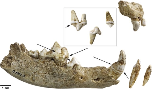
The teeth and jaw from the younger dog in the grave: This pup likely had canine distemper.
By Laura Geggel
Ancient people likely cared for a sick, domesticated pup for weeks on end before it died about 14,000 years ago during the Paleolithic era, a new study finds.
After it died, the dog was buried with the remains of another dog and an adult man and woman — making it not only the oldest burial of a domestic dog on record, but also the oldest known grave to contain both dogs and people, the researchers said.
This discovery suggests that even though the dog was young, sick and likely untrained as a result, ancient people still had an emotional bond with it, the researchers wrote in the study. This may explain why the people buried the animal with two of their own, the researchers said.
The grave itself was found in 1914 in Oberkassel, a suburb of Bonn in western Germany. Until now, however, researchers thought the burial contained two humans and just one dog. But a new analysis of the canid bones and teeth revealed that two dogs were in fact buried there: an older dog and a younger dog, which likely had a serious case of morbillivirus, better known as canine distemper.
The younger dog was about 28 weeks old when it died, the study’s lead researcher, Luc Janssens, a veterinarian and doctoral student of archaeology at Leiden University in the Netherlands, said in a statement. A dental analysis showed that the pup likely contracted the disease at around 3 to 4 months of age, and likely had two or even three periods of serious illness, each lasting up to six weeks, Janssens said.
Canine distemper is a serious illness that has three phases. During the first week, infected dogs can show signs of high fever, lack of appetite, dehydration, tiredness, diarrhea and vomiting, the researchers wrote in the study. Up to 90 percent of dogs with distemper die during the second phase, when they can develop a stuffy nose, laryngitis and pneumonia. In the third phase, dogs experience neurological problems, including seizures.
There is now a vaccine for canine distemper, but unvaccinated dogs, as well as tigers and Amur leopards, can still die from the virus.
Given the severity of the disease, the ancient pup would have likely died right away unless it received intensive human care, the researchers said. “This would have consisted of keeping the dog warm and clean [from] diarrhea, urine, vomit [and] saliva,” as well as giving the pup water and possibly food, the researchers wrote in the study.
“While it was sick, the dog would not have been of any practical use as a working animal,” Janssens said. “This, together with the fact that the dogs were buried with people, who[m] we may assume were their owners, suggests that there was a unique relationship of care between humans and dogs as long as 14,000 years ago.”
The humans buried with the dogs had medical problems of their own. The roughly 40-year-old man had two healed bones, one on his arm and the other by his clavicle. He and the roughly 25-year-old woman also had moderate-to-severe dental disease, the researchers noted.
The grave also contained several artifacts, including a bone pin, a sculpture of an elk made from elk antlers, the penis bone of a bear and a red-deer tooth.
Although this finding is the oldest known domestic dog burial, it’s not the only ancient one. Other dog burials have been dated to about 11,600 years ago in the Near East, and archaeologists have found others dating to about 8,500 to 6,500 years ago in Scandinavia and about 8,000 years ago at the Koster Site in Illinois, the researchers said.
The study was published online Feb. 3 in the Journal of Archaeological Science.
NASA’s New Horizons spacecraft is now one of the most distant human-made objects, and it just took the most distant photograph ever. The image of an icy rock in the Kuiper belt has had colour added to increase the contrast.
After its visit to Pluto, the spacecraft headed out toward the Kuiper belt on its way to its next target, a Kuiper belt object (KBO) called 2014 MU69. It is now about 41 times as far from Earth as Earth is from the sun. There are only four spacecraft that have ever traveled that far from home: Voyager 1 and 2, and Pioneer 10 and 11.
But New Horizons is the first to send back a picture for so far afield. Its four predecessors did not send back images because their cameras were shut down before they got that far away.
New Horizons is still on an active mission to visit the Kuiper Belt. During its voyage to the outer reaches of the solar system, the spacecraft usually stays in hibernation mode to conserve energy. Every once in a while, its mission operators turn on its camera to take a few pictures and calibrations and beam them back to Earth.
The last time they did this was 5 December, when New Horizons took a routine calibration image of a cluster of stars, breaking a record for the most distant photograph ever taken. Two hours later, it broke the record again with two images of KBOs that are also the closest-up image ever taken of any such object. From here on out, every image it sends back will be the most distant image ever sent back.