Personal subjective reports of memory ability may be a reliable early marker of Alzheimer’s disease.


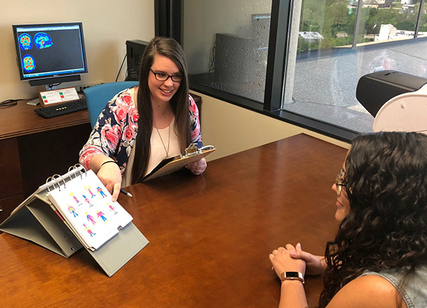
Psychological sciences doctoral student Marci Horn (left) conducts a name-face memory test as part of a study at the Center for Vital Longevity.

New research from the Center for Vital Longevity (CVL) at The University of Texas at Dallas suggests that subjective complaints about poor memory performance, especially in people over 60, could be a useful early marker for the onset of mild cognitive decline, which sometimes foreshadows Alzheimer’s disease.

Subjective memory is a person’s unscientific self-evaluation of how good his or her memory is, and whether, in that person’s opinion, there has been any worsening of memory through age. While some changes may be undetectable to others and are often too subtle to register on cognitive tests, the person subjectively believes that memory is slipping.

Published recently in Psychology and Aging, the research from Dr. Karen Rodrigue’s lab at CVL examined subjective memory complaints in nearly 200 healthy adults, ages 20 to 94. Previous studies suggest that subjective memory complaints are not necessarily indicative of cognitive decline, and may stem from underlying conditions such as anxiety and depression, which have been shown to impede memory.

The current study measured mood and screened out depressed individuals. Researchers also measured participants for known risk factors for memory loss and Alzheimer’s, such as higher levels of beta-amyloid in the brain and the presence of a gene variant called ApoE4. These factors were taken into account to examine whether subjective memory alone was a reliable correlate of actual memory ability.

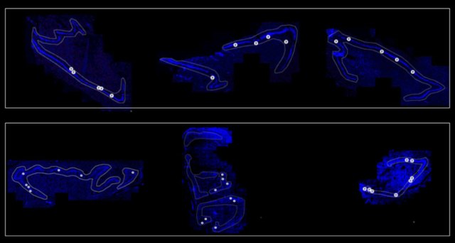
The study focused on associative memory — for example, remembering word pairs and name-face pairs. This type of memory is particularly sensitive to age-related decline, and the most common complaint of aging individuals.

The study found that a person’s intuitive or intrinsic assessment of his or her own memory was actually a reliable predictor of performance on the laboratory memory assessment. This result was particularly true for individuals with genetic risk for memory loss.

“Our findings show that subjective memory can be a reliable indicator of memory performance, even in cognitively healthy adults,” said psychological sciences doctoral student Marci Horn, the lead author of the study. “The same people who self-report memory problems may also have other risk factors associated with increased risk of Alzheimer’s disease.”

The researchers also found that men who had higher amyloid levels reported the most subjective memory complaints in the study. Previous studies had not uncovered a sex-specific relationship, nor did they account for the genetic and amyloid risk factors in these associations, the researchers said.

The strongest correlation of subjective memory complaints with actual cognitive performance was in study participants older than 60, when people are generally at greater risk for Alzheimer’s disease.

“It seems that awareness of memory changes may be a reliable indicator of one’s current memory ability, and may serve as another harbinger of future loss, as this relationship was strongest in those with known risk factors for Alzheimer’s disease, namely ApoE4 genotype and beta-amyloid burden in the brain,” said Rodrigue, the senior author of the study and assistant professor in the School of Behavioral and Brain Sciences (BBS). “We are following these individuals over time to further test this idea.”

Dr. Kristen Kennedy, an assistant professor in BBS, also was an author of the study. The research was funded in part by grants from the National Institutes of Health.

https://www.utdallas.edu/news/2018/4/30-32929_Subjective-Memory-May-Play-Role-in-Signaling-Cogni_story-wide.html?WT.mc_id=NewsHomePageCenterColumn

University of Michigan scientists develop a pill that may be superior to mammogram from detecting breast cancer

As many as one in three women treated for breast cancer undergo unnecessary procedures, but a new method for diagnosing it could do a better job distinguishing between benign and aggressive tumors.

Researchers at the University of Michigan are developing a pill that makes tumors light up when exposed to infrared light, and they have demonstrated that the concept works in mice.

Mammography is an imprecise tool. About a third of breast cancer patients treated with surgery or chemotherapy have tumors that are benign or so slow-growing that they would never have become life-threatening, according to a study out of Denmark last year. In other women, dense breast tissue hides the presence of lumps and results in deaths from treatable cancers. All that, and mammograms are notoriously uncomfortable.

“We overspend $4 billion per year on the diagnosis and treatment of cancers that women would never die from,” said Greg Thurber, U-M assistant professor of chemical engineering and biomedical engineering, who led the team. “If we go to molecular imaging, we can see which tumors need to be treated.”

The move could also catch cancers that would have gone undetected. Thurber’s team uses a dye that responds to infrared light to tag a molecule commonly found on tumor cells, in the blood vessels that feed tumors and in inflamed tissue. By providing specific information on the types of molecules on the surface of the tumor cells, physicians can better distinguish a malignant cancer from a benign tumor.

Compared to visible light, infrared light penetrates the body easily—it can get to all depths of the breast without an X-ray’s tiny risk of disrupting DNA and seeding a new tumor. Using a dye delivered orally rather than directly into a vein also improves the safety of screening, as a few patients in 10,000 can have severe reactions to intravenous dyes. These small risks turn out to be significant when tens of millions of women are screened every year in the U.S. alone.

But it’s not easy to design a pill that can carry the dye to the tumor.

“To get a molecule absorbed into the bloodstream, it needs to be small and greasy. But an imaging agent needs to be larger and water-soluble. So you need exact opposite properties,” Thurber said.

Fortunately, they weren’t the only people looking for a molecule that could get from the digestive system to a tumor. The pharmaceutical company Merck was working on a new treatment for cancer and related diseases. They got as far as phase II clinical trials demonstrating its safety, but unfortunately, it wasn’t effective.

“It’s actually based on a failed drug,” Thurber said. “It binds to the target, but it doesn’t do anything, which makes it perfect for imaging.”

The targeting molecule has already been shown to make it through the stomach unscathed, and the liver also gives it a pass, so it can travel through the bloodstream. The team attached a molecule that fluoresces when it is struck with infrared light to this drug. Then, they gave the drug to mice that had breast cancer, and they saw the tumors light up.

The research is described in a study in the journal Molecular Pharmaceutics, titled, “Oral administration and detection of a near-infrared molecular imaging agent in an orthotopic mouse model for breast cancer screening.”

This work was done in collaboration with David Smith, the John G. Wagner Collegiate Professor of Pharmaceutical Sciences at the U-M College of Pharmacy. It was supported by the Foundation for Studying and Combating Cancer and the National Institutes of Health.
Bhatnagar, S., Verma, K. D., Hu, Y., Khera, E., Priluck, A., Smith, D., & Thurber, G. M. (2018). Oral Administration and Detection of a Near-Infrared Molecular Imaging Agent in an Orthotopic Mouse Model for Breast Cancer Screening. Molecular Pharmaceutics. doi:10.1021/acs.molpharmaceut.7b00994

Frequently heading the ball in soccer is more dangerous to the brain than acute concussions

Frequent soccer ball heading is a common and under recognized cause of concussion symptoms, according to a study of amateur players led by Albert Einstein College of Medicine researchers. The findings run counter to earlier soccer studies suggesting concussion injuries mainly result from inadvertent head impacts, such as collisions with other players or a goalpost. The study was published in Neurology, the medical journal of the American Academy of Neurology.

“The prevailing wisdom is that routine heading in soccer is innocuous and we need only worry about players when they have unintentional head collisions,” says study leader Michael L. Lipton, M.D., Ph.D., professor of radiology and of psychiatry and behavioral sciences at Einstein and director of MRI Services at Montefiore. “But our study suggests that you don’t need an overt collision to warrant this type of concern. Many players who head the ball frequently are experiencing classic concussion symptoms such as headache, confusion, and dizziness during games and practice, even though they are not actually diagnosed with concussion. Concussion sufferers should avoid additional collisions or head impacts during the following days or weeks, when their risk of incurring a second concussion is extremely high. Because these injuries go unrecognized and unmanaged, there may be important clinical consequences for the short and long term.”

Studies clearly show that single or repeated concussion causes neurologic problems. But little is known about the effects of frequent but lesser impacts, such as those experienced while heading a soccer ball. Some research, notably a recent study of adolescent players published in JAMA Pediatrics, suggest that heading is not a common cause of concussion. “However, these studies did not actually measure heading, and thus they were unable to separate the relative contributions of intentional and unintentional head impacts,” says Dr. Lipton.

In the current study, a part of the Einstein Soccer Study, Dr. Lipton and his colleagues asked 222 adult amateur soccer players (80 percent men, ages 18 to 55) to fill out online questionnaires on their soccer-related activities during the previous two weeks, including details about heading and other unintentional head impacts and any resulting headaches, pain and dizziness as well as more severe symptoms, such as feeling dazed, needing medical attention, and becoming unconscious. Some of the 222 players filled out questionnaires for more than a single two-week span, resulting in a total of 470 questionnaires during a nine-month period in 2013-2014.

Approximately 35 percent of the participants reported one unintentional head impact, and 16 percent reported more than one such impact. The median number of headings during the two-week reporting period for all respondents was 40.5. Twenty percent of the participants reported experiencing moderate-to-very severe concussion symptoms, with 18 percent reporting severe and 7 percent very severe symptoms. Although these symptoms were more strongly connected with unintentional head impacts, heading was shown to be an independent risk factor for concussion symptoms.

“This finding is consistent with one of our previous studies, where 30 percent of soccer players who’d had more than 1,000 headings per year had a higher risk of microstructural changes in the brain’s white matter, typical of traumatic brain injury, and worse cognitive performance,” says Dr. Lipton

In the new study, players who headed the most were the most susceptible to concussion. “The extent to which lesser degrees of exposure to heading lead to cumulative injury over time is not known and deserves further study,” Dr. Lipton says. “Our findings certainly indicate that heading is more than just a ‘sub-concussive’ impact, and that heading-related concussions are common. We need to give people who have these injuries proper care and make efforts to prevent multiple head impacts, which are particularly dangerous.”

The study is titled, “Symptoms from Repeated Intentional and Unintentional Head Impact in Soccer Players.” Other contributors are Walter F. Stewart, Ph.D., M.P.H., at Sutter Health Research, Walnut Creek, CA; Namhee Kim, Chloe Ifrah, Richard B. Lipton, M.D., Tamar Glattstein, and Mimi Kim, Sc.D., all at Einstein; and Molly E. Zimmerman, Ph.D., at Einstein and Fordham University, Bronx, NY.

http://www.einstein.yu.edu/news/releases/1218/soccer-ball-heading-may-commonly-cause-concussion-symptoms/

Laboratory mouse studies suggest that long-term, low dose caffeine worsens anxiety and emotional and cognitive flexibility in people with Alzheimer’s disease, while providing only little benefit to learning and memory.


The study simulated long-term consumption of three cups of coffee a day.

It is well known that memory problems are the hallmarks of Alzheimer’s disease. However, this dementia is also characterized by neuro-psychiatric symptoms, which may be strongly present already in the first stages of the disorder. Known as Behavioural and Psychological Symptoms of Dementia (BPSD), this array of symptoms — including anxiety, apathy, depression, hallucinations, paranoia and sundowning (or late-day confusion) — are manifested in different manners depending on the individual patient, and are considered the strongest source of distress for patients and caregivers.


Coffee and caffeine: good or bad for dementia?

Caffeine has recently been suggested as a strategy to prevent dementia, both in patients with Alzheimer’s disease and in normal ageing processes. This is due to its action in blocking molecules — adenosine receptors — which may cause dysfunctions and diseases in old age. However, there is some evidence that once cognitive and neuro-psychiatric symptoms develop, caffeine may exert opposite effects.

To investigate this further, researchers from Spain and Sweden conducted a study with normal ageing mice and familial Alzheimer’s models. The research, published in Frontiers in Pharmacology, was conducted from the onset of the disease up to more advanced stages, as well as in healthy age-matched mice.

“The mice develop Alzheimer’s disease in a very close manner to human patients with early-onset form of the disease,” explains first author Raquel Baeta-Corral, from Universitat Autònoma de Barcelona, Spain. “They not only exhibit the typical cognitive problems but also a number of BPSD-like symptoms. This makes them a valuable model to address whether the benefits of caffeine will be able to compensate its putative negative effects.”

“We had previously demonstrated the importance of the adenosine A1 receptor as the cause of some of caffeine’s adverse effects,” explains Dr. Björn Johansson, a researcher and physician at the Karolinska University Hospital, Sweden.

“In this study, we simulated a long oral treatment with a very low dose of caffeine (0.3 mg/mL) — equivalent to three cups of coffee a day for a human — to answer a question which is relevant for patients with Alzheimer’s, but also for the ageing population in general, and that in people would take years to be solved since we would need to wait until the patients were aged.”

Worsened Alzheimer’s symptoms outweigh cognition benefits

The results indicate that caffeine alters the behavior of healthy mice and worsens the neuropsychiatric symptoms of mice with Alzheimer’s disease. The researchers discovered significant effects in the majority of the study variables — and especially in relation to neophobia (a fear of everything new), anxiety-related behaviors, and emotional and cognitive flexibility.

In mice with Alzheimer’s disease, the increase in neophobia and anxiety-related behaviours exacerbates their BPSD-like profile. Learning and memory, strongly influenced by anxiety, got little benefit from caffeine.

“Our observations of adverse caffeine effects in an Alzheimer’s disease model, together with previous clinical observations, suggest that an exacerbation of BPSD-like symptoms may partly interfere with the beneficial cognitive effects of caffeine. These results are relevant when coffee-derived new potential treatments for dementia are to be devised and tested,” says Dr. Lydia Giménez-Llort, researcher from the INc-UAB Department of Psychiatry and Legal Medicine, Universitat Autònoma de Barcelona, and lead researcher of the project.

The results of the study form part of the PhD thesis of Raquel Baeta-Corral, first author of the article, and are the product of a research led by Lydia Giménez-Llort, Director of the Medical Psychology Unit, Department of Psychiatry and Legal Medicine and researcher at the UAB Institute of Neuroscience, together with Dr Björn Johansson, Researcher at the Department of Molecular Medicine and Surgery, Karolinska Institutet and the Department of Geriatrics, Karolinska University Hospital, Sweden, under the framework of the Health Research Fund project of the Institute of Health Carlos III.

Long-term caffeine worsens symptoms associated with Alzheimer’s disease

What Blind People Experience When They Take LSD

By Bahar Gholipour

The consciousness-altering drug LSD is best known for its bizarre visual effects: even a small dose of the drug can turn the flat walls of your living room into something out of Wonderland. Objects bend, colors blend and intricate patterns cast a shimmer on everything you see. But what would LSD feel like if you couldn’t see?

In an unusual case report published in the April issue of the journal Cognition and Consciousness, a blind 70-year-old former rock musician has some answers.

The man, who is referred to as “Mr. Blue Pentagon” after his favorite kind of LSD, gave researchers a detailed account of what he experienced when taking the drug during his music career in the 1970s. Mr. Pentagon was born blind. He did not perceive vision, with or without LSD. Instead, under the influence of psychedelics, he had strong auditory and tactile hallucinations, including an overlap of the two in a form of synesthesia, according to the report.

“I never had any visual images come to me. I can’t see or imagine what light or dark might look like,” Mr. Blue Pentagon told the researchers. But under the influence of LSD (lysergic acid diethylamide, also known as acid), sounds felt unique and listening to music felt like being immersed in a waterfall, he said. “The music of Bach’s third Brandenburg concerto brought on the waterfall effect. I could hear violins playing in my soul and found myself having a one hour long monologue using different tones of voices … LSD gave everything ‘height.’ The sounds coming from songs I would normally listen to became three dimensional, deep and delayed.”

Mr. Blue Pentagon’s account is a rare glimpse into how LSD may feel in the absence of vision. Beyond a few Q&A threads on Reddit, the only other resource is a 1963 study of 24 blind people, which was actually conducted by an ophthalmologist to test whether a functioning retina (the part of the eye that senses light) is enough for visual hallucinations (it’s not), and didn’t include the participants’ psychological experiences beyond vision.

Understanding Mr. Blue Pentagon’s experience with the drugmay give unique insights about how novel synesthetic experiences through multiple senses are concocted by the brain — especially a brain that is wired differently due to lack of vision, according to the researchers from the University of Bath in the U.K. who published the report. Synesthesia is a rare condition in which one sense is perceived in the form of another; for example, a person may “hear” colors or “taste” sounds. This overlap of senses may ocurr because of cross communication between brain networks processing each sense, scientists have proposed.

As numerous anecdotal reports suggest and a few studies have documented, LSD causes auditory-visual synesthesia, an experience in which sounds and sights influence each other. Mr. Blue Pentagon appeared to experience a similar phenomenon, but rather than mixing sound and sight, it involved the senses that were available to him: sound and touch, the researchers suggested.

Still, there’s only so much to be gleaned from a qualitative report based on a single person.

“It is next to impossible to gain ‘general’ insights from individual narratives,” said Ilsa Jerome, a clinical researcher for the Multidisciplinary Association for Psychedelic Studies (MAPS) who was not involved with the report.

Jerome, who is visually impaired herself, said she is unconvinced that having a visual impairment provides any special insight on how LSD alters sensory processes. “But it might provide greater motivation or interest in the sensory impact of psychedelic compounds,” she told Live Science.

The brain in blindness
The details of what exactly LSD does in the brain are still unclear, but research suggests that the drug’s psychedelic effects occur because LSD alters neuronal communication in the brain. Specifically, LSD latches onto receptors for serotonin, one of the neurotranmitters neurons use to communicate. The visual hallucinations are likely a result of LSD stimulating these receptors in the visual cortex, the part of the brain that processes light, color and other visual information. [10 Things You Didn’t Know About the Brain]

The first studyto look at the brain effects of LSD using modern technology was published recently, in 2016, in the journal Proceedings of the National Academy of Sciences. In that study, when people took LSD, the researchers observed that the visual cortex was unusually activeand showed greater synchronous activity with many areas of the brain. This connectivity was correlated with the complex visual hallucinations reported by the participants.

The visual cortex develops into a fully functioning system during early life in response to sensory information from the eyes. But in the absence of early visual experience, which is the case for people born blind, the visual cortex doesn’t develop normally. Instead, it rewires to process sound and touch.

This could explain the nature of Mr. Blue Pentagon’s experience with LSD.

“I expect that the cortical ‘real estate’ that would have housed vision does not do so in Mr. Pentagon’s case,” Jerome said. “So LSD may be doing the same thing with that area of cortex, but since that area is, for him, connected with other senses, those experiences — such as sound, touch or sense of self in space — are altered.”

Visual or other sensory hallucinations are only one part of LSD’s effects. The compound can cause profound changes in emotions and consciousness, all of which are reported by both blind and sighted people. The few studies that exist on the subject suggest LSD may be doing this by lowering the barriers between brain networks, allowing them to communicate in a more flexible way.

Original article on Live Science.

https://www.livescience.com/62343-psychedelics-lsd-effects-blind-people.html

Exploding Ants Kill Foes, and Themselves, with a Blast of Toxic Goo

By Mindy Weisberger

Treetop-dwelling ants from Southeast Asia have an explosive defensive move: The insects take down their foes by blowing themselves up. If that sounds gut-wrenching to you, just imagine what it feels like to the ant.

Commonly known as “exploding ants,” workers in this group respond to threats by deliberately (and fatally) rupturing their body walls, spattering rivals with toxic fluid.

Exploding ants are typically lumped together into a species group called Colobopsis cylindrical, but researchers recently determined that there are at least 15 species of these self-sacrificing insects — including one previously unknown species in Borneo, which they described in a new study.

Many animals engage in chemical warfare, stewing toxic brews in their own bodies to subdue prey or scare off enemies. Venomous creatures — which include snakes, spiders, insects, fish, cephalopods, amphibians, reptiles and even some types of mammals — deliver their toxins with stings, stabs or bites.

But others, such as skunks, venom-squirting scorpions and bombardier beetles, opt to spray their chemicals. In fact, bombardier beetles can emit their heated, poisonous blasts even after they’ve been swallowed, with unfortunate results for their predator’s digestion (and a sticky escape for the beetle).

However, defensively rupturing one’s own body — a process called autothysis, from the Greek words for “self” and “sacrifice” — is somewhat more unusual, and is known only in ants and termites, the scientists reported.

Tick, tick, boom!
The new ant species — Colobopsis explodens — was formerly called “yellow goo,” after the brightly colored gunk produced by its exploding worker ants. Their colonies can contain thousands of individuals, inhabiting the leafy canopies of trees that stand as tall as 197 feet (60 meters), and covering an area of at least 26,900 square feet (2,500 square meters), the study authors reported.

The researchers decided to make C. explodens a model species — one that scientists look at to draw conclusions about a larger group; in this case, exploding ants. They noted that C. explodens ants were “particularly prone to self-sacrifice” in the presence of threats — which included intruding researchers.

To blow themselves up, the reddish-brown minor workers — all sterile females — contracted a part of their abdomens called the gaster. They clenched it so tightly that it ruptured, spewing a yellow secretion that was manufactured in the ants’ jaw glands and had “a distinctive spice-like odor,” according to the study.

And suicidal explosions aren’t the only weird adaptation in C. explodens. Major workers — the bigger “soldier” ants that are also sterile females — have enlarged heads with raised shield-like sections that are circular and flattened at the top. The oddly shaped heads create a perfect plug that the ants use to temporarily block openings into their nests, the scientists wrote.

The findings were published online today (April 19) in the journal ZooKeys.

Original article on Live Science.

https://www.livescience.com/62354-exploding-ants-new-species.html?utm_source=notification

Food that helps battle depression

By Elizabeth Bernstein

You’re feeling depressed. What have you been eating?

Psychiatrists and therapists don’t often ask this question. But a growing body of research over the past decade shows that a healthy diet—high in fruits, vegetables, whole grains, fish and unprocessed lean red meat—can prevent depression. And an unhealthy diet—high in processed and refined foods—increases the risk for the disease in everyone, including children and teens.

Now recent studies show that a healthy diet may not only prevent depression, but could effectively treat it once it’s started.

Researchers, led by epidemiologist Felice Jacka of Australia’s Deakin University, looked at whether improving the diets of people with major depression would help improve their mood. They chose 67 people with depression for the study, some of whom were already being treated with antidepressants, some with psychotherapy, and some with both. Half of these people were given nutritional counseling from a dietitian, who helped them eat healthier. Half were given one-on-one social support—they were paired with someone to chat or play cards with—which is known to help people with depression.

After 12 weeks, the people who improved their diets showed significantly happier moods than those who received social support. And the people who improved their diets the most improved the most. The study was published in January 2017 in BMC Medicine. A second, larger study drew similar conclusions and showed that the boost in mood lasted six months. It was led by researchers at the University of South Australia and published in December 2017 in Nutritional Neuroscience.

And later this month in Los Angeles at the American Academy of Neurology’s annual meeting, researchers from Rush University Medical Center in Chicago will present results from their research that shows that elderly adults who eat vegetables, fruits and whole grains are less likely to develop depression over time.

The findings are spurring the rise of a new field: nutritional psychiatry. Dr. Jacka helped to found the International Society for Nutritional Psychiatry Research in 2013. It held its first conference last summer. She’s also launched Deakin University’s Food & Mood Centre, which is dedicated to researching and developing nutrition-based strategies for brain disorders.

The annual American Psychiatric Association conference has started including presentations on nutrition and psychiatry, including one last year by chef David Bouley on foods that support the peripheral nervous system. And some medical schools, including Columbia University’s Vagelos College of Physicians and Surgeons, are starting to teach psychiatry residents about the importance of diet on mental health.

Depression has many causes—it may be genetic, triggered by a specific event or situation, such as loneliness, or brought on by lifestyle choices. But it’s really about an unhealthy brain, and too often people forget this. “When we think of cardiac health, we think of strengthening an organ, the heart,” says Drew Ramsey, a psychiatrist in New York, assistant clinical professor of psychiatry at Columbia and author of “Eat Complete.” “We need to start thinking of strengthening another organ, the brain, when we think of mental health.”

A bad diet makes depression worse, failing to provide the brain with the variety of nutrients it needs, Dr. Ramsey says. And processed or deep-fried foods often contain trans fats that promote inflammation, believed to be a cause of depression. To give people evidenced-based information, Dr. Ramsey created an e-course called “Eat to Beat Depression.”

A bad diet also affects our microbiome—the trillions of micro-organisms that live in our gut. They make molecules that can alter the production of serotonin, a neurotransmitter found in the brain, says Lisa Mosconi, a neuroscientist, nutritionist and associate director of the Alzheimer’s Prevention Clinic at Weill Cornell Medical College in New York. The good and bad bacteria in our gut have complex ways to communicate with our brain and change our mood, she says. We need to maximize the good bacteria and minimize the bad.

So what should we eat? The research points to a Mediterranean-style diet made up primarily of fruits and vegetables, extra-virgin olive oil, yogurt and cheese, legumes, nuts, seafood, whole grains and small portions of red meat. The complexity of this diet will provide the nutrition our brain needs, regulate our inflammatory response and support the good bacteria in our gut, says Dr. Mosconi, author of “Brain Food: The Surprising Science of Eating for Cognitive Power.”

Can a good diet replace medicine or therapy? Not for everyone. But people at risk for depression should pay attention to the food they eat. “It really doesn’t matter if you need Prozac or not. We know that your brain needs nutrients,” Dr. Ramsey says. A healthy diet may work even when other treatments fail. And at the very least, it can serve as a supplemental treatment—one with no bad side effects, unlike antidepressants—that also has a giant upside. It can prevent other health problems, such as heart disease, obesity and diabetes.

Loretta Go, a 60-year-old mortgage consultant in Ballwin, Mo., suffered from depression for decades. She tried multiple antidepressants and cognitive behavioral therapy, but found little relief from symptoms including insomnia, crying jags and feelings of hopelessness. About five years ago, after her doctor wanted to prescribe yet another antidepressant, she refused the medicine and decided to look for alternative treatments.

Ms. Go began researching depression and learned about the importance of diet. When she read that cashews were effective in reducing depression symptoms, she ordered 100 pounds, stored them in the freezer, and started putting them in all her meals.

She also ditched processed and fried foods, sugar and diet sodas. In their place, she started to eat primarily vegetables and fruits, eggs, turkey and a lot of tofu. She bought a Vitamix blender and started making a smoothie with greens for breakfast each morning.

Within a few months, Ms. Go says she noticed a difference in her mood. She stopped crying all the time. Her insomnia went away and she had more energy. She also began enjoying activities again that she had given up when she was depressed, such as browsing in bookstores and volunteering at the animal shelter.

Ms. Go’s depression has never come back. “This works so well,” she says. “How come nobody else talks about this?”

https://www.wsj.com/articles/the-food-that-helps-battle-depression-1522678367

Brain-boosting prosthesis moves from mice to humans

by Robbie Gonzalez

THE SHAPE ON the screen appears only briefly—just long enough for the test subject to commit it to memory. At the same time, an electrical signal snakes past the bony perimeter of her skull, down through a warm layer of grey matter toward a batch of electrodes near the center of her brain. Zap zap zap they go, in a carefully orchestrated pattern of pulses. The picture disappears from the screen. A minute later, it reappears, this time beside a handful of other abstract images. The patient pauses, recognizes the shape, then points to it with her finger.

What she’s doing is remarkable, not for what she remembers, but for how well she remembers. On average, she and seven other test subjects perform 37 percent better at the memory game with the brain pulses than they do without—making them the first humans on Earth to experience the memory-boosting benefits of a tailored neural prosthesis.

If you want to get technical, the brain-booster in question is a “closed-loop hippocampal neural prosthesis.” Closed loop because the signals passing between each patient’s brain and the computer to which it’s attached are zipping back and forth in near-real-time. Hippocampal because those signals start and end inside the test subject’s hippocampus, a seahorse-shaped region of the brain critical to the formation of memories. “We’re looking at how the neurons in this region fire when memories are encoded and prepared for storage,” says Robert Hampson, a neuroscientist at Wake Forest Baptist Medical Center and lead author of the paper describing the experiment in the latest issue of the Journal of Neural Engineering.

By distinguishing the patterns associated with successfully encoded memories from unsuccessful ones, he and his colleagues have developed a system that improves test subjects’ performance on visual memory tasks. “What we’ve been able to do is identify what makes a correct pattern, what makes an error pattern, and use microvolt level electrical stimulations to strengthen the correct patterns. What that has resulted in is an improvement of memory recall in tests of episodic memory.” Translation: They’ve improved short-term memory by zapping patients’ brains with individualized patterns of electricity.

Today, their proof-of-concept prosthetic lives outside a patient’s head and connects to the brain via wires. But in the future, Hampson hopes, surgeons could implant a similar apparatus entirely within a person’s skull, like a neural pacemaker. It could augment all manner of brain functions—not just in victims of dementia and brain injury, but healthy individuals, as well.

If the possibility of a neuroprosthetic future strikes you as far-fetched, consider how far Hampson has come already. He’s been studying the formation of memories in the hippocampus since the 1980s. Then, about two decades ago, he connected with University of Southern California neural engineer Theodore Berger, who had been working on ways to model hippocampal activity mathematically. The two have been collaborating ever since. In the early aughts, they demonstrated the potential of a neuroprosthesis in slices of brain tissue. In 2011 they did it in live rats. A couple years later, they pulled it off in live monkeys. Now, at long last, they’ve done it in people.

“In one sense, that makes this prosthesis a culmination,” Hampson says. “But in another sense, it’s just the beginning. Human memory is such a complex process, and there is so much left to learn. We’re only at the edge of understanding it.”

To test their system in human subjects, the researchers recruited people with epilepsy; those patients already had electrodes implanted in their hippocampi to monitor for seizure-related electrical activity. By piggybacking on the diagnostic hardware, Hampson and his colleagues were able to record, and later deliver, electrical activity.

You see, the researchers weren’t just zapping their subjects’ brains willy nilly. They determined where and when to deliver stimulation by first recording activity in the hippocampus as each test subject performed the visual memory test described above. It’s an assessment of working memory—the short-term mental storage bin you use to stash, say, a two-factor authentication code, only to retrieve it seconds later.

All the while, electrodes were recording the brain’s activity, tracking the firing patterns in the hippocampus when the patient guessed right and wrong. From those patterns, Berger, together with USC biomedical engineer Dong Song, created a mathematical model that could predict how neurons in each subject’s hippocampus would fire during successful memory-formation. And if you can predict that activity, that means you can stimulate the brain to mimic that memory formation.

Stimulating the patients’ hippocampi had a similar effect on longer-term memory retention—like your ability to remember where you parked when you leave the grocery store. In a second test, Hampson’s team introduced a 30- to 60-minute delay between displaying an image and asking the subjects to pull it out of a lineup. On average, test subjects performed 35 percent better in the stimulated trials.

The effect came as a shock to the researchers. “We weren’t surprised to see improvement, because we’d had success in our preliminary animal studies. We were surprised by the amount of improvement,” Hampson says. “We could tell, as we were running the patients, that they were performing better. But we didn’t appreciate how much better until we went back and analyzed the results.”

The results have impressed other researchers, as well. “The loss of one’s memories and the ability to encode new memories is devastating—we are who we are because of the memories we have formed throughout our lifetimes,” Rob Malenka, a psychiatrist and neurologist at Stanford University who was unaffiliated with the study, said via email. In that light, he says, “this very exciting neural prosthetic approach, which borders on science fiction, has great potential value. (Malenka has expressed cautious optimism about neuroprosthetic research in the past, noting as recently as 2015 that the translation of the technology from animal to human subjects would constitute “a huge leap.”) However, he says, it’s important to be remain clear-headed. “This kind of approach is certainly worth pursuing with vigor but I think it will still be decades before this kind of approach will ever be used routinely in large numbers of patient populations.”

Then again, with enough support, it could happen sooner than that. Facebook is working on brain computer interfaces; so is Elon Musk. Berger himself briefly served as the chief science officer of Kernel, an ambitious neurotechnology startup led by entrepreneur Bryan Johnson. “Initially, I was very hopeful about working with Bryan,” Berger says now. “We were both excited about the possibility of the work, and he was willing to put in the kind of money that would be required to see it thrive.”

But the partnership crumbled, right in the middle of Kernel’s first clinical test. Berger declines to go into details, except to say that Johnson—either out of hubris or ignorance—wanted to move too fast. (Johnson declined to comment for this story.)

https://www.wired.com/story/hippocampal-neural-prosthetic?mbid=nl_040618_daily_list3_p1&CNDID=50678559

New research shows that human make lots of new nerve cells in the brain well into old age.


Roughly the same number of new nerve cells (dots) exist in the hippocampus of people in their 20s (three hippocampi shown, top row) as in people in their 70s (bottom). Blue marks the dentate gyrus, where new nerve cells are born.

BY LAUREL HAMERS

Healthy people in their 70s have just as many young nerve cells, or neurons, in a memory-related part of the brain as do teenagers and young adults, researchers report in the April 5 Cell Stem Cell. The discovery suggests that the hippocampus keeps generating new neurons throughout a person’s life.

The finding contradicts a study published in March, which suggested that neurogenesis in the hippocampus stops in childhood (SN Online: 3/8/18). But the new research fits with a larger pile of evidence showing that adult human brains can, to some extent, make new neurons. While those studies indicate that the process tapers off over time, the new study proposes almost no decline at all.

Understanding how healthy brains change over time is important for researchers untangling the ways that conditions like depression, stress and memory loss affect older brains.

When it comes to studying neurogenesis in humans, “the devil is in the details,” says Jonas Frisén, a neuroscientist at the Karolinska Institute in Stockholm who was not involved in the new research. Small differences in methodology — such as the way brains are preserved or how neurons are counted — can have a big impact on the results, which could explain the conflicting findings. The new paper “is the most rigorous study yet,” he says.

Researchers studied hippocampi from the autopsied brains of 17 men and 11 women ranging in age from 14 to 79. In contrast to past studies that have often relied on donations from patients without a detailed medical history, the researchers knew that none of the donors had a history of psychiatric illness or chronic illness. And none of the brains tested positive for drugs or alcohol, says Maura Boldrini, a psychiatrist at Columbia University. Boldrini and her colleagues also had access to whole hippocampi, rather than just a few slices, allowing the team to make more accurate estimates of the number of neurons, she says.

To look for signs of neurogenesis, the researchers hunted for specific proteins produced by neurons at particular stages of development. Proteins such as GFAP and SOX2, for example, are made in abundance by stem cells that eventually turn into neurons, while newborn neurons make more of proteins such as Ki-67. In all of the brains, the researchers found evidence of newborn neurons in the dentate gyrus, the part of the hippocampus where neurons are born.

Although the number of neural stem cells was a bit lower in people in their 70s compared with people in their 20s, the older brains still had thousands of these cells. The number of young neurons in intermediate to advanced stages of development was the same across people of all ages.

Still, the healthy older brains did show some signs of decline. Researchers found less evidence for the formation of new blood vessels and fewer protein markers that signal neuroplasticity, or the brain’s ability to make new connections between neurons. But it’s too soon to say what these findings mean for brain function, Boldrini says. Studies on autopsied brains can look at structure but not activity.

Not all neuroscientists are convinced by the findings. “We don’t think that what they are identifying as young neurons actually are,” says Arturo Alvarez-Buylla of the University of California, San Francisco, who coauthored the recent paper that found no signs of neurogenesis in adult brains. In his study, some of the cells his team initially flagged as young neurons turned out to be mature cells upon further investigation.

But others say the new findings are sound. “They use very sophisticated methodology,” Frisén says, and control for factors that Alvarez-Buylla’s study didn’t, such as the type of preservative used on the brains.

M. Boldrini et al. Human hippocampal neurogenesis persists throughout aging. Cell Stem Cell. Vol. 22, April 5, 2018, p. 589. doi:10.1016/j.stem.2018.03.015.

S.F. Sorrells et al. Human hippocampal neurogenesis drops sharply in children to undetectable levels in adults. Nature. Vol. 555, March 15, 2018, p. 377. doi: 10.1038/nature25975.

Human brains make new nerve cells — and lots of them — well into old age

Lab mini-brains now growing their own blood vasculature systems

THE FIRST HUMAN brain balls—aka cortical spheroids, aka neural organoids—agglomerated into existence just a few short years ago. In the beginning, they were almost comically crude: just stem cells, chemically coerced into proto-neurons and then swirled into blobs in a salty-sweet bath. But still, they were useful for studying some of the most dramatic brain disorders, like the microcephaly caused by the Zika virus.

Then they started growing up. The simple spheres matured into 3D structures, fusing with other types of brain balls and sparking with electricity. The more like real brains they became, the more useful they were for studying complex behaviors and neurological diseases beyond the reach of animal models. And now, in their most human act yet, they’re starting to bleed.

Neural organoids don’t yet, even remotely, resemble adult brains; developmentally, they’re just pushing second trimester tissue organization. But the way Ben Waldau sees it, brain balls might be the best chance his stroke patients have at making a full recovery—and a homegrown blood supply is a big step toward that far-off goal. A blood supply carries oxygen and nutrients, allowing brain balls to grow bigger, complex networks of tissues, those that a doctor could someday use to shore up malfunctioning neurons.

“The whole idea with these organoids is to one day be able to develop a brain structure the patient has lost made with the patient’s own cells,” says Waldau, a vascular neurosurgeon at UC Davis Medical Center. “We see the injuries still there on the CT scans, but there’s nothing we can do. So many of them are left behind with permanent neural deficits—paralysis, numbness, weakness—even after surgery and physical therapy.”

Last week, it was Waldau’s group at UC Davis that published the first results of vascularized human neural organoids. Using brain membrane cells taken from one of his patients during a routine surgery, the team coaxed them first into stem cells, then some of them into the endothelial cells that line blood vessels’ insides. The stem cells they grew into brain balls, which they incubated in a gel matrix coated with those endothelial cells. After incubating for three weeks, they took a single organoid and transplanted it into a tiny cavity carefully carved into a mouse’s brain. Two weeks later the organoid was alive, well—and, critically, had grown capillaries that penetrated all the way to its inner layers.

Waldau got the idea from his work treating a rare disorder called Moyamoya disease. Patients have blocked arteries at the base of their brain, keeping blood from reaching the rest of the organ. “We sometimes lay a patient’s own artery on top of the brain to get the blood vessels to start growing in,” says Waldau. “When we replicated that process on a miniaturized scale we saw these vessels self-assemble.”

While it wasn’t clear from this experiment whether or not there was rodent blood coursing through its capillaries—the scientists had to flush them to accomplish fluorescent staining—the UC Davis team did demonstrate that the blood vessels themselves were comprised of human cells. Other research groups at the Salk Institute and the University of Pennsylvania have successfully transplanted human organoids into the brains of mice, but in both cases, blood vessels from the rodent host spontaneously grew into the transplanted tissue. When brain balls make their own blood vessels, they can potentially live much longer by hooking them up to microfluidic pumps—no rodent required.

That might give them a chance to actually mature into a complex computational organ. “It’s a big deal,” says Christof Koch, president of the Allen Institute for Brain Science in Seattle, “but it’s still early days.” The next problem will be getting these cells wired into circuits that can receive and process information. “The fact that I can look out at the world and see it as spatially organized—left, right, near, far— is all due to the organization of my cortex that reflects the regularity of the world,” says Koch. “There’s nothing like that in these organoids yet.”

Not yet, maybe, but it’s not too soon to start asking what happens when they do. How large do they have to be before society has a moral mandate to provide them some kind of special protections? If an organoid comes from your cells, are you then its legal guardian? Can a brain ball give its consent to be studied?

Just last week the National Institutes of Health convened a neuroethics workshop to confront some of these thorny questions. Addressing a room filled with neuroscientists, doctors, and philosophers, Walter Koroshetz, director of the NIH’s National Institute of Neurological Disorders and Stroke, said the time for involving the public was now, even if the technology takes a century to become reality. “The question here is, as those cells come together to form information processing units, when do they get to the point where they’re as good as what we do now in a mouse? When does it go beyond that, to information processing you only see in a human? And what type of information processing would be to a point where you would say, ‘I don’t think we should go there’?”

https://www.wired.com/story/mini-brains-just-got-creepiertheyre-growing-their-own-veins/