Pupil size changes with different stages of sleep, getting smaller as sleep gets deeper, in mice

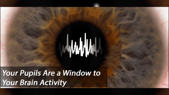
When people are awake, their pupils regularly change in size. Those changes are meaningful, reflecting shifting attention or vigilance, for example. Now, researchers reporting in Current Biology on January 18 have found in studies of mice that pupil size also fluctuates during sleep. They also show that pupil size is a reliable indicator of sleep states.

“We found that pupil size rhythmically fluctuates during sleep,” says Daniel Huber of the University of Geneva in Switzerland. “Intriguingly, these pupil fluctuations follow the sleep-related brain activity so closely that they can indicate with high accuracy the exact stage of sleep—the smaller the pupil, the deeper the sleep.”

Studies of pupil size had always been a challenge for an obvious reason: people and animals generally sleep with their eyes closed. Huber says that he and his colleagues were inspired to study pupil size in sleep after discovering that their laboratory mice sometimes sleep with their eyes open. They knew that pupil size varies strongly during wakefulness. What, they wondered, happened during sleep?

To investigate this question, they developed a novel optical pupil-tracking system for mice. The device includes an infrared light positioned close to the head of the animal. That invisible light travels through the skull and brain to illuminate the back of the eye. When the eyes are imaged with an infrared camera, the pupils appear as bright circles. Thanks to this new method, it was suddenly possible to track changes in pupil size accurately, particularly when the animals snoozed naturally with their eyelids open.

Their images show that mouse pupils rhythmically fluctuate during sleep and that those fluctuations are not at all random; they correlate with changes in sleep states.

Further experiments showed that changes in pupil size are not just a passive phenomenon, either. They are actively controlled by the parasympathetic autonomic nervous system. The evidence suggests that in mice, at least, pupils narrow in deep sleep to protect the animals from waking up with a sudden flash of light.

“The common saying that ‘the eyes are the window to the soul’ might even hold true behind closed eyelids during sleep,” Özge Yüzgeç, the student conducting the study, says. “The pupil continues to play an important role during sleep by blocking sensory input and thereby protecting the brain in periods of deep sleep, when memories should be consolidated.”

Huber says they would like to find out whether the findings hold in humans and whether their new method can be adapted in the sleep clinic. “Inferring brain activity by non-invasive pupil tracking might be an interesting alternative or complement to electrode recordings,” he says.

Reference:

Yüzgeç, Ö., Prsa, M., Zimmermann, R., & Huber, D. (2018). Pupil Size Coupling to Cortical States Protects the Stability of Deep Sleep via Parasympathetic Modulation. Current Biology. doi:10.1016/j.cub.2017.12.049

https://www.technologynetworks.com/neuroscience/news/pupil-size-couples-to-cortical-states-to-protect-deep-sleep-stability-296519?utm_campaign=NEWSLETTER_TN_Neuroscience_2017&utm_source=hs_email&utm_medium=email&utm_content=60184122&_hsenc=p2ANqtz-_uyMIjTK1pmq-79zMcyJIvQNsa8i7gH9l8Tn-_75Taz2opCD4t1otYN6OBmeI-iAKoenGO8wKWNZ7VV6E_JcYum4fHlA&_hsmi=60184122

Pupil response to negative facial expressions predicts risk for depression relapse

Pupil dilation in reaction to negative emotional faces predicts risk for depression relapse, according to new research from Binghamton University, State University of New York.

Researchers at Binghamton University, led by PhD student Anastacia Kudinova, aimed to examine whether physiological reactivity to emotional stimuli, assessed via pupil dilation, served as a biological marker of risk for depression recurrence among individuals who are known to be at a higher risk due to having previous history of depression. Participants were 57 women with a history of major depressive disorder (MDD). The researchers recorded the change in pupil dilation in response to angry, happy, sad and neutral faces. The team found that women’s pupillary reactivity to negative (sad or angry faces) but not positive stimuli prospectively predicted MDD recurrence.

“The study focuses on trying to identify certain markers of depression risk using measures that are readily accessible, reliable and less expensive,” said Kudinova. “It is something we can put in any doctor’s office that gives us a quick and easy objective measure of risk.”

Additionally, the researchers found that both high and low reactivity to angry faces predicted risk for MDD recurrence. These findings suggest that disrupted physiological response to negative stimuli indexed via pupillary dilation could serve as a physiological marker of MDD risk, thus presenting clinicians with a convenient and inexpensive method to predict which of the at-risk women are more likely to experience depression recurrence.

“It’s a bit complicated because different patterns of findings were found for pupil reactivity to angry versus sad faces. Specifically, really high or really low pupil dilation to angry faces was associated with increased risk whereas only low dilation to sad faces was associated with risk (high dilation to sad faces was actually protective),” said Brandon Gibb, professor of psychology at Binghamton University and director of the Mood Disorders Institute and Center for Affective Science.

Other contributors to this research include Katie Burkhouse and Mary Woody, both PhD students; Max Owens, assistant professor of psychology at the University of South Florida, St. Petersburg; and Greg Siegle, associate professor of psychiatry at the University of Pittsburgh School of Medicine.
The paper, “Pupillary reactivity to negative stimuli prospectively predicts recurrence of major depressive disorder in women,” was published in Psychophysiology.

https://www.binghamton.edu/mpr/news-releases/news-release.html?id=2448

Pupil Response Predicts Depression Risk in Kids

Emerging research suggests pupil dilation in children of depressed mothers when seeing an emotional image can help predict his or her risk of depression over the next two years.

Dr. Brandon Gibb, a professor of psychology at Binghamton University in New York, said the new findings suggest physiological reactivity to sad stimuli can be a potential biomarker of depression risk for some kids.

An important aspect of this finding is that pupillometry is an inexpensive tool that could be administered in family practice or pediatricians’ offices.

The simple test can help identify which children of depressed mothers are at highest risk for developing depression themselves.

“We think this line of research could eventually lead to universal screenings in pediatricians’ offices to assess future depression risk in kids,” said Gibb.

Gibb recruited children whose mothers had a history of major depressive disorder and measured their pupil dilation as they viewed angry, happy, and sad faces.

Follow-up assessments occurred over the next two years, during which structured interviews were used to assess for the children’s level of depressive symptoms, as well as the onset of depressive diagnoses.

Researchers found that a child’s reaction to faces can help predict the risk of developing short-term depression.

Specifically, children exhibiting relatively greater pupil dilation to sad faces experienced higher levels of depressive symptoms during the follow-up period. They also displayed a shorter time to the onset of a clinically significant depressive episode.

Interestingly, the type of emotions displayed by faces were a significant predictor of future depression. That is, the findings were specific to children’s pupil responses to sad faces and were not observed for children’s pupillary reactivity to angry or happy faces.

http://psychcentral.com/news/2015/07/09/new-predictive-test-for-childhood-depression/86632.html