Daily step counts between 5,000 to 10,000 or more reduced depression symptoms across 33 studies.
The associations may be due to several mechanisms, like improvement in sleep quality and inflammation.
Daily step counts of 5,000 or more corresponded with fewer depressive symptoms among adults, results of a systematic review and meta-analysis published in JAMA Network Open suggested.
The results are consistent with previous studies linking exercise to various risk reductions for mental health disorders and show that setting step goals “may be a promising and inclusive public health strategy for the prevention of depression,” the researchers wrote.
According to Bruno Bizzozero-Peroni, PhD, MPH, from Universidad De Castilla-La Mancha in Spain, and colleagues, daily step counts are a “simple and intuitive objective measure” of physical activity, while tracking such counts has become increasingly feasible for the general population thanks to the availability of fitness trackers.
“To our knowledge, the association between the number of daily steps measured
with wearable trackers and depression has not been previously examined through a meta-analytic approach,” they wrote.
The researchers searched multiple databases for analyses assessing the effects of daily step counts on depressive symptoms, ultimately including a total of 27 cross-sectional studies and six longitudinal studies comprising 96,173 adults aged 18 years or older.
They found that in the cross-sectional studies, daily step counts of 10,000 or more (standardized mean difference [SMD] = 0.26; 95% CI, 0.38 to 0.14), 7,500 to 9,999 (SMD = 0.27; 95% CI, 0.43 to 0.11) and 5,000 to 7,499 (SMD = 0.17; 95% CI, 0.3 to 0.04) corresponded with reduced depressive symptoms vs. daily step counts less than 5,000.
In the prospective cohort studies, people with 7,000 or more steps a day had a reduced risk for depression vs. with people with fewer than 7,000 daily steps (RR = 0.69; 95% CI, 0.62-0.77), whereas an increase of 1,000 steps a day suggested an association with a lower risk for depression (RR = 0.91; 95% CI, 0.87-0.94).
There were a couple study limitations. The researchers noted that reverse associations are possible, while they could not rule out residual confounders.
They also pointed out that there are some remaining questions, such as whether there is a ceiling limit after which further step counts would no longer reduce the risk for depression.
Bizzozero-Peroni and colleagues highlighted several possible biological and psychosocial mechanisms behind the associations, like changes in sleep quality, inflammation, social support, self-esteem, neuroplasticity and self-efficacy.
They concluded that “a daily active lifestyle may be a crucial factor in regulating and reinforcing these pathways” regardless of the exact combination of mechanisms responsible for the positive link.
“Specifically designed experimental studies are still needed to explore whether there are optimal and maximal step counts for specific population subgroups,” they wrote.
The genetic mutation that causes Huntington’s disease (HD)—a devastating brain disease that disrupts mobility and diminishes cognitive ability—may also enhance early brain development and play a role in promoting human intelligence.
This revelation comes from more than 10 years of brain imaging and brain function data, including motor, cognitive, and behavioral assessments, collected from a unique population—children and young adults who carry the gene for HD. While an HD mutation will eventually cause fatal brain disease in adulthood, the study finds that early in life, children with the HD mutation have bigger brains and higher IQ than children without the mutation.
“The finding suggests that early in life, the gene mutation is actually beneficial to brain development, but that early benefit later becomes a liability,” says Peg Nopoulos, MD, professor and head of psychiatry at the UI Carver College of Medicine, and senior author on the study published in The Annals of Neurology.
The finding may also have implications for developing effective treatments for HD. If the gene’s early action is beneficial, then simply aiming to knock out the gene might result in loss of the developmental benefit, too. Creating therapies that can disrupt the gene’s activity later in the patient’s lifetime might be more useful.
The new data about the gene’s positive effect on early brain development is also exciting to Nopoulos for another reason.
“We are very interested in the fact that this appears to be a gene that drives IQ,” she says. “No previous study has found any gene of significant effect on IQ, even though we know intelligence is heritable.”
HD gene linked to better brain development in early life
Huntington’s disease is caused by a mutation in the huntingtin (HTT) gene. The protein produced by the HTT gene is necessary for normal development, but variations within a segment of the protein have a profound effect on the brain.
The segment in question is a long repeat of one amino acid called glutamine. More repeats are associated with bigger, more complex brains. For example, species such as sea urchins or fish have no repeats, but these repeats start to appear higher up the evolutionary ladder. Rodents have a few repeats, while apes (our closest relatives) have even more repeats; and humans have the most.
Most people have repeats in the range of 10–26, but if a person has 40 or more repeats, then they develop HD. Although the gene expansion is present before birth, HD symptoms do not appear until middle age. Nopoulos’s team at the University of Iowa has a long history of studying how the HTT gene expansion affects brain development in the decades before disease onset.
“We know that the expanded gene causes a horrible degenerative disease later in life, but we also know it is a gene that is crucial for general development,” she says.
“We were surprised to find that it does have a positive effect on brain development early in life. Those who have the gene expansion have an enhanced brain with larger volumes of the cerebrum and higher IQ compared to those who don’t.”
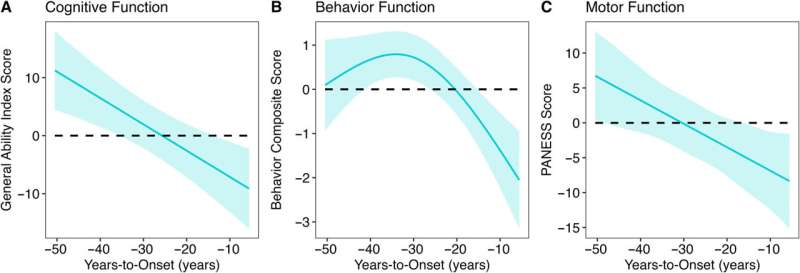
In particular, the study found that decades before HD symptoms appeared, children with the HD gene expansion showed significantly better cognitive, behavioral, and motor scores compared to children with repeats within the normal range. Children with the expanded gene also had larger cerebral volumes and greater cortical surface area and folding. After this initial peak, a prolonged deterioration was seen in both brain function and structure.
The study gathered this data by following almost 200 participants in the Kids-HD study, the only longitudinal study of children and young adults at risk for HD due to having a parent or grandparent with the disease.
Evolutionary benefit comes at a cost
Although surprising, the findings are in line with studies by evolutionary biologists who believe that genes like HTT may have been “positively selected” for human brain evolution. This theory, known as antagonistic pleiotropy, suggests that certain genes can produce a beneficial effect early in life, but come at a cost later in life.
The finding also challenges the idea that the protein produced by the HD gene is solely a toxic protein that causes brain degeneration.
“Overall, our study suggests that we should rethink the notion of the toxic protein theory,” says Nopoulos, who is also a member of the Iowa Neuroscience Institute.
“Instead, we should consider the theory of antagonistic pleiotropy—a theory that suggests that genes like HTT build a better brain early in life, but the cost of the superior brain is that it isn’t built to last and may be prone to premature or accelerating aging.
“This means that instead of knocking down the gene for therapy, drugs that slow the aging process may be more effective.”
Next steps
Nopoulos’s team is already making progress extending the research from the Kids-HD program. Nopoulos has established the Children to Adult Neurodevelopment in Gene-Expanded Huntington’s Disease (ChANGE-HD), a multi-site study that aims to recruit hundreds of participants for a total of over 1,200 assessments to validate the key findings from the Kids-HD study and to enhance future research on HD.
A primary area of focus will be understanding how an enlarged brain can later lead to degeneration. One hypothesis Nopoulos and her team will explore involves the idea that an enlarged cortex might produce excess glutamate (an important neurotransmitter), which is beneficial in early brain development, but later leads to neurotoxicity and brain degeneration.
In addition to Nopoulos, the UI team included Mohit Neema, MD, UI research scientist and first author of the study; Jordan Schultz, PharmD; Douglas Langbehn, MD, Ph.D.; Amy Conrad, Ph.D.; Eric Epping, MD, Ph.D.; and Vincent Magnotta, Ph.D.
More information: Mohit Neema et al, Mutant Huntingtin Drives Development of an Advantageous Brain Early in Life: Evidence in Support of Antagonistic Pleiotropy, Annals of Neurology (2024). DOI: 10.1002/ana.27046
Scientists at the University of Copenhagen have discovered a new weight loss drug target that reduces appetite, increases energy expenditure, and improves insulin sensitivity without causing nausea or loss of muscle mass. The discovery was reported in the journal Nature and could lead to a new therapy for millions of people with both obesity and type 2 diabetes who do not respond well to current treatments.
Millions of people around the world benefit from weight-loss drugs based on the incretin hormone GLP-1. These drugs also improve kidney function, reduce the risk of fatal cardiac events, and are linked to protection against neurodegeneration.
However, many people stop taking the drugs due to common side effects, including nausea and vomiting. Studies also show that incretin-based therapies like Wegovy and Mounjaro are much less effective at lowering weight in people living with both obesity and type 2 diabetes—a group numbering more than 380 million people globally.
In the study, scientists from the University of Copenhagen describe a powerful new drug candidate that lowers appetite without loss of muscle mass or side effects like nausea and vomiting. And, unlike the current generation of treatments, the drug also increases the body’s energy expenditure—the capacity of the body to burn calories.
“While GLP-1-based therapies have revolutionized patient care for obesity and type 2 diabetes, safely harnessing energy expenditure and controlling appetite without nausea remain two Holy Grails in this field. By addressing these needs, we believe our discovery will propel current approaches to make more tolerable, effective treatments accessible to millions more individuals,” says Associate Professor Zach Gerhart-Hines from the NNF Foundation Center for Basic Metabolic Research (CBMR) at the University of Copenhagen.
NK2R activation lowers body weight and reverses diabetes
Our weight is largely determined by the balance between the energy we consume and the amount of energy we expend. Eating more and burning less creates a positive energy balance leading to weight gain, while eating less and burning more creates a negative balance, resulting in weight loss.
The current generation of incretin-based therapies tip the scales toward a negative energy balance by lowering appetite and the total calories a person consumes. But scientists have also recognized the potential on the other side of the equation—increasing the calories the body burns.
This approach is especially relevant, given recent research that has shown that our bodies seem to be burning fewer calories at rest than they did a few decades ago. However, there are currently no clinically approved ways to safely increase energy expenditure, and few options are in development.
This was the starting point when scientists at the University of Copenhagen decided to test the effect of activating the neurokinin 2 receptor (NK2R) in mice. The Gerhart-Hines Group identified the receptor through genetic screens that suggested NK2R played a role in maintaining energy balance and glucose control.
They were astonished by the results of the studies—not only did activating the receptor safely increase calorie-burning, it also lowered appetite without any signs of nausea.
Further studies in non-human primates with type 2 diabetes and obesity showed that NK2R activation lowered body weight and reversed their diabetes by increasing insulin sensitivity and lowering blood sugar, triglycerides, and cholesterol.
“One of the biggest hurdles in drug development is translation between mice and humans. This is why we were excited that the benefits of NK2R agonism translated to diabetic and obese nonhuman primates, which represents a big step towards clinical translation,” says Ph.D. Student Frederike Sass from CBMR at the University of Copenhagen, and first author of the study.
The discovery could result in the next generation of drug therapies that bring more efficacious and tolerable treatments for the almost 400 million people globally who live with both type 2 diabetes and obesity.
The University of Copenhagen holds the patent rights for targeting NK2R. To date, research by the Gerhart-Hines lab has led to the creation of three biotech companies—Embark Biotech, Embark Laboratories, and Incipiam Pharma.
In 2023, Embark Biotech was acquired by Novo Nordisk to develop next generation therapeutics for cardiometabolic disease.
Researchers have developed an AI-powered model that—in 10 seconds—can determine during surgery if any part of a cancerous brain tumor that could be removed remains, a study published in Nature suggests.
The technology, called FastGlioma, outperformed conventional methods for identifying what remains of a tumor by a wide margin, according to the research team led by University of Michigan and University of California San Francisco.
“FastGlioma is an artificial intelligence-based diagnostic system that has the potential to change the field of neurosurgery by immediately improving comprehensive management of patients with diffuse gliomas,” said senior author Todd Hollon, M.D., a neurosurgeon at University of Michigan Health and assistant professor of neurosurgery at U-M Medical School.
“The technology works faster and more accurately than current standard of care methods for tumor detection and could be generalized to other pediatric and adult brain tumor diagnoses. It could serve as a foundational model for guiding brain tumor surgery.”
When a neurosurgeon removes a life threatening tumor from a patient’s brain, they are rarely able to remove the entire mass.
Commonly, the tumor is missed during the operation because surgeons are not able to differentiate between healthy brain and residual tumor in the cavity where the mass was removed. Residual tumor’s ability to resemble healthy brain tissue remains a major challenge in surgery.
Neurosurgical teams employ different methods to locate that residual tumor during a procedure.
They may get MRI imaging, which requires intraoperative machinery that is not available everywhere. The surgeon might also use a fluorescent imaging agent to identify tumor tissue, which is not applicable for all tumor types. These limitations prevent their widespread use.
In this international study of the AI-driven technology, neurosurgical teams analyzed fresh, unprocessed specimens sampled from 220 patients who had operations for low- or high-grade diffuse glioma.
FastGlioma detected and calculated how much tumor remained with an average accuracy of approximately 92%.
In a comparison of surgeries guided by FastGlioma predictions or image- and fluorescent-guided methods, the AI technology missed high-risk, residual tumor just 3.8% of the time—compared to a nearly 25% miss rate for conventional methods.
“This model is an innovative departure from existing surgical techniques by rapidly identifying tumor infiltration at microscopic resolution using AI, greatly reducing the risk of missing residual tumor in the area where a glioma is resected,” said co-senior author Shawn Hervey-Jumper, M.D., professor of neurosurgery at University of California San Francisco and a former neurosurgery resident at U-M Health.
“The development of FastGlioma can minimize the reliance on radiographic imaging, contrast enhancement or fluorescent labels to achieve maximal tumor removal.”
How it works
To assess what remains of a brain tumor, FastGlioma combines microscopic optical imaging with a type of artificial intelligence called foundation models. These are AI models, such as GPT-4 and DALL·E 3, trained on massive, diverse datasets that can be adapted to a wide range of tasks.
After large scale training, foundation models can classify images, act as chatbots, reply to emails and generate images from text descriptions.
To build FastGlioma, investigators pre-trained the visual foundation model using over 11,000 surgical specimens and 4 million unique microscopic fields of view.
The tumor specimens are imaged through stimulated Raman histology, a method of rapid, high resolution optical imaging developed at U-M. The same technology was used to train DeepGlioma, an AI based diagnostic screening system that detects a brain tumor’s genetic mutations in under 90 seconds.
“FastGlioma can detect residual tumor tissue without relying on time-consuming histology procedures and large, labeled datasets in medical AI, which are scarce,” said Honglak Lee, Ph.D., co-author and professor of computer science and engineering at U-M.
Full resolution images take around 100 seconds to acquire using stimulated Raman histology; a “fast mode” lower resolution image takes just 10 seconds.
Researchers found that the full resolution model achieved accuracy up to 92%, with the fast mode slightly lower at approximately 90%.
“This means that we can detect tumor infiltration in seconds with extremely high accuracy, which could inform surgeons if more resection is needed during an operation,” Hollon said.
AI’s future in cancer
Over the last 20 years, the rates of residual tumor after neurosurgery have not improved.
Not only is FastGlioma an accessible and affordable tool for neurosurgical teams operating on gliomas, but researchers say, it can also accurately detect residual tumor for several non-glioma tumor diagnoses, including pediatric brain tumors, such as medulloblastoma and ependymoma, and meningiomas.
“These results demonstrate the advantage of visual foundation models such as FastGlioma for medical AI applications and the potential to generalize to other human cancers without requiring extensive model retraining or fine-tuning,” said co-author Aditya S. Pandey, M.D., chair of the Department of Neurosurgery at U-M Health.
“In future studies, we will focus on applying the FastGlioma workflow to other cancers, including lung, prostate, breast, and head and neck cancers.”
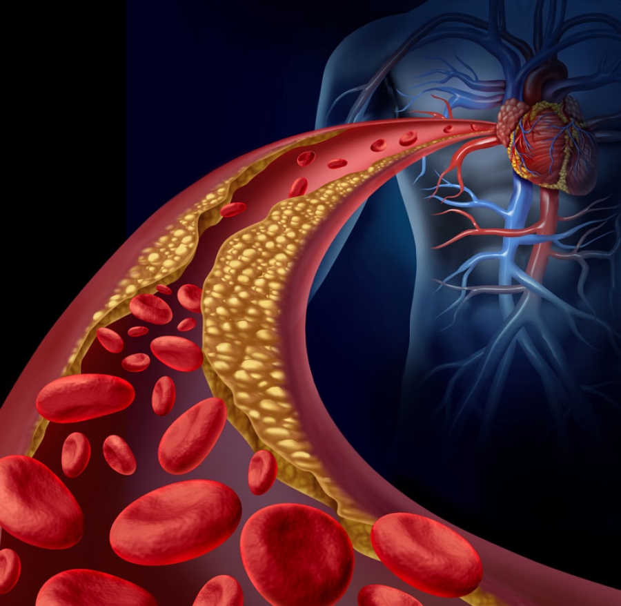
Researchers at Case Western Reserve University have identified a new target to treat atherosclerosis, a condition where plaque clogs arteries and causes major cardiac issues, including stroke and heart attack.
In a new study, published in the journal Cell Reports, the team identified an inflammation-reducing molecule—called itaconate (ITA)—that could be the foundation of a new approach to treat such a common and deadly disease.
Heart disease is the leading cause of death for men, women and people of most racial and ethnic groups, according to the U.S. Centers for Disease Control and Prevention.
Medications help but don’t completely protect patients from cardiovascular risk. So, doctors also recommend lifestyle changes, such as a low-cholesterol/low-fat diet (LCLFD), to further reduce plaque and inflammation that increase the risk of cardiovascular disease. Yet many patients find it challenging to follow diet restrictions long-term.
Identifying the role ITA plays in diet and heart disease may help address this.
“We’ve found that itaconate is crucial to the diet’s ability to stabilize plaques and reduce inflammation, which has been a mystery until now,” said Andrei Maiseyeu, associate professor at the Cardiovascular Research Institute and Department of Biomedical Engineering at Case Western Reserve’s School of Medicine.
“This discovery marks a major leap forward in the understanding of how diet-induced plaque resolution occurs at a molecular level.”
Based on their discovery, Maiseyeu and his team have developed a new treatment: ITA-conjugated lipid nanoparticles. This new therapeutic approach allows ITA to accumulate in plaque and bone marrow, where it reduces inflammation and mimics the beneficial effects of LCLFD without requiring drastic lifestyle changes.
“We have already seen its effectiveness in multiple models of atherosclerosis,” Maiseyeu said. “We are optimistic that this will result in better treatments that will greatly lower the long-term risk of heart attacks and strokes while also improving patients’ quality of life.”
Maiseyeu and his team are now taking steps to translate ITA-LNP to the clinic, including engineering a pill form of the treatment, which they believe will not only be convenient for patients, but also transformative.
More information: Natalie E. Hong et al, Nanoparticle-based itaconate treatment recapitulates low-cholesterol/low-fat diet-induced atherosclerotic plaque resolution, Cell Reports (2024). DOI: 10.1016/j.celrep.2024.114911
Life and death are traditionally viewed as opposites. But the emergence of new multicellular life-forms from the cells of a dead organism introduces a “third state” that lies beyond the traditional boundaries of life and death.
Usually, scientists consider death to be the irreversible halt of functioning of an organism as a whole. However, practices such as organ donation highlight how organs, tissues and cells can continue to function even after an organism’s demise. This resilience raises the question: What mechanisms allow certain cells to keep working after an organism has died?
The third state challenges how scientists typically understand cell behavior. While caterpillars metamorphosing into butterflies, or tadpoles evolving into frogs, may be familiar developmental transformations, there are few instances where organisms change in ways that are not predetermined. Tumors, organoids and cell lines that can indefinitely divide in a petri dish, like HeLa cells, are not considered part of the third state because they do not develop new functions.
However, researchers found that skin cells extracted from deceased frog embryos were able to adapt to the new conditions of a petri dish in a lab, spontaneously reorganizing into multicellular organisms called xenobots. These organisms exhibited behaviors that extend far beyond their original biological roles. Specifically, these xenobots use their cilia – small, hair-like structures – to navigate and move through their surroundings, whereas in a living frog embryo, cilia are typically used to move mucus.
Xenobots are also able to perform kinematic self-replication, meaning they can physically replicate their structure and function without growing. This differs from more common replication processes that involve growth within or on the organism’s body.
Researchers have also found that solitary human lung cells can self-assemble into miniature multicellular organisms that can move around. These anthrobots behave and are structured in new ways. They are not only able to navigate their surroundings but also repair both themselves and injured neuron cells placed nearby.
Taken together, these findings demonstrate the inherent plasticity of cellular systems and challenge the idea that cells and organisms can evolve only in predetermined ways. The third state suggests that organismal death may play a significant role in how life transforms over time.
Postmortem conditions
Several factors influence whether certain cells and tissues can survive and function after an organism dies. These include environmental conditions, metabolic activity and preservation techniques.
Different cell types have varying survival times. For example, in humans, white blood cells die between 60 and 86 hours after organismal death. In mice, skeletal muscle cells can be regrown after 14 days postmortem, while fibroblast cells from sheepandgoats can be cultured up to a month or so postmortem.
Metabolic activity plays an important role in whether cells can continue to survive and function. Active cells that require a continuous and substantial supply of energy to maintain their function are more difficult to culture than cells with lower energy requirements. Preservation techniques such as cryopreservation can allow tissue samples such as bone marrow to function similarly to that of living donor sources.
Factors such as age, health, sex and type of species further shape the postmortem landscape. This is seen in the challenge of culturing and transplanting metabolically active islet cells, which produce insulin in the pancreas, from donors to recipients. Researchers believe that autoimmune processes, high energy costs and the degradation of protective mechanisms could be the reason behind many islet transplant failures.
How the interplay of these variables allows certain cells to continue functioning after an organism dies remains unclear. One hypothesis is that specialized channels and pumps embedded in the outer membranes of cells serve as intricate electrical circuits. These channels and pumps generate electrical signals that allow cells to communicate with each other and execute specific functions such as growth and movement, shaping the structure of the organism they form.
The extent to which different types of cells can undergo transformation after death is also uncertain. Previous research has found that specific genes involved in stress, immunity and epigenetic regulation are activated after death in mice, zebrafishand people, suggesting widespread potential for transformation among diverse cell types.
Implications for biology and medicine
The third state not only offers new insights into the adaptability of cells. It also offers prospects for new treatments.
For example, anthrobots could be sourced from an individual’s living tissue to deliver drugs without triggering an unwanted immune response. Engineered anthrobots injected into the body could potentially dissolve arterial plaque in atherosclerosis patients and remove excess mucus in cystic fibrosis patients.
Importantly, these multicellular organisms have a finite life span, naturally degrading after four to six weeks. This “kill switch” prevents the growth of potentially invasive cells.
A better understanding of how some cells continue to function and metamorphose into multicellular entities some time after an organism’s demise holds promise for advancing personalized and preventive medicine.
Transcripts circulating in the blood provide real-time information about maternal, fetal, and placental health.
by Amanda Heidt
Preeclampsia, a potentially fatal complication that affects roughly 5 percent of pregnancies worldwide, can only be diagnosed after the onset of symptoms such as high blood pressure, so treatment is always reactive. “The next really big need is better methods to diagnose or predict risk of pregnancy complications such as preeclampsia,” says Fiona Kaper, a senior director of scientific research at the biotech company Illumina.
To identify possible biomarkers of the condition, Kaper and her colleagues drew blood from 40 pregnant women with early-onset severe preeclampsia and 73 unaffected expecting mothers. Circulating in the blood of each mom-to-be is her own RNA, as well as transcripts from the placenta and the fetus. Studying these circulating RNAs (cRNAs), the team identified 30 maternal, fetal, or placental genes with altered expression patterns in women with preeclampsia compared with controls. A machine algorithm also identified 49 genes with altered expression, including 12 that overlapped with the earlier list, suspected of being linked to preeclampsia.
To test the ability of the 49 suspect genes to predict preeclampsia, the researchers classified an independent cohort of two dozen women, half with early-onset preeclampsia and half without signs of the condition. The model predicted which women had preeclampsia with 85 percent to 89 percent accuracy.
While large-scale, prospective studies are still needed, cRNA screening represents a step toward earlier preemptive diagnosis, says Kathryn Gray, an obstetrician at Brigham and Women’s Hospital who was not involved in the study. She notes that researchers have been doing something similar in detecting circulating tumor DNA for cancer screening. “It’s really exciting that we’re applying some of these . . . strategies that have been used in cancer to pregnancy. We’re always a bit behind in women’s health and pregnancy in applying the most cutting-edge technologies.”
S. Munchel et al., “Circulating transcripts in maternal blood reflect a molecular signature of early-onset preeclampsia,” Sci Transl Med, 12:eaaz0131, 2020.
Eugenia Trushina, PhD: Mitochondria as a Therapeutic Target for Alzheimer
The Alzheimer’s Drug Discovery Foundation (ADDF) and Harrington Discovery Institute at University Hospitals have granted Eugenia Trushina, Ph.D., of Mayo Clinic Rochester, the ADDF-Harrington Scholar Award.
Dr. Trushina has been awarded $600,000 for her late stage preclinical research on new drug candidates that show promise in restoring mitochondrial function. In addition to funding, she will receive in-depth drug development support to help maximize her project’s potential for clinical success.
“The ADDF-Harrington partnership helps scientists move academic discoveries from their labs toward clinical studies, and eventually into the clinic to improve the lives of people living with and at risk of Alzheimer’s,” said Dr. Howard Fillit, the ADDF’s Founding Executive Director and Chief Science Officer. “The mitochondria, which are the powerhouses of the cell, are a promising new target in the fight to combat this devastating disease.”
Dr. Trushina has shown that restoring function in mitochondria may delay the onset or slow the progression of Alzheimer’s disease. The compounds she and her team have developed have shown a positive effect in both symptomatic and pre-symptomatic models of Alzheimer’s.
“Supporting this innovative therapeutic approach for patients with Alzheimer’s disease represents our dedication to developing new classes of medicines that are not otherwise traditionally pursued in the field,” said Dr. Andrew Pieper, Director of the Neurotherapeutics Center of the Harrington Discovery Institute and University Hospitals Morley-Mather Chair in Neuropsychiatry.
Dr. Trushina’s research was selected through a competitive process, based on its potential to advance towards the clinic as a novel approach to treat, prevent, or cure Alzheimer’s disease and related dementias. Collaboration between the ADDF and Harrington Discovery Institute for this award provides recipients with both research funding and expert guidance in order to efficiently bridge the gap between academia and pharma.
“We are in our seventh year of collaborating with the ADDF to address this major unmet medical need,” said Jonathan Stamler, MD, President, Harrington Discovery Institute and Robert S. and Sylvia K. Reitman Family Foundation Distinguished Chair of Cardiovascular Innovation and Professor of Medicine at University Hospitals and Case Western Reserve University. “This partnership leverages our combined expertise and resources to give the science the best chance of advancing towards a cure for Alzheimer’s disease.”
###
About the Alzheimer’s Drug Discovery Foundation (ADDF)
The Alzheimer’s Drug Discovery Foundation is dedicated to rapidly accelerating the discovery of drugs to prevent, treat and cure Alzheimer’s disease. The ADDF is the only public charity solely focused on funding the development of drugs for Alzheimer’s, employing a venture philanthropy model to support research in academia and the biotech industry. Through the generosity of its donors, the ADDF has awarded more than $150 million to fund over 626 Alzheimer’s drug discovery programs and clinical trials in 19 countries. To learn more, please visit: https://www.alzdiscovery.org/
About the Harrington Discovery Institute
The Harrington Discovery Institute at University Hospitals in Cleveland, Ohio–part of The Harrington Project for Discovery & Development–aims to advance medicine and society by enabling our nation’s most inventive scientists to turn their discoveries into medicines that improve human health. The institute was created in 2012 with a $50 million founding gift from the Harrington family and instantiates the commitment they share with University Hospitals to a Vision for a “Better World.”
About the Harrington Project for Discovery & Development
The Harrington Project for Discovery & Development (The Harrington Project), founded in late February 2012 by the Harrington Family and University Hospitals of Cleveland, is a $300 million national initiative built to bridge the translational valley of death. It includes the Harrington Discovery Institute and BioMotiv, a for-profit, mission-aligned drug development company that accelerates early discovery into pharma pipelines.
An unusual new treatment for Tourette’s syndrome involves applying an electrical current to the wrist, which travels up nerves to the brain and changes brainwaves. The approach, which moderately reduced the number of tics in volunteers with Tourette’s, suggests the condition is linked with an underactivity in brainwaves that normally keep us still.
People with Tourette’s syndrome make frequent involuntary jerks, facial twitches and noises. Tics usually arise around the age of 6, and while they often fade with time, for some they continue and can be debilitating. “Sometimes children literally break bones because they’re flinging themselves around so much,” says Stephen Jackson at the University of Nottingham in the UK.
In most people, when they are motionless, brainwaves cycle at about 12 times a second in part of the brain called the motor cortex, located at the top of the head. “It’s like the handbrake on a car – it maintains a stable posture,” says Jackson.
Previous research has shown that stimulating brainwaves in this area by using a strong oscillating magnetic field above the head can reduce tics in people with Tourette’s. Jackson wondered if there was a way to get this effect more easily.
His group placed an electrode on the wrist to deliver a mild current with a frequency of 12 times a second; the current was noticeable but not uncomfortable. The idea is that this current travels via nerves to the sensory cortex of the brain and induces oscillations at the same frequency in the neighbouring motor cortex.
When 19 people with Tourette’s tried out the electrode, it reduced the frequency of their tics by a third, compared with when the electrode was turned on for the same length of time but had no regular frequency. Voluntary movements were only slowed a little.
Normally people with Tourette’s feel an urge to tic slowly building until it becomes irresistable. Many of the volunteers reported that when the electrodes were at the right frequency, their urges reduced. Charlie, a 21-year-old with severe tics, said in a statement: “When the electrical pulses on the wrist started to increase, the tic urges decreased, which was a completely shocking experience for me, I was silent and still. I wanted to cry with happiness.”
Jackson’s group is developing a watch-like device that people can turn on to deliver the stimulation when it is needed.
Journal reference: Current Biology, DOI: 10.1016/j.cub.2020.04.044
Human cell types within corresponding organs that express the genes for both ACE2 and CTSL (green dot) or both ACE2 and TMPRSS2 (orange dot).
by Chris Baraniuk
When the SARS-CoV-2 virus enters the human body, it breaks into cells with the help of two proteins that it finds there, ACE2 and TMPRSS2. While there has been much discussion of viral infection in gut and lung cells, researchers have dug into massive gene expression datasets to show that other potential target cells also producing ACE2 and TMPRSS2 are scattered throughout the body—including in the heart, bladder, pancreas, kidney, and nose. There are even some in the eye and brain.
The results, published in a preprint on bioRxiv April 21, show that such cells are strikingly abundant. Many are epithelial cells, which line the outer surface of organs. The new findings add to an emerging picture of SARS-CoV-2 as a virus that can target cells in many places in the human body, rather than being focused on a particular organ or part of the respiratory tract.
Cardiologist Frank Ruschitzka at the University Hospital of Zürich and colleagues separately published a letter in The Lancet April 17 in which they described how virus particles had been found in the vascular endothelium, a thin layer of cells lining blood vessels in various organs of the body, for instance.
“This is not just a virus pneumonia,” Ruschitzka, who was not involved in the latest study, tells The Scientist, referring to COVID-19. “This is a disease like we have never seen before—it is not an influenza, it hits the vessels all over, it hits the heart as well.”
To uncover the locations of cells bearing ACE2 and TMPRSS2, the preprint researchers turned to the Human Cell Atlas, a project that has allowed scientists to pool together data on human cells since 2016.
By scouring single-cell sequencing records of around 1.2 million individual cells from human tissue samples, the team was able to find out which of those cells produce both ACE2 and TMPRSS2, and note their locations in the body. The analysis used 16 unpublished datasets of lung and airway cells and 91 published datasets spanning a range of human organs.
Coauthor Christoph Muus, a graduate student at Harvard University and the Broad Institute, explains that while the data show cells in many locations in the body produce SARS-CoV-2 receptors, it’s not certain that the virus can infect all of those tissues.
“Expressing the receptor is a necessary condition but not necessarily a sufficient condition,” he says. For example, potential target cells were found in the testes, but scientists still don’t know if SARS-CoV-2 infects and replicates in that part of the body.
Jeremy Kamil, a virologist at Louisiana State University Health Shreveport, says the preprint provides important details about the human body that may help scientists understand how SARS-CoV-2 infects hosts. By finding viral protein fragments in tissue samples from patients who died because of COVID-19, scientists might be able to firm up which organs are genuine sites of infection, he adds.
“I’d say this paper gives people a roadmap at where you might want to look in the body to understand where this virus is going,” he says.
One limitation of the work is that relatively little metadata about the people who donated tissue samples were available for the various datasets, though information about age and gender were included in many. The researchers don’t know, for example, whether there was an ethnicity bias in the data, whether patients had pre-existing conditions, or whether they were taking any medications. All of these things could affect gene expression in particular cells.
Smoking status was available for a subset of the data, and the team used this to show that smoking is correlated with a greater expression of the ACE2 gene in the upper airway, but lower expression in certain lung cells. Further research is needed to understand whether this affects smokers’ susceptibility to COVID-19. Data from China suggest that smokers are 14 times more likely to develop a severe form of the disease.
Some researchers from the same group using similar data have also recently published papers in Cell and Nature. In those cases, the researchers focused on certain groups of cells. The study reported in Nature examined cells potentially involved in viral transmission and found that nasal epithelial cells, in particular, were associated with expression of ACE2 and TMPRSS2. The authors report that the virus might exploit cells that secrete fluids in the nasal passage, which might help it spread from one person to another in droplets released, say, when someone sneezes.
The Cell study, meanwhile, also found ACE2 and TMPRSS2 transcripts in nasal, gut, and lung cells but the researchers also found that the protein interferon activated ACE2 expression in vitro. The human body uses interferon to fight infections, so it is not clear whether the protein is of overall benefit or detriment to COVID-19 patients.
The use of so many different data sources backs up the validity of the preprint authors’ findings, says Marta Gaglia, a molecular biologist at Tufts University. She agrees with the researchers that discovering ACE2- and TMPRSS2-producing cells in various places around the body does not prove the virus can always infect such cells.
“I think the reality is that most of the problems come from the lung,” she adds. Plus, while doctors treating COVID-19 patients may detect problems in multiple organs, those issues might not necessarily be caused directly by SARS-CoV-2 infection, says Gaglia. A problematic immune system response, for instance, could damage certain tissues in the body as an indirect consequence of viral infection.