Immune cells invade aging brains, disrupt new nerve cell formation

A study by Stanford University School of Medicine investigators has revealed that immune cells infiltrate the rare newborn nerve-cell nurseries of the aging brain. There’s every reason to think those interlopers are up to no good. Experiments in a dish and in living animals indicate they’re secreting a substance that chokes off new nerve cell production.

While most of the experiments in the study were carried out in mice, the central finding—the invasion, by immune cells called killer T cells, of neurogenic niches (specialized spots in the brain where new nerve cells, or neurons, are generated)—was corroborated in tissue excised from autopsied human brains.

The findings could accelerate progress in hunting down the molecules in the body that promote the common deterioration of brain function in older individuals and in finding treatments that might stall or even reverse that deterioration. They also signify a crack in the wall of dogma that’s deemed the healthy brain impervious to invasion by the body’s immune cells, whose unbridled access to the organ could cause damage.

“The textbooks say that immune cells can’t easily get into the healthy brain, and that’s largely true,” said Anne Brunet, Ph.D., professor of genetics and senior author of the study. “But we’ve shown that not only do they get into otherwise healthy aging brains—including human brains—but they reach the very part of the brain where new neurons arise.”

Lead authorship of the study, to be published online July 3 in Nature, is shared by medical student Ben Dulken, Ph.D., graduate student Matthew Buckley and postdoctoral scholar Paloma Navarro Negredo, Ph.D.

The cells that aid memory

Many a spot in a young mammal’s brain is bursting with brand new neurons. But for the most part, those neurons have to last a lifetime. Older mammals’ brains retain only a couple of neurogenic niches, consisting of several cell types whose mix is critical for supporting neural stem cells that can both differentiate into neurons and generate more of themselves. New neurons spawned in these niches are considered essential to forming new memories and to learning, as well as to odor discrimination.

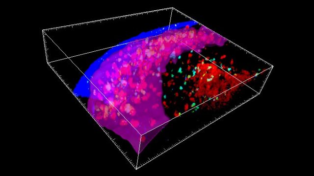
In order to learn more about the composition of the neurogenic niche, the Stanford researchers catalogued, one cell at a time, the activation levels of the genes in each of nearly 15,000 cells extracted from the subventricular zone (a neurogenic niche found in mice and human brains) of healthy 3-month-old mice and healthy 28- or 29-month-old mice.

This high-resolution, single-cell analysis allowed the scientists to characterize each cell they looked at and see what activities it was engaged in. Their analysis confirmed the presence of nine familiar cell types known to compose the neurogenic niche. But when Brunet and her colleagues compared their observations in the brains of young mice (equivalent in human years to young adults) with what they saw in the brains of old mice (equivalent to people in their 80s), they identified a couple of cell types in the older mice not typically expected to be there—and barely present in the young mice. In particular, they found immune cells known as killer T cells lurking in the older mice’s subventricular zone.

The healthy brain is by no means devoid of immune cells. In fact, it boasts its own unique version of them, called microglia. But a much greater variety of immune cells abounding in the blood, spleen, gut and elsewhere in the body are ordinarily denied entry to the brain, as the blood vessels pervading the brain have tightly sealed walls. The resulting so-called blood-brain barrier renders a healthy brain safe from the intrusion of potentially harmful immune cells on an inflammatory tear as the result of a systemic illness or injury.

“We did find an extremely sparse population of killer T cells in the subventricular zone of young mice,” said Brunet, who is the Michele and Timothy Barakett Endowed Professor. “But in the older mice, their numbers were expanded by 16-fold.”

That dovetailed with reduced numbers of proliferation-enabled neural stem cells in the older mice’s subventricular zone. Further experiments demonstrated several aspects of the killer T cells’ not-so-mellow interaction with neural stem cells. For one thing, tests in laboratory dishware and in living animals indicated that killer T cells isolated from old mice’s subventricular zone were far more disposed than those from the same mice’s blood to pump out an inflammation-promoting substance that stopped neural stem cells from generating new nerve cells.

Second, killer T cells were seen nestled next to neural stem cells in old mice’s subventricular zones and in tissue taken from the corresponding neurogenic niche in autopsied brains of old humans; where this was the case, the neural stem cells were less geared up to proliferate.

Possible brain-based antigens

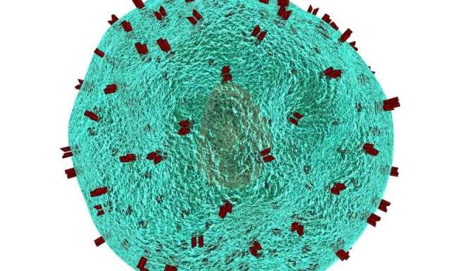
A third finding was especially intriguing. Killer T cells’ job is to roam through the body probing the surfaces of cells for biochemical signs of a pathogen’s presence or of the possibility that a cell is becoming, or already is, cancerous. Such telltale biochemical features are called antigens. The tens of billions of killer T cells in a human body are able to recognize a gigantic range of antigens by means of receptors on their own surfaces. That’s because every unexposed, or naïve, killer T cell has its own unique receptor shape.

When an initially naïve killer T cell is exposed to an unfamiliar antigen that fits its uniquely shaped receptor, it reacts by undergoing multiple successive rounds of replication, culminating in a large set of warlike cells all sharing the same receptor and all poised to destroy any cells bearing the offending antigen. This process is called clonal expansion.

The killer T cells found in old mice’s brains had undergone clonal expansion, indicating likely exposure to triggering antigens. But the receptors on those killer T cells differed from the ones found in the old mice’s blood, suggesting that the brain-localized killer T cells hadn’t just traipsed through a disrupted blood-brain barrier via passive diffusion but were, rather, reacting to different, possibly brain-based, antigens.

Brunet’s group is now trying to determine what those antigens are. “They may bear some responsibility for the disruption of new neuron production in the aging brain’s neurogenic niches,” she said.

Single cell analysis reveals T cell infiltration in old neurogenic niches, Nature (2019). DOI: 10.1038/s41586-019-1362-5 , https://www.nature.com/articles/s41586-019-1362-5

https://medicalxpress.com/news/2019-07-immune-cells-invade-aging-brains.html

He Got Schizophrenia. He Got Cancer. And Then He Got Cured.

merlin_144488709_dd45e03b-d06e-4037-9c40-c8353cdb464b-superJumbo

By Moises Velasquez-Manoff

The man was 23 when the delusions came on. He became convinced that his thoughts were leaking out of his head and that other people could hear them. When he watched television, he thought the actors were signaling him, trying to communicate. He became irritable and anxious and couldn’t sleep.

Dr. Tsuyoshi Miyaoka, a psychiatrist treating him at the Shimane University School of Medicine in Japan, eventually diagnosed paranoid schizophrenia. He then prescribed a series of antipsychotic drugs. None helped. The man’s symptoms were, in medical parlance, “treatment resistant.”

A year later, the man’s condition worsened. He developed fatigue, fever and shortness of breath, and it turned out he had a cancer of the blood called acute myeloid leukemia. He’d need a bone-marrow transplant to survive. After the procedure came the miracle. The man’s delusions and paranoia almost completely disappeared. His schizophrenia seemingly vanished.

Years later, “he is completely off all medication and shows no psychiatric symptoms,” Dr. Miyaoka told me in an email. Somehow the transplant cured the man’s schizophrenia.

A bone-marrow transplant essentially reboots the immune system. Chemotherapy kills off your old white blood cells, and new ones sprout from the donor’s transplanted blood stem cells. It’s unwise to extrapolate too much from a single case study, and it’s possible it was the drugs the man took as part of the transplant procedure that helped him. But his recovery suggests that his immune system was somehow driving his psychiatric symptoms.

At first glance, the idea seems bizarre — what does the immune system have to do with the brain? — but it jibes with a growing body of literature suggesting that the immune system is involved in psychiatric disorders from depression to bipolar disorder.

The theory has a long, if somewhat overlooked, history. In the late 19th century, physicians noticed that when infections tore through psychiatric wards, the resulting fevers seemed to cause an improvement in some mentally ill and even catatonic patients.

Inspired by these observations, the Austrian physician Julius Wagner-Jauregg developed a method of deliberate infection of psychiatric patients with malaria to induce fever. Some of his patients died from the treatment, but many others recovered. He won a Nobel Prize in 1927.

One much more recent case study relates how a woman’s psychotic symptoms — she had schizoaffective disorder, which combines symptoms of schizophrenia and a mood disorder such as depression — were gone after a severe infection with high fever.

Modern doctors have also observed that people who suffer from certain autoimmune diseases, like lupus, can develop what looks like psychiatric illness. These symptoms probably result from the immune system attacking the central nervous system or from a more generalized inflammation that affects how the brain works.

Indeed, in the past 15 years or so, a new field has emerged called autoimmune neurology. Some two dozen autoimmune diseases of the brain and nervous system have been described. The best known is probably anti-NMDA-receptor encephalitis, made famous by Susannah Cahalan’s memoir “Brain on Fire.” These disorders can resemble bipolar disorder, epilepsy, even dementia — and that’s often how they’re diagnosed initially. But when promptly treated with powerful immune-suppressing therapies, what looks like dementia often reverses. Psychosis evaporates. Epilepsy stops. Patients who just a decade ago might have been institutionalized, or even died, get better and go home.

Admittedly, these diseases are exceedingly rare, but their existence suggests there could be other immune disorders of the brain and nervous system we don’t know about yet.

Dr. Robert Yolken, a professor of developmental neurovirology at Johns Hopkins, estimates that about a third of schizophrenia patients show some evidence of immune disturbance. “The role of immune activation in serious psychiatric disorders is probably the most interesting new thing to know about these disorders,” he told me.

Studies on the role of genes in schizophrenia also suggest immune involvement, a finding that, for Dr. Yolken, helps to resolve an old puzzle. People with schizophrenia tend not to have many children. So how have the genes that increase the risk of schizophrenia, assuming they exist, persisted in populations over time? One possibility is that we retain genes that might increase the risk of schizophrenia because those genes helped humans fight off pathogens in the past. Some psychiatric illness may be an inadvertent consequence, in part, of having an aggressive immune system.

Which brings us back to Dr. Miyaoka’s patient. There are other possible explanations for his recovery. Dr. Andrew McKeon, a neurologist at the Mayo Clinic in Rochester, Minn., a center of autoimmune neurology, points out that he could have suffered from a condition called paraneoplastic syndrome. That’s when a cancer patient’s immune system attacks a tumor — in this case, the leukemia — but because some molecule in the central nervous system happens to resemble one on the tumor, the immune system also attacks the brain, causing psychiatric or neurological problems. This condition was important historically because it pushed researchers to consider the immune system as a cause of neurological and psychiatric symptoms. Eventually they discovered that the immune system alone, unprompted by malignancy, could cause psychiatric symptoms.

Another case study from the Netherlands highlights this still-mysterious relationship. In this study, on which Dr. Yolken is a co-author, a man with leukemia received a bone-marrow transplant from a schizophrenic brother. He beat the cancer but developed schizophrenia. Once he had the same immune system, he developed similar psychiatric symptoms.

The bigger question is this: If so many syndromes can produce schizophrenia-like symptoms, should we examine more closely the entity we call schizophrenia?

Some psychiatrists long ago posited that many “schizophrenias” existed — different paths that led to what looked like one disorder. Perhaps one of those paths is autoinflammatory or autoimmune.

If this idea pans out, what can we do about it? Bone marrow transplant is an extreme and risky intervention, and even if the theoretical basis were completely sound — which it’s not yet — it’s unlikely to become a widespread treatment for psychiatric disorders. Dr. Yolken says that for now, doctors treating leukemia patients who also have psychiatric illnesses should monitor their psychiatric progress after transplantation, so that we can learn more.

And there may be other, softer interventions. A decade ago, Dr. Miyaoka accidentally discovered one. He treated two schizophrenia patients who were both institutionalized, and practically catatonic, with minocycline, an old antibiotic usually used for acne. Both completely normalized on the antibiotic. When Dr. Miyaoka stopped it, their psychosis returned. So he prescribed the patients a low dose on a continuing basis and discharged them.

Minocycline has since been studied by others. Larger trials suggest that it’s an effective add-on treatment for schizophrenia. Some have argued that it works because it tamps down inflammation in the brain. But it’s also possible that it affects the microbiome — the community of microbes in the human body — and thus changes how the immune system works.

Dr. Yolken and colleagues recently explored this idea with a different tool: probiotics, microbes thought to improve immune function. He focused on patients with mania, which has a relatively clear immunological signal. During manic episodes, many patients have elevated levels of cytokines, molecules secreted by immune cells. He had 33 mania patients who’d previously been hospitalized take a probiotic prophylactically. Over 24 weeks, patients who took the probiotic (along with their usual medications) were 75 percent less likely to be admitted to the hospital for manic attacks compared with patients who didn’t.

The study is preliminary, but it suggests that targeting immune function may improve mental health outcomes and that tinkering with the microbiome might be a practical, cost-effective way to do this.

Watershed moments occasionally come along in medical history when previously intractable or even deadly conditions suddenly become treatable or preventable. They are sometimes accompanied by a shift in how scientists understand the disorders in question.

We now seem to have reached such a threshold with certain rare autoimmune diseases of the brain. Not long ago, they could be a death sentence or warrant institutionalization. Now, with aggressive treatment directed at the immune system, patients can recover. Does this group encompass a larger chunk of psychiatric disorders? No one knows the answer yet, but it’s an exciting time to watch the question play out.

Scientists Have Discovered a Brand New ‘Micro-Organ’ in The Human Immune System: subcapsular proliferative foci

LymphSlide_web_1024

Researchers have identified a brand new ‘micro-organ’ inside the immune system of mice and humans – the first discovery of its kind for decades – and it could put scientists on the path to developing more effective vaccines in the future.

Vaccines are based on centuries of research showing that once the body has encountered a specific type of infection, it’s better able to defend against it next time. And this new research suggests this new micro-organ could be key to how our body ‘remembers’ immunity.

The researchers from the Garvan Institute of Medical Research in Australia spotted thin, flat structures on top of the immune system’s lymph nodes in mice, which they’ve dubbed “subcapsular proliferative foci” (or SPFs for short).

These SPFs appear to work like biological headquarters for planning a counter-attack to infection.

lymph-node-spf-2
Immune cells gathering at the SPF, with the purple band showing the SPF surface.

These SPFs only appear when the mice immune systems are fighting off infections that have been encountered before.

What’s more, the researchers detected SPFs in human lymph nodes too, suggesting our bodies react in the same way.

“When you’re fighting bacteria that can double in number every 20 to 30 minutes, every moment matters,” says senior researcher Tri Phan. “To put it bluntly, if your immune system takes too long to assemble the tools to fight the infection, you die.”

“This is why vaccines are so important. Vaccination trains the immune system, so that it can make antibodies very rapidly when an infection reappears. Until now we didn’t know how and where this happened.”

Traditional microscopy approaches analyse thin 2D slices of tissue, and the researchers think that’s why SPFs haven’t been spotted before – they themselves are very thin, and they only appear temporarily.

In this case the team made the equivalent of a 3D movie of the immune system in action, which revealed the collection of many different types of immune cell in these SPFs. The researchers describe them as a “one-stop shop” for fighting off remembered infections, and fighting them quickly.

Crucially, the collection of immune cells spotted by the researchers included Memory B type cells – cells which tell the immune system how to fight off a particular infection. Memory B cells then turn into plasma cells to produce antibodies and do the actual work of tackling the threat.

“It was exciting to see the memory B cells being activated and clustering in this new structure that had never been seen before,” says one of the team, Imogen Moran.

“We could see them moving around, interacting with all these other immune cells and turning into plasma cells before our eyes.”

According to the researchers, the positioning of the SPF structures on top of lymph nodes makes them perfectly positioned for fighting off infections – and fast.

They’re strategically placed at points where bacteria would invade, and contain all the ingredients required to keep the infection at bay.

Now we know how the body does it, we might be able to improve vaccine techniques – vaccines currently focus on making memory B cells, but this study suggests the process could be made more efficient by also looking at how they transform into plasma cells through the inner workings of an SPF.

“So this is a structure that’s been there all along, but no one’s actually seen it yet, because they haven’t had the right tools,” says Phan.

“It’s a remarkable reminder that there are still mysteries hidden within the body – even though we scientists have been looking at the body’s tissues through the microscope for over 300 years.”

The research has been published in Nature Communications.

https://www.sciencealert.com/researchers-identify-new-lymph-node-structures-powering-immunity

Stem cell transplants to be used in treating Crohn’s disease

Crohn’s disease is a long-term condition that causes inflammation of the lining of the digestive system, and results in diarrhoea, abdominal pain, extreme tiredness and other symptoms that significantly affect quality of life.

Current treatments include drugs to reduce inflammation but these have varying results, and surgery is often needed to remove the affected part of the bowel. In extreme cases, after multiple operations over the years, patients may require a final operation to divert the bowel from the anus to an opening in the stomach, called a stoma, where stools are collected in a pouch.

Chief investigator Professor James Lindsay from Queen Mary’s Blizard Institute and a consultant at Barts Health NHS Trust said: “Despite the introduction of new drugs, there are still many patients who don’t respond, or gradually lose response, to all available treatments. Although surgery with the formation of a stoma may be an option that allows patients to return to normal daily activities, it is not suitable in some and others may not want to consider this approach.

“We’re hoping that by completely resetting the patient’s immune system through a stem cell transplant, we might be able to radically alter the course of the disease. While it may not be a cure, it may allow some patients to finally respond to drugs which previously did not work.”

Helen Bartlett, a Crohn’s disease patient who had stem cell therapy at John Radcliffe Hospital, Oxford, said: “Living with Crohn’s is a daily struggle. You go to the toilet so often, you bleed a lot and it’s incredibly tiring. You also always need to be careful about where you go. I’ve had to get off trains before because there’s been no toilet, and I needed to go there and then.

“I’ve been in and out of hospital for the last twenty years, operation after operation, drug after drug, to try to beat this disease. It’s frustrating, it’s depressing and you just feel so low.

“When offered the stem cell transplant, it was a complete no brainer as I didn’t want to go through yet more failed operations. I cannot describe how much better I feel since the treatment. I still have problems and I’m always going to have problems, but I’m not in that constant pain.”

The use of stem cell transplants to wipe out and replace patients’ immune systems has recently been found to be successful in treating multiple sclerosis. This new trial will investigate whether a similar treatment could reduce gut inflammation and offer hope to people with Crohn’s disease.

In the trial, patients undergo chemotherapy and hormone treatment to mobilise their stem cells, which are then harvested from their blood. Further chemotherapy is then used to wipe out their faulty immune system. When the stem cells are re-introduced back into the body, they develop into new immune cells which give the patient a fresh immune system.

In theory, the new immune system will then no longer react adversely to the patient’s own gut to cause inflammation, and it will also not act on drug compounds to remove them from their gut before they have had a chance to work.

Professor Tom Walley, Director of the NIHR Evaluation, Trials and Studies programmes, which funded the trial, said: “Stem cell therapies are an important, active and growing area of research with great potential. There are early findings showing a role for stem cells in replacing damaged tissue. In Crohn’s disease this approach could offer real benefits for the clinical care and long term health of patients.”

The current clinical trial, called ‘ASTIClite’, is a follow up to the team’s 2015 ‘ASTIC’ trial, which investigated a similar stem cell therapy. Although the therapy in the original trial did not cure the disease, the team found that many patients did see benefit from the treatment, justifying a further clinical trial. There were also some serious side effects from the doses of drugs used, so this follow-up trial will be using a lower dose of the treatment to minimise risks due to toxicity.

Patients will be recruited to the trial through Barts Health NHS Trust, Cambridge University Hospitals NHS Foundation Trust, Guy’s & St Thomas’ NHS Foundation Trust, NHS Lothian, Nottingham University Hospitals NHS Trust, Oxford University Hospitals NHS Foundation Trust, Royal Liverpool and Broadgreen University Hospital NHS Trust and Sheffield Teaching Hospitals NHS Foundation Trust.

The trial will involve academics from the University of Manchester, University of Nottingham, University of Sheffield, Nottingham Trent University, University of Edinburgh, University of Oxford, King’s College London, as well as Queen Mary University of London.

The study was funded by a Medical Research Council and NIHR partnership created to support the evaluation of interventions with potential to make a step-change in the promotion of health, treatment of disease and improvement of rehabilitation or long-term care.

https://www.qmul.ac.uk/media/news/2018/smd/stem-cell-transplants-to-be-used-in-treating-crohns-disease.html

‘Fat-clogged’ Immune Cells Fail to Fight Tumors

by Laura Elizabeth Lansdowne

Researchers have gained a new understanding of the link between obesity and cancer. In the presence of excess fat, the immune surveillance system fails due to an obesity-fueled lipid accumulation in natural killer (NK) cells, preventing their cellular metabolism and trafficking. The new findings were published in Nature Immunology.1

More than 1.9 billion adults are either overweight or obese and a growing amount of evidence proposes that numerous cancer types (including liver, kidney, endometrial and pancreatic cancers)2 are more common in overweight or obese people. Cancer risk is increased in those with higher body fat, with up to 49% of certain types attributed to obesity.3

Previous findings from the GLOBOCAN project indicate that, in 2012 in the United States, approximately 28,000 new cases of cancer in men and approximately 72,000 in women were associated with being obese or overweight.4

The 2018 study1 investigated the effect of obesity on the cellular metabolism, gene expression, and function of NK cells, and its influence on cancer development.

NK cells are cells of the innate immune system that limit the spread of tumors – numerous in vitro models have shown that tumor cells are recognized as ‘targets’ by NK cells.5 These cells destroy their targets by secreting lytic granules containing perforin and apoptosis-inducing granzymes. NK cells require a greater amount of energy to support their anti-tumor activity, therefore they switch their metabolic activity from oxidative phosphorylation (OXPHOS) to glycolysis to meet the increased demand for ATP.1

The researchers discovered that NK cells in an ‘obese environment’ display increased lipid accumulation which affects their cellular bioenergetics, resulting in ‘metabolic paralysis’. This lipid-induced metabolic paralysis led to loss of anti-tumor activity both in vitro and in vivo models. They were able to mimic obesity through fatty-acid administration and by using PPARα/δ agonists, which inhibited mechanistic target of rapamycin (mTOR)-mediated glycolysis.1

However, the researchers also discovered that it was possible to reverse the metabolic paralysis by either inhibiting PPARα/δ or by blocking lipid transport, suggesting that metabolic reprogramming of NK cells could restore their anti-tumor activity in human obesity.1

Corresponding author of the study, Lydia Lynch commented on the importance of the findings in a recent press release: “Despite increased public awareness, the prevalence of obesity and related diseases continue. Therefore, there is increased urgency to understand the pathways whereby obesity causes cancer and leads to other diseases, and to develop new strategies to prevent their progression.”

References

1. Michelet, X., et al. Metabolic reprogramming of natural killer cells in obesity limits antitumor responses. Nature Immunology. (2018) https://www.nature.com/articles/s41590-018-0251-7
2. Mason, L. E., The Link Between Cancer and Obesity. Technology Networks. Available at: https://www.technologynetworks.com/cancer-research/articles/the-link-between-cancer-and-obesity-298207. Accessed: November 12, 2018
3. Renehan, A. G., et al. Body-mass index and incidence of cancer: a systematic review and meta-analysis of prospective observational studies. Lancet. (2008) 371, 569–578
4. Arnold, M., et al. Global burden of cancer attributable to high body-mass index in 2012: a population-based study. Lancet Oncol. (2015) 16(1): 36–46
5. Vivier, E., et al. Functions of natural killer cells. Nature Immunology. (2008) 9, 503–510

https://www.technologynetworks.com/cancer-research/news/obesity-and-cancer-fat-clogged-immune-cells-fail-to-fight-tumors-311744?utm_campaign=NEWSLETTER_TN_Breaking%20Science%20News&utm_source=hs_email&utm_medium=email&utm_content=68543465&_hsenc=p2ANqtz–8ZbNt7sDLF6bujB3qX9CeJA-hpSQwPHeSLoR5Ju1WYXA6SnOEepdO0o-J7qw_1aGB3nfwldpf30hV3pAvVi7SzJa8fw&_hsmi=68543465