For The First Time, Scientists Have Captured Video of Brains Clearing Out Dead Neurons

by DAVID NIELD

We already know that our brains have a waste disposal system that keeps dead and toxic neurons from clogging up our biological pathways. Now, scientists have managed to capture a video of the process for the first time, in laboratory tests on mice.

There’s still a lot we don’t know about how dead neurons are cleared out, and how the brain reacts to them, so the new research could be a significant step forward in figuring some of that out – even if we’ve not yet confirmed that human brains work in the exact same way.

“This is the first time the process has ever been seen in a live mammalian brain,” says neurologist Jaime Grutzendler from the Yale School of Medicine in Connecticut.

Further down the line, these findings might even inform treatments for age-related brain decline and neurological disorders – once we know more about how brain clean-up is supposed to work, scientists can better diagnose what happens when something goes wrong.

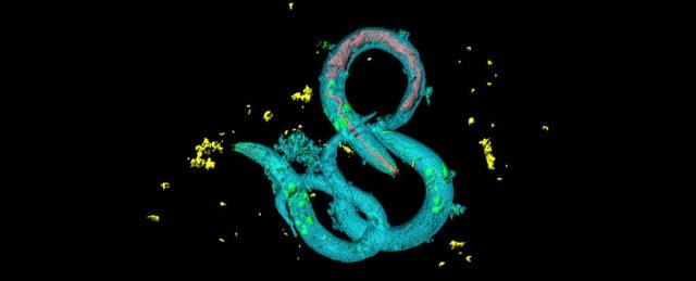
The team focussed in on the glial cells responsible for doing the clean-up work in the brain; they used a technique called 2Phatal to target a single brain cell for apoptosis (cell death) in a mouse and then followed the route of glial cells using fluorescent markers.

“Rather than hitting the brain with a hammer and causing thousands of deaths, inducing a single cell to die allows us to study what is happening right after the cells start to die and watch the many other cells involved,” says Grutzendler.

“This was not possible before. We are able to show with great clarity what exactly is going on and understand the process.”

Three types of glial cells – microglia, astrocytes, and NG2 cells – were shown to be involved in a highly coordinated cell removal process, which removed both the dead neuron and any connecting pathways to the rest of the brain. The researchers observed one microglia engulf the neuron body and its main branches (dendrites), while astrocytes targeted smaller connecting dendrites for removal. They suspect NG2 may help prevent the dead cell debris from spreading.

The researchers also demonstrated that if one type of glial cell missed the dead neuron for whatever reason, other types of cells would take over their role in the waste removal process – suggesting some sort of communication is occurring between the glial cells.

Another interesting finding from the research was that older mouse brains were less efficient at clearing out dead neural cells, even though the garbage removal cells seemed to be just as aware that a dying cell was there.

This is a good opportunity for future research, and could give experts insight into how older brains start to fail in various ways, as the garbage disposal service starts to slow down or even breaks.

New treatments might one day be developed that can take over this clearing process on the brain’s behalf – not just in elderly people, but also those who have suffered trauma to the head, for example.

“Cell death is very common in diseases of the brain,” says neurologist Eyiyemisi Damisah, from the Yale School of Medicine.

“Understanding the process might yield insights on how to address cell death in an injured brain from head trauma to stroke and other conditions.”

The research has been published in Science Advances.

https://www.sciencealert.com/for-the-first-time-scientists-capture-video-of-brains-clearing-out-dead-neurons

Fascinating Study Finds That Stressed Out Baby Worms Tend to Live Longer

by David Nield

Scientists researching a key aspect of biochemistry in living creatures have been taking a very close look at the tiny Caenorhabditis elegans roundworm. Their latest results show that when these nematodes get put under more biochemical stress early in their lives, they somehow tend to live longer.

This type of stress, called oxidative stress – an imbalance of oxygen-containing molecules that can result in cellular and tissue damage – seems to better prepare the worms for the strains of later life, along the same lines as the old adage that whatever doesn’t kill you, makes you stronger.

You might think that worm lifespans have no bearing on human life. And surely, until we have loads more research done in this field, it would be a big leap to say the same principles of prolonging one’s lifespan might hold true for human beings.

But there’s good reason to put C. elegans through the paces. This model organism has proven immensely helpful for researchers trying to better understand key biological functions present in worm and human alike – and oxidative stress is one such function.

The little wriggly creatures are known to have significant variations in their lifespan even when the whole population is genetically identical and grows up in the exact same conditions. So the team went looking for other factors that affect C. elegans’ longevity.

“The general idea that early life events have such profound, positive effects later in life is truly fascinating,” says biochemist Ursula Jakob from the University of Michigan.

Jakob and her colleagues sorted thousands of C. elegans larvae based on the oxidative stress levels they experienced during development – this stress arises when cells produce more oxidants and free radicals than they can handle. It’s a normal part of the ageing process, but it’s also triggered by exercise and a limited food supply.

One way to measure this stress is by the levels of reactive oxygen species (ROS) molecules an organism produces – simply put, this measurement indicates the biochemical stress an organism is under. In the case of these roundworms, the more ROS were produced during development, the longer their lifespans turned out to be.

To explain how this effect of ROS might come about, the researchers went looking for changes in the worms’ genetic regulation, specifically those genes that are known to be involved in dealing with oxidative stress.

While doing so, they detected a key difference – the nematodes exposed to more ROS during development appeared to have undergone an epigenetic change (a gene expression switch that can happen due to environmental influences) that increased the oxidative stress resistance of their body’s cells.

There are still a lot of questions to answer, but the researchers think their results identify one of the stochastic – or random – influences on the lifespan of organisms; it’s something that has been hypothesised in the field of the genetics of ageing. And down the line, it may turn out to be relevant for ageing humans, too.

“This study provides a foundation for future work in mammals, in which very early and transient metabolic events in life seem to have equally profound impacts on lifespan,” the researchers conclude.

The study has been published in Nature.

https://www.sciencealert.com/biological-stress-in-early-life-could-be-one-of-the-keys-to-a-long-lifespan?perpetual=yes&limitstart=1

These 6 Specific Exercises Can Cut Body Weight, Even if You’re Predisposed to Obesity

by DAVID NIELD

We know that a range of factors influence weight, including those related to lifestyle and genetics, but researchers have now identified six specific exercises that seem to offer the best chance of keeping your weight down – even if your genes don’t want you to.

Based on an analysis of 18,424 Han Chinese adults in Taiwan, aged between 30 and 70 years old, the best ways of reducing body mass index (BMI) in individuals predisposed to obesity are: regular jogging, mountain climbing, walking, power walking, dancing (to an “international standard”), and lengthy yoga practices.

But interestingly, many popular exercise types weren’t shown to do much good for those who’s genetic risk score makes them more likely to be obese.

Specifically, exercises including cycling, stretching, swimming and legendary console game Dance Dance Revolution don’t appear to be able to counteract genetic bias (though are beneficial in many other ways).

“Our findings show that the genetic effects on obesity measures can be decreased to various extents by performing different kinds of exercise,” write the researchers in their paper published in PLOS Genetics.

“The benefits of regular physical exercise are more impactful in subjects who are more predisposed to obesity.”

Besides BMI, the team also looked at four other obesity measures for a more complete picture: body fat percentage (BFP), waist circumference (WC), hip circumference (HC), and waist-to-hip ratio (WHR).

Regular jogging – 30 minutes, three times a week – turned out to be the most effective way of counteracting obesity genes across all of them.

The researchers also suggest, based on the information dug up in the Taiwan BioBank database, that the less effective forms of exercise typically don’t use up as much energy, which is why they don’t work quite so well.

The researchers specifically noted that activities in cold water, such as swimming, could make people hungrier and cause them to eat more.

The study was able to succeed in one of its main aims, which was to show that having a genetic disposition towards obesity doesn’t mean that obesity is inevitable – the right type of exercise, carried out regularly, can fight back against that built-in genetic coding.

“Obesity is caused by genetics, lifestyle factors, and the interplay between them,” epidemiologist Wan-Yu Lin, from the National Taiwan University, told Newsweek. “While hereditary materials are inborn, lifestyle factors can be determined by oneself.”

It’s worth noting that not every type of exercise was popular enough within the sample population to be included: activities like weight training, table tennis, badminton or basketball may or may not be helpful, too. There wasn’t enough data to assess.

But with obesity numbers rising sharply across the world – and 13 percent of the global population now thought to quality as being obese – it’s clear that measures need to be taken to reverse the trend.

Being obese affects our physiological health in the way it increases the risk of cardiovascular disease, some cancers, and other issues; and there’s evidence that being seriously overweight can have a negative effect on our brains too.

Studies like this latest one can point towards ways of sticking at a healthy weight, even when the genetic cards are stacked against it. In some cases all it takes is a few minutes of exertion per day.

“Previous studies have found that performing regular physical exercise could blunt the genetic effects on BMI,” conclude the researchers.

“However, few studies have investigated BFP or measures of central obesity. These obesity measures are even more relevant to health than BMI.”

The research has been published in PLOS Genetics.

https://www.sciencealert.com/these-six-exercises-can-keep-weight-down-even-with-genetic-tendencies-for-obesity

Gut microbes can impact the efficacy of Parkinson’s disease medications

by DAVID NIELD

Dosing medicines can be a tricky process: How much of a medication actually ends up hitting its target can vary a lot between patients, sometimes for mysterious reasons. As it turns out, even the things living in our bodies could be gobbling up our drugs.

In a series of experiments with levodopa (L-dopa) drug treatments for Parkinson’s, a new study has found that the gut microbes Enterococcus faecalis and Eggerthella lenta can intercept L-dopa and chemically transform it before it reaches the brain.

While this research only focuses on a specific treatment for one condition, the team behind the work thinks we might be underappreciating the role that our gut microbiome plays in controlling the efficacy and potency of medicines.

“Maybe the drug is not going to reach its target in the body, maybe it’s going to be toxic all of a sudden, maybe it’s going to be less helpful,” says chemical biologist Maini Rekdal from Harvard University.

The job of L-dopa is to deliver dopamine to the brain, replacing the dopamine eaten up by Parkinson’s. However, since the introduction of L-dopa in the 1960s, scientists have known that enzymes in the gut can stop this delivery from happening, leading to some nasty side effects as dopamine “spills out” before reaching the brain.

A second drug, carbidopa, was introduced to keep L-dopa intact, but it doesn’t always seem to help. Even with this additional drug, the effectiveness of L-dopa can vary between patients. What this new research does is identify the specific bacteria to blame, out of trillions of potential species.

With reference to the Human Microbiome Project, the team found that not only our own gut enzymes can wreak havoc on the medication, but the bacterium E. faecalis can also convert L-dopa to dopamine before it reaches the brain. Sure enough, it ate up all the L-dopa in lab tests.

Using faecal samples and supplies of dopamine, the researchers identified that another strain of gut bacteria, E. lenta, then consumes the converted dopamine and produces the neuromodulator meta-thyramine as a byproduct.

Thus, E. faecalis and E. lenta are apparently working as a sort of microbe tag team, preventing the medication from reaching its target. Furthermore, while carbidopa is used to stop a human gut enzyme from converting L-dopa to dopamine in the digestive system, it doesn’t seem to work on the E. faecalis enzyme that’s doing the same.

The good news is that the researchers have already found a molecule, alpha-fluoromethyltyrosine (AFMT), that can stop E. faecalis from breaking down L-dopa without destroying the bacterium itself, by targeting a non-essential enzyme.

Ultimately, we might end up with a way of making L-dopa significantly more effective as a Parkinson’s treatment, without as many of the side effects – but that’s still a long way off.

“All of this suggests that gut microbes may contribute to the dramatic variability that is observed in side effects and efficacy between different patients taking L-dopa,” says chemical biologist Emily Balskus from Harvard University.

Even if we can’t fix the problem just yet, we now have a proof of concept that particular combinations of gut microbes can indeed cause havoc with our meds. Hopefully, this will give other researchers food for thought and we might see similar investigations of other medicines, too.

The research has been published in Science.

https://www.sciencealert.com/gut-microbes-could-be-eating-up-our-meds-before-they-get-chance-to-work

Scientists Identify The Location of ‘Taste’ in Your Head, And It’s Not The Tongue

by David Nield

How exactly do our brains sort between the five taste groups: sweet, sour, salty, bitter and umami? We’ve now got a much better idea, thanks to research that has pinned down where in the brain this taste processing happens.

Step forward: the insular cortex. Already thought to be responsible for everything from motor control to social empathy, we can now add flavour identification to its list of jobs.

It’s an area of the brain scientists have previously suspected could be responsible for sorting tastes, and which has been linked to taste in rodents, but this new study is much more precise in figuring out the role it plays in decoding what our tongues are telling us.

“We have known that tastes activate the human brain for some time, but not where primary taste types such as sweet, sour, salty, and bitter are distinguished,” says one of the team, Adam Anderson from Cornell University in New York.

“By using some new techniques that analyse fine-grained activity patterns, we found a specific portion of the insular cortex – an older cortex in the brain hidden behind the neocortex – represents distinct tastes.”

Anderson and his team used detailed fMRI scans of 20 adults as well as a new statistical model to dig deeper than previous studies into the link between the insular cortex and taste. This helped separate the taste response from other related responses – like the disgust we might feel when eating something sour or bitter.

Part of the problem in pinning down the taste-testing parts of the brain is that multiple regions of neurons get busy whenever we’re eating something. However, this study helps to cut through some of that noise.

In particular, it seems that different tastes don’t necessarily affect different parts of the insular cortex, but rather prompt different patterns of activity. Those patterns help the brain determine what it’s tasting.

For example, one particular section of the insular cortex was found to light up – in terms of neural activity – whenever something sweet was tasted. It’s a literal sweet spot, in other words, but it also showed that different brains have different wiring.

“While we identified a potential sweet spot, its precise location differed across people and this same spot responded to other tastes, but with distinct patterns of activity,” says Anderson.

“To know what people are tasting, we have to take into account not only where in the insula is stimulated, but also how.”

The work follows on from previous research showing just how big a role the brain plays in perceiving taste. It used to be thought that receptors on the tongue did most of the taste testing, but now it seems the brain is largely in charge of the process.

That prior study showed how switching certain brain cells on and off in mice was enough to prevent them from distinguishing between sweet and bitter. The conclusion is that while the tongue does identify certain chemicals, it’s the brain that interprets them.

The new research adds even more insight into what’s going on in the brain in humans when we need to work out what we’re tasting – and shows just how important a job the insular cortex is doing.

“The insular cortex represents experiences from inside our bodies,” says Anderson. “So taste is a bit like perceiving our own bodies, which is very different from other external senses such as sight, touch, hearing or smell.”

The research has been published in Nature Communications.

https://www.sciencealert.com/now-we-know-the-part-of-the-brain-that-tells-us-what-we-re-tasting

The World Just Got Closer to a Controversial Mosquito ‘Wipe Out’ Experiment

by David Nield

Scientists are genetically modifying mosquitoes in a high-security lab – and they’re hoping the insects will help wipe out some of the mosquito-borne diseases that continue to plague communities worldwide.

It’s known as a gene drive: where mosquitoes modified to be incapable of passing on a particular virus are used to replace the existing population of insects over several generations, with the modified genes being passed on to all their offspring.

The idea has attracted controversy because it messes with the fundamentals of nature, but it’s now under consideration by the World Health Organisation (WHO). This particular testing has entered a new phase, NPR reports, with a large-scale release of genetically modified mozzies inside a facility in Terni, Italy.

“This will really be a breakthrough experiment,” entomologist Ruth Mueller, who runs the lab, told Rob Stein at NPR. “It’s a historic moment. It’s very exciting.”

Using the ‘molecular scissor’ editing technique CRISPR, a gene known as “doublesex” in the bugs has been altered. The gene transforms female mosquitoes, taking away their biting ability and making them infertile.

At the moment, the bugs are being released in cages designed to replicate their natural environments, with hot and humid air, and places to shelter. Artificial lights are used to simulate sunrise and sunset.

The idea is to see if the mosquitoes with CRISPR-edited genetic code can wipe out the unmodified insects inside the cages. It follows on from previous proof-of-concept studies that we’ve seen before.

Ultimately these mosquitoes could be released in areas hit by malaria, bringing the local mozzie population crashing down and saving human lives. The disease is responsible for more than 400,000 deaths every year – mostly young children.

Reducing those figures sounds like a great idea, so why the controversy? Well, many scientists are urging caution when it comes to altering genetic code at this fundamental level – we just don’t know what impact these genetically edited mosquitoes will have on the world around them.

For that reason the lab has been designed to minimise any chance that the specially engineered mosquitoes could escape. The testing has also been specifically located in Italy, where this mosquito species – Anopheles gambiae – wouldn’t be able to survive outside in the natural climate.

“This is a technology where we don’t know where it’s going to end,” Nnimmo Bassey, director of the Health of Mother Earth Foundation in Nigeria, told NPR. “We need to stop this right where it is. They’re trying to use Africa as a big laboratory to test risky technologies.”

Some experts think adding genetically modified mosquitoes to natural ecosystems could harm other plants and animals that depend on them. There are a lot of unknowns.

The team behind the new experiments counters the critique by saying they’re working slowly and methodically – and that the potential side effects are outweighed by the benefits of eradicating malaria.

At the moment scientists are targeting just one species of mosquito out of hundreds, and several more years of research and consultation are planned before genetically edited mozzies would ever be released.

“There’s going to be concerns with any technology,” one of the research team, Tony Nolan from Imperial College London in the UK, told NPR.

“But I don’t think you should throw out a technology without having done your best to understand what its potential is to be transformative for medicine. And, were it to work, this would be transformative.”

https://www.sciencealert.com/scientists-take-first-step-in-controversial-mosquito-gene-drive-experiment

The reason that sonar makes whales beach themselves

by David Nield

Since the 1980s scientists have spotted a link between naval sonar systems and beaked whales seemingly killing themselves – by deliberately getting stranded on beaches. Now, researchers might have revealed the horrifying reason why.

In short, the sound pulses appear to scare the whales to death, acting like a shot of adrenaline might in a human, and causing deadly changes in their otherwise perfectly calibrated diving techniques.

By studying mass stranding events (MSEs) from recent history, the team found that beaked whales bring a sort of decompression sickness (also known as ‘the bends’ or ‘divers’ disease’) on themselves when they sense sonar. When panicked, their veins fill up with nitrogen gas bubbles, their brains suffer severe haemorrhaging, and other organs get damaged.

“In the presence of sonar they are stressed and swim vigorously away from the sound source, changing their diving pattern,” one of the researchers, Yara Bernaldo de Quiros from the University of Las Palmas de Gran Canaria in Spain, told AFP.

“The stress response, in other words, overrides the diving response, which makes the animals accumulate nitrogen.”

The end result is these poor creatures die in agony after getting the whale version of the bends – not something you would normally expect from whales that are so adept at navigating deep underwater.

Typically, these animals naturally lower their heart rate to reduce oxygen use and prevent nitrogen build-up when they plunge far below the surface. Tragically, it appears that a burst of sonar actually overrides these precautions.

The researchers weighed up the evidence from some 121 MSEs between the years 1960 and 2004, and particularly focussed on the autopsies of 10 dead whales stranded in the Canary Islands in 2002 after a nearby naval exercise.

It’s here that the decompression sickness effects were noticed, as they have been in other stranding events that the researchers looked at.

While the team notes that the effects of sonar on whales seem to “vary among individuals or populations”, and “predisposing factors may contribute to individual outcomes”, there does seem to be a common thread in terms of what happens to these unsuspecting mammals.

That’s especially true for Cuvier’s beaked whale (Ziphius cavirostris) – of the 121 MSEs we’ve mentioned, 61 involved Cuvier’s beaked whales, and the researchers say they appear particularly vulnerable to sonar.

There’s also a particular kind of sonar to be worried about: mid-frequency active sonar (MFAS), in the range of about 5 kilohertz.

Now the researchers behind the new report want to see the use of such sonar technology banned in areas where whales are known to live – such a ban has been in place in the Canary Islands since the 2002 incident.

“Up until then, the Canaries were a hotspot for this kind of atypical stranding,” de Quiros told AFP. “Since the moratorium, none have occurred.”

The research has been published in the Royal Society Journal Proceedings B.

https://www.sciencealert.com/this-is-the-horrifying-reason-why-sonar-makes-beaked-whales-beach-themselves

Scientists Have Detected an Entirely New Visual Phenomenon in The Human Eye

ghosted-images-1_1024

by DAVID NIELD

New research suggests the human eye and brain are capable of seeing ghosted images, a new type of visual phenomenon that scientists previously thought could only be detected by a computer. It turns out our eyes are more powerful than we thought.

The discovery could teach us more about the inner workings of the eye and brain and how they process information, as well as changing our thinking on what we human beings can truly see of the world around us.

Having been developed as a way of low-cost image capture for light outside the visible spectrum, the patterns produced by these ghosted images are usually processed by software algorithms – but, surprisingly, our eyes have the same capabilities.

“Ghost-imaging with the eye opens up a number of completely novel applications such as extending human vision into invisible wavelength regimes in real-time, bypassing intermediary screens or computational steps,” write the researchers.

“Perhaps even more interesting are the opportunities that ghost imaging offers for exploring neurological processes.”

Ghost imaging works using a camera with a single pixel, rather than the millions of pixels used by the sensors inside today’s digital cameras and smartphones. When it comes to capturing light beyond the visible spectrum, it’s even a more cost-effective method.

These single pixel cameras capture light as it reflects from an object – by watching different random patterns of bouncing light, and crunching through some calculations, the camera can gradually build up a picture of something even with just one pixel.

In some setups, the single pixel camera is used in combination with a second light, modulated in response to the first, and beamed back on the original random patterns. The advantage is that fewer patterns are needed to produce an image.

In this case a second camera using some smart algorithms can pick up the image without having looked at the object at all – just by looking at the patterns being cast and the light being produced from them.

That’s the ghosted image that was previously thought to only be visible to computers running specialist software. However, the new study shows the human visual perception can make sense of these patterns, called Hadamard patterns.

This diagram from the research paper should give you an idea of what’s happening:

ghosted-images-2

It’s a little bit like when our eyes and brains look at a series of still images and treat them as a moving picture – the same sort of subconscious processing seems to be going on.

Of the four volunteers who took part in the study, all four could make out an image of Albert Einstein sticking out his tongue from the Hadamard patterns. Interestingly, though, the illusion only appeared when the patterns were projected quickly enough.

If the rate dropped below 200 patterns per 20 milliseconds, the image couldn’t be seen by the study participants.

As the researchers point out, this is potentially hugely exciting – it means we might be able to devise simple systems to see light outside the visible spectrum, with no computer processing required in the middle.

That’s all to come – and this is really preliminary stuff, so we can’t get too carried away. For now, the team of researchers is using the findings to explore more about how our visual systems work, and whether our eyes and brains have yet-undiscovered superpowers for looking at the world around us.

The research has yet to be peer-reviewed, but you can read it on the pre-print resource Arxiv.

https://www.sciencealert.com/human-eye-sees-ghosted-images-reflected-light

Scientists Say They’ve Found The Driver of False Beliefs, And It’s Not a Lack of Intelligence

flatearthbeliefsconfidence_Web_1024

by DAVID NIELD

Why is it sometimes so hard to convince someone that the world is indeed a globe, or that climate change is actually caused by human activity, despite the overwhelming evidence?

Scientists think they might have the answer, and it’s less to do with lack of understanding, and more to do with the feedback they’re getting.

Getting positive or negative reactions to something you do or say is a greater influence on your thinking than logic and reasoning, the new research suggests – so if you’re in a group of like-minded people, that’s going to reinforce your thinking.

Receiving good feedback also encourages us to think we know more than we actually do.

In other words, the more sure we become that our current position is right, the less likely we are to take into account other opinions or even cold, hard scientific data.

“If you think you know a lot about something, even though you don’t, you’re less likely to be curious enough to explore the topic further, and will fail to learn how little you know,” says one of the team members behind the new study, Louis Marti from the University of California, Berkeley.

For the research, more than 500 participants were recruited and shown a series of colored shapes. As each shape appeared, the participants got asked if it was a “Daxxy” – a word made up for these experiments.

The test takers had no clues as to what a Daxxy was or wasn’t, but they did get feedback after guessing one way or the other – the system would tell them if the shape they were looking at qualified as a Daxxy or not. At the same time they were also asked how sure they were about what a Daxxy actually was.

In this way the researchers were able to measure certainty in relation to feedback. Results showed the confidence of the participants was largely based on the results of their last four or five guesses, not their performance overall.

You can see the researchers explain the experiment in the video below:

The team behind the tests says this plays into something we already know about learning – that for it to happen, learners need to recognise that there is a gap between what they currently know and what they could know. If they don’t think that gap is there, they won’t take on board new information.

“What we found interesting is that they could get the first 19 guesses in a row wrong, but if they got the last five right, they felt very confident,” says Marti. “It’s not that they weren’t paying attention, they were learning what a Daxxy was, but they weren’t using most of what they learned to inform their certainty.”

This recent feedback is having more of an effect than hard evidence, the experiments showed, and that might apply in a broader sense too. It could apply to learning something new or trying to differentiate between right and wrong.

And while in this case the study participants were trying to identify a made-up shape, the same cognitive processes could be at work when it comes to echo chambers on social media or on news channels – where views are constantly reinforced.

“If you use a crazy theory to make a correct prediction a couple of times, you can get stuck in that belief and may not be as interested in gathering more information,” says one of the team, psychologist Celeste Kidd from UC Berkeley.

So if you think vaccinations are harmful, for example, the new study suggests you might be basing that on the most recent feedback you’ve had on your views, rather than the overall evidence one way or the other.

Ideally, the researchers say, learning should be based on more considered observations over time – even if that’s not quite how the brain works sometimes.

“If your goal is to arrive at the truth, the strategy of using your most recent feedback, rather than all of the data you’ve accumulated, is not a great tactic,” says Marti.

The research has been published in Open Mind.

https://www.sciencealert.com/feedback-study-explains-why-false-beliefs-stick

Scientists Think They’ve Found The Part of The Brain That Makes People Pessimistic

\

by DAVID NIELD

A specific part of the brain called the caudate nucleus could control pessimistic responses, according to animal tests, a finding which might help us unlock better treatments for mental disorders like anxiety and depression.

These disorders often come with negative moods triggered by a pessimistic reaction, and if scientists can figure out how to control that reaction, we might stand a better chance of dealing with the neuropsychiatric problems that affect millions of people worldwide – and maybe discover the difference between glass half full and glass half empty people along the way.

The research team from MIT found that when the caudate nucleus was artificially stimulated in macaques, the animals were more likely to make negative decisions, and consider the potential drawback of a decision rather than the potential benefit.

This pessimistic decision-making continued right through the day after the original stimulation, the researchers found.

“We feel we were seeing a proxy for anxiety, or depression, or some mix of the two,” says lead researcher Ann Graybiel. “These psychiatric problems are still so very difficult to treat for many individuals suffering from them.”

The caudate nucleus has previously been linked to emotional decision-making, and the scientists stimulated it with a small electrical current while the monkeys were offered a reward (juice) and an unpleasant experience (a puff of air to the face) at the same time.

In each run through the amount of juice and the strength of the air blast varied, and the animals could choose whether or not to accept the reward – essentially measuring their ability to weigh up the costs of an action against the benefits.

When the caudate nucleus was stimulated, this decision-making got skewed, so the macaques started rejecting juice/air ratios they would have previously accepted. The negative aspects apparently began to seem greater, while the the rewards became devalued.

“This state we’ve mimicked has an overestimation of cost relative to benefit,” says Graybiel. After a day or so, the effects gradually disappeared.

The researchers also found brainwave activity in the caudate nucleus, part of the basal ganglia, changed when decision-making patterns changed. This might give doctors a marker to indicate whether someone would be responsive to treatment targeting this part of the brain or not.

The next stage is to see whether the same effect can be noticed in human beings – scientists have previously linked abnormal brain activity in people with mood disorders to regions connected to the caudate nucleus, but there’s a lot more work to be done to confirm these neural connections.

Making progress isn’t easy because of the incredibly complexity of the brain, but the researchers think their results show the caudate nucleus could be disrupting dopamine activity in the brain, controlling mood and our sense of reward and pleasure.

“There must be many circuits involved,” says Gabriel. “But apparently we are so delicately balanced that just throwing the system off a little bit can rapidly change behaviour.”

The research has been published in Neuron.

https://www.sciencealert.com/we-found-the-brain-region-for-pessimism