Case Western Reserve University-led team develops new approach to treat certain neurological diseases


Paul Tesar, professor of genetics and genome sciences, School of Medicine


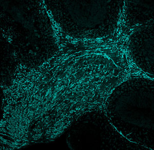
Regeneration of myelin in the brain, shown in blue, after ASO drug treatment

A team led by Case Western Reserve University medical researchers has developed a potential treatment method for Pelizaeus-Merzbacher disease (PMD), a fatal neurological disorder that produces severe movement, motor and cognitive dysfunction in children. It results from genetic mutations that prevent the body from properly making myelin, the protective insulation around nerve cells.

Using mouse models, the researchers identified and validated a new treatment target—a toxic protein resulting from the genetic mutation. Next, they successfully used a family of drugs known as ASOs (antisense oligonucleotides) to target the ribonucleic acid (RNA) strands that created the abnormal protein to stop its production. This treatment reduced PMD’s hallmark symptoms and extended lifespan, establishing the clinical potential of this approach.

By demonstrating effective delivery of the ASOs to myelin-producing cells in the nervous system, researchers raised the prospect for using this method to treat other myelin disorders that result from dysfunction within these cells, including multiple sclerosis (MS).

Their research was published online July 1 in the journal Nature.

“The pre-clinical results were profound. PMD mouse models that typically die within a few weeks of birth were able to live a full lifespan after treatment,” said Paul Tesar, principal investigator on the research, a professor in the Department of Genetics and Genome Sciences at the School of Medicine and the Dr. Donald and Ruth Weber Goodman Professor of Innovative Therapeutics. “Our results open the door for the development of the first treatment for PMD as well as a new therapeutic approach for other myelin disorders.”

Study co-authors include an interdisciplinary team of researchers from the medical school, Ionis Pharmaceuticals Inc., a Carlsbad, California-based pioneer developer of RNA-targeted therapies, and Cleveland Clinic. First author Matthew Elitt worked in Tesar’s lab as a Case Western Reserve medical and graduate student.

PMD attacks the young

PMD is a rare, genetic condition involving the brain and spinal cord that primarily affects boys. Symptoms can appear in early infancy and begin with jerky eye movements and abnormal head movements. Over time, children develop severe muscle weakness and stiffness, cognitive dysfunction, difficulty walking and fail to reach developmental milestones such as speaking. The disease shortens life-expectancy, and people with the most severe cases die in childhood.

The disease results from errors in a gene called proteolipid protein 1 (PLP1). Normally, this gene produces proteolipid protein (PLP) a major component of myelin, which wraps and insulates nerve fibers to allow proper transmission of electrical signals in the nervous system. But a faulty PLP1 gene produces toxic proteins that kill myelin producing cells and prevent myelin from developing and functioning properly—resulting in the severe neurological dysfunction in PMD patients.

PMD impacts a few thousand people around the world. So far, no therapy has lessened symptoms or extended lifespans.

For nearly a decade, Tesar and his team have worked to better understand and develop new therapies for myelin disorders. They have had a series of successes, and their myelin-regenerating drugs for MS are now in commercial development.

Latest research

In the current laboratory work, the researchers found that suppressing mutant PLP1 and its toxic protein restored myelin-producing cells, produced functioning myelin, reduced disease symptoms and extended lifespans.

After validating that PLP1 was their therapeutic target, the researchers pursued pre-clinical treatment options. They knew mutations in the PLP1 gene produced faulty RNA strands that, in turn, created the toxic PLP protein.

So they teamed with Ionis Pharmaceuticals, a leader in RNA-targeted therapeutics and pioneer of ASOs. These short strings of chemically modified DNA can be designed to bind to a specific RNA target and block production of its protein product.

And that’s exactly what happened in their studies. The result was improved myelin and locomotion, and substantial extension of lifespan. “ASOs provided an opportunity to cut the disease-causing protein off at its source,” Elitt said.

The successful clinical use of ASOs is relatively new, yet recent developments seem promising. In 2016, the U.S. Food and Drug Administration approved the first ASO drug for a neurological disorder, spinal muscular atrophy. The drug, Spinraza, was developed by Ionis and commercialized by Biogen Inc. More ASO therapies are in development, and clinical trials and hold promise for addressing many neurological diseases that as of now have no effective treatment options.

Tesar said that ongoing and planned experiments in his laboratory will help guide future clinical development of ASO therapy for PMD. For example, researchers want to understand more about how well the treatment works after the onset of symptoms, how long it lasts, how often treatment needs to be given and whether it might be effective for all PMD patients, regardless of their specific form of the disease.

“While important research questions remain, I’m cautiously optimistic about the prospect for this method to move into clinical development and trials for PMD patients,” Tesar said. “I truly hope our work can make a difference for PMD patients and families.”

Case Western Reserve University-led team develops new approach to treat certain neurological diseases

New Medical Device to Detect Precancerous Lesions in the Esophagus Wins 2020 Edison Award

A medical device based on technology developed by three faculty members from Case Western Reserve University and University Hospitals Cleveland Medical Center (UH) has won a prestigious 2020 Edison Best New Product Award.

EsoCheck, a device designed to help detect precancerous changes in the esophagus, was named a “Silver” winner of the 2020 Edison Best New Product Awards in the “Medical/Dental – Testing Solutions” subcategory.

Esophageal adenocarcinomas have increased more than five-fold in recent years and are a highly lethal cancer, with less than 20% 5-year survival. These cancers arise from a precursor lesion of Barrett’s esophagus (BE), which is an abnormal cell type that arises in the lower esophagus.

EsoCheck is a swallowable balloon-based device that, in a simple five-minute outpatient exam, can collect cells from the lower region of the esophagus to help determine if Barrett’s disease is present. Unlike endoscopy, the current method for examining the esophagus, EsoCheck does not require a patient to undergo sedation, lose a day of work or need a companion for transportation.

The EsoCheck device works together with EsoGuard, a companion molecular assay that tests the DNA from the cells retrieved by EsoCheck for the presence of genetic changes indicative of the presence or absence of Barrett’s disease.

Lucid Diagnostics, a subsidiary of New York-based PAVmed Inc., licensed the EsoCheck and EsoGuard technology through the Case Western Reserve University Technology Transfer Office in 2018.

The EsoCheck device and EsoGuard DNA test were co-invented by Amitabh Chak, MD, (Professor of Medicine at the Case Western Reserve School of Medicine and gastroenterologist at the University Hospitals Digestive Health Institute); Sanford Markowitz, MD, PhD, (Ingalls Professor of Cancer Genetics and Medicine at the School of Medicine and an oncologist at University Hospitals Seidman Cancer Center); and Joseph Willis, MD,(Professor of Pathology at the School of Medicine and Pathology Vice-Chair for translational research at UH).

The technology was developed as part of the Case Comprehensive Cancer Center’s GI SPORE (Gastrointestinal Specialized Program of Research Excellence) and BETRNet (Barrett’s Esophagus Translational Research Network) programs led by Markowitz and Chak, and was first tested in humans in a clinical trial led by Chak at University Hospitals.

Further support for the clinical assay development was derived from a National Cancer Institute award led by Willis. The development was also supported by the Case-Coulter partnership and the State of Ohio Third Frontier Technology Validation Start-up Fund.

Last fall, the new EsoCheck method for examining the esophagus received clearance from the U.S. Food and Drug Administration for clinical use, and, this February, the companion EsoGuard DNA test for Barrett’s detection received breakthrough designation from the FDA.

Since 1987, the Edison Awards, named after Thomas Alva Edison, have recognized some of the most innovative products and business leaders in the world. They’re among the most prestigious accolades, honoring excellence in new product and service development, marketing, design and innovation.

About University Hospitals / Cleveland, Ohio

Founded in 1866, University Hospitals serves the needs of patients through an integrated network of 18 hospitals, more than 50 health centers and outpatient facilities, and 200 physician offices in 16 counties throughout northern Ohio. The system’s flagship academic medical center, University Hospitals Cleveland Medical Center, located in Cleveland’s University Circle, is affiliated with Case Western Reserve University School of Medicine. The main campus also includes University Hospitals Rainbow Babies & Children’s Hospital, ranked among the top children’s hospitals in the nation; University Hospitals MacDonald Women’s Hospital, Ohio’s only hospital for women; University Hospitals Harrington Heart & Vascular Institute, a high-volume national referral center for complex cardiovascular procedures; and University Hospitals Seidman Cancer Center, part of the NCI-designated Case Comprehensive Cancer Center. UH is home to some of the most prestigious clinical and research programs in the nation, including cancer, pediatrics, women’s health, orthopedics, radiology, neuroscience, cardiology and cardiovascular surgery, digestive health, transplantation and urology. UH Cleveland Medical Center is perennially among the highest performers in national ranking surveys, including “America’s Best Hospitals” from U.S. News & World Report. UH is also home to Harrington Discovery Institute at University Hospitals – part of The Harrington Project for Discovery & Development. UH is one of the largest employers in Northeast Ohio with 28,000 physicians and employees. Advancing the Science of Health and the Art of Compassion is UH’s vision for benefitting its patients into the future, and the organization’s unwavering mission is To Heal. To Teach. To Discover. Follow UH on LinkedIn, Facebook @UniversityHospitals and Twitter @UHhospitals. For more information, visit UHhospitals.org.

https://finance.yahoo.com/news/medical-device-developed-cwru-uh-123000489.html

Using artificial intelligence to determine whether immunotherapy is working


Case Western Reserve researchers use AI with routine CT scans to predict how well lung cancer patients will respond to expensive treatment based off changes in texture patterns inside and outside the tumor.

Scientists from the Case Western Reserve University digital imaging lab, already pioneering the use of artificial intelligence (AI) to predict whether chemotherapy will be successful, can now determine which lung-cancer patients will benefit from expensive immunotherapy.

And, once again, they’re doing it by teaching a computer to find previously unseen changes in patterns in CT scans taken when the lung cancer is first diagnosed compared to scans taken after the first two to three cycles of immunotherapy treatment. And, as with previous work, those changes have been discovered both inside—and outside—the tumor, a signature of the lab’s recent research.

“This is no flash in the pan—this research really seems to be reflecting something about the very biology of the disease, about which is the more aggressive phenotype, and that’s information oncologists do not currently have,” said Anant Madabhushi, whose Center for Computational Imaging and Personalized Diagnostics (CCIPD) has become a global leader in the detection, diagnosis and characterization of various cancers and other diseases by meshing medical imaging, machine learning and AI.

Currently, only about 20% of all cancer patients will actually benefit from immunotherapy, a treatment that differs from chemotherapy in that it uses drugs to help your immune system fight cancer, while chemotherapy uses drugs to directly kill cancer cells, according to the National Cancer Institute.

Madabhushi said the recent work by his lab would help oncologists know which patients would actually benefit from the therapy, and who would not.

“Even though immunotherapy has changed the entire ecosystem of cancer, it also remains extremely expensive—about $200,000 per patient, per year,” Madabhushi said. “That’s part of the financial toxicity that comes along with cancer and results in about 42% of all new diagnosed cancer patients losing their life savings within a year of diagnosis.”

Having a tool based on the research being done now by his lab would go a long way toward “doing a better job of matching up which patients will respond to immunotherapy instead of throwing $800,000 down the drain,” he added, referencing the four patients out of five who will not benefit, multiplied by annual estimated cost.

Case Western Reserve researchers use AI with routine CT scans to predict how well lung cancer patients will respond to expensive treatment based off changes in texture patterns inside and outside the tumor
Scientists from the Case Western Reserve University digital imaging lab, already pioneering the use of artificial intelligence (AI) to predict whether chemotherapy will be successful, can now determine which lung-cancer patients will benefit from expensive immunotherapy.

And, once again, they’re doing it by teaching a computer to find previously unseen changes in patterns in CT scans taken when the lung cancer is first diagnosed compared to scans taken after the first two to three cycles of immunotherapy treatment. And, as with previous work, those changes have been discovered both inside—and outside—the tumor, a signature of the lab’s recent research.

“This is no flash in the pan—this research really seems to be reflecting something about the very biology of the disease, about which is the more aggressive phenotype, and that’s information oncologists do not currently have,” said Anant Madabhushi, whose Center for Computational Imaging and Personalized Diagnostics (CCIPD) has become a global leader in the detection, diagnosis and characterization of various cancers and other diseases by meshing medical imaging, machine learning and AI.

Currently, only about 20% of all cancer patients will actually benefit from immunotherapy, a treatment that differs from chemotherapy in that it uses drugs to help your immune system fight cancer, while chemotherapy uses drugs to directly kill cancer cells, according to the National Cancer Institute.

Madabhushi said the recent work by his lab would help oncologists know which patients would actually benefit from the therapy, and who would not.

“Even though immunotherapy has changed the entire ecosystem of cancer, it also remains extremely expensive—about $200,000 per patient, per year,” Madabhushi said. “That’s part of the financial toxicity that comes along with cancer and results in about 42% of all new diagnosed cancer patients losing their life savings within a year of diagnosis.”

Having a tool based on the research being done now by his lab would go a long way toward “doing a better job of matching up which patients will respond to immunotherapy instead of throwing $800,000 down the drain,” he added, referencing the four patients out of five who will not benefit, multiplied by annual estimated cost.

New research published
The figure above shows differences in CT radiomic patterns before and after initiation of checkpoint inhibitor therapy.

The new research, led by co-authors Mohammadhadi Khorrami and Prateek Prasanna, along with Madabhushi and 10 other collaborators from six different institutions was published in November in the journal Cancer Immunology Research.

Khorrami, a graduate student working at the CCIPD, said one of the more significant advances in the research was the ability of the computer program to note the changes in texture, volume and shape of a given lesion, not just its size.

“This is important because when a doctor decides based on CT images alone whether a patient has responded to therapy, it is often based on the size of the lesion,” Khorrami said. “We have found that textural change is a better predictor of whether the therapy is working.

“Sometimes, for example, the nodule may appear larger after therapy because of another reason, say a broken vessel inside the tumor—but the therapy is actually working. Now, we have a way of knowing that.”

Prasanna, a postdoctoral research associate in Madabhushi’s lab, said the study also showed that the results were consistent across scans of patients treated at two different sites and with three different types of immunotherapy agents.

“This is a demonstration of the fundamental value of the program, that our machine-learning model could predict response in patients treated with different immune checkpoint inhibitors,” he said. “We are dealing with a fundamental biological principal.”

Prasanna said the initial study used CT scans from 50 patients to train the computer and create a mathematical algorithm to identify the changes in the lesion. He said the next step will be to test the program on cases obtained from other sites and across different immunotherapy agents. This research recently won an ASCO 2019 Conquer Cancer Foundation Merit Award.

Additionally, Madabhushi said, researchers were able show that the patterns on the CT scans which were most associated with a positive response to treatment and with overall patient survival were also later found to be closely associated with the arrangement of immune cells on the original diagnostic biopsies of those patients.

This suggests that those CT scans actually appear to capturing the immune response elicited by the tumors against the invasion of the cancer—and that the ones with the strongest immune response were showing the most significant textural change and most importantly, would best respond to the immunotherapy, he said.

Madabhushi established the CCIPD at Case Western Reserve in 2012. The lab now includes nearly 60 researchers.

Some of the lab’s most recent work, in collaboration with New York University and Yale University, has used AI to predict which lung cancer patients would benefit from adjuvant chemotherapy based on tissue-slide images. That advancement was named by Prevention Magazine as one of the top 10 medical breakthroughs of 2018.

Other authors on the paper were: Germán Corredor, Mehdi Alilou and Kaustav Bera from biomedical engineering, Case Western Reserve University; Pingfu Fu from population and quantitative health sciences, Case Western Reserve University; Amit Gupta of University Hospitals Cleveland Medical Center; Pradnya Patil of Cleveland Clinic; Priya D. Velu of Weill Cornell Medicine; Rajat Thawani of Maimonides Medical Center; Michael Feldman from Perelman School of Medicine of the University of Pennsylvania; and Vamsidhar Velcheti from NYU-Langone Medical Center.

For more information, contact Mike Scott at mike.scott@case.edu.

Using artificial intelligence to determine whether immunotherapy is working

Sweating is a clue into who develops PTSD—and who doesn’t

Within four hours of a traumatic experience, certain physiological markers—namely, sweating—are higher in people who go on to develop posttraumatic stress disorder (PTSD), according to a new study by a researcher at Case Western Reserve University and other institutions.

Around 90% of people who experience a traumatic event do not develop PTSD, according to existing data and research, making the medical community eager for better insights into the 10% who do—and for how to best treat these patients.

The study, conducted at Atlanta’s Grady Memorial Hospital, found that micro perspirations—detected non-invasively by a mobile device in an emergency department—can be plugged into a new mathematical model developed by the researchers to help predict who may be more at risk for developing PTSD.

The findings are especially important for targeting early treatment efforts and prevention of the disorder, said Alex Rothbaum, a pre-doctoral researcher in the Department of Psychological Sciences in the College of Arts and Sciences at Case Western Reserve.

“With PTSD, there is a need for more reliable and immediate patient information, especially in situations where research suggests people may underreport their own symptoms, such as with men, and those who live in violent neighborhoods or are on active duty,” said Rothbaum, a co-author of the study, which was published in the journal Chronic Stress.

“While skin is always secreting sweat, our method can discern meaningful, actionable information from perspirations too small for the naked eye to see,” he added.

The measurement differs from traditional practices to diagnose PTSD, which look for psychological differences in patients based on self-reported data and clusters of symptoms defined by the Diagnostic and Statistical Manual of Mental Disorders (often referred to as the DSM) published by the American Psychiatric Association.

“Eventually, this finding may help contribute to changes in how we diagnose and treat PTSD, pointing us toward which patients would do better in therapy, with medication, or a combination of the two—or no treatment at all,” said Rothbaum.

New testing device: less expensive, more accessible
Researchers hope the PTSD test can become available and standard in emergency departments, aided by the recent development of a practical and inexpensive device that can plug into common tablets and can measure “skin conductance response”—a measure of sweating.

Before, such tests could only be conducted on a large stand-alone machine costing upwards of $10,000. While the new device lacks the sensitivity of its more expensive counterpart, the readings it provides can be used to determine who should continue with additional testing and who is not at risk for developing PTSD.

The study—which included nearly 100 patients—was prompted, in part, by recent research showing the ineffectiveness of current methods practiced with patients immediately after traumas, known as critical incident stress debriefing and psychological debriefing.

Both the new method and model created by researchers will need to be further validated by a larger study underway with a National Institutes of Health grant.

The research
The study was co-authored with researchers at Emory University School of Medicine: Rebecca Hinrichs, Sanne J. H. van Rooij, Jennifer Stevens, Jessica Maples-Keller and Barbara O. Rothbaum; Vasiliki Michopoulos of Emory and Yerkes National Primate Research Center; Katharina Schultebraucks and Isaac Galatzer-Levy of New York University School of Medicine; Sterling Winters of Wayne State University; Tanja Jovanovic of Emory and Wayne State; and Kerry J. Ressler of Emory and Harvard/ McLean Hospital.

The research was supported by the National Institute of Mental Health and a Brain and Behavior Research Foundation NARSAD Independent Investigator Award.

Sweating is a clue into who develops PTSD—and who doesn’t

Case Western Reserve University Scientists generate first microscopic image of full length serotonin receptor


3D reconstruction of a serotonin receptor generated by cryo-electron microscopy

by Rebecca Pool

Claiming a world first and using cryo-electron microscopy, researchers from Case Western Reserve University School of Medicine, US, have observed full-length serotonin receptors. The proteins are common drug targets, and the new images provide details about molecular binding sites that could lead to more precise drug design. Serotonin receptors, which reside in cell membranes throughout the body, are highly dynamic and difficult to image. In the past, the receptors have been sectioned into pieces to study, but by capturing full-length samples, researchers have revealed how different portions interact.

Dr Sandip Basak from Physiology and Biophysics, and colleagues, describe ‘a finely tuned orchestration of three domain movements’ that allows the receptors to elegantly control passageways across cell membranes. “The serotonin receptor acts as a gateway, or channel, from outside the cell to inside,” he says. “When serotonin binds onto the receptor, the channel switches conformation from closed to open. It eventually twists into a ‘desensitized’ state, where the channel closes but serotonin remains attached,” he adds. “This prevents it from being reactivated.”

For this study, the researchers used a FEI Titan Krios microscope, operating at 300 kV, and equipped with a Gatan K2-Summit direct detector camera, at the National Cryo-Electron Microscopy Facility in Frederick, Maryland.

“Successful design of safer therapeutics [for cancer therapies and gastrointestinal diseases] has slowed because there is currently a limited understanding of the structure of the serotonin receptor itself, and what happens after serotonin binds,” says research leader, Professor Sudha Chakrapani. “Our new structure of the serotonin receptor in the resting state has the potential to serve as a structural blueprint to drive targeted drug design and better therapeutic strategies.”

This research is published in Nature Communications.

https://microscopy-analysis.com/editorials/editorial-listings/first-images-full-length-receptor-structure