New type of ‘extinction neuron’ in the brain may fight off fearful memories

Summary: Newly identified ‘extinction neurons’ in the hippocampus suppress fearful memories when activated, and allow the memories to return when deactivated. The findings may provide new treatment avenues for PTSD, phobias, and anxiety.

Neuroscientists at The University of Texas at Austin have discovered a group of cells in the brain that are responsible when a frightening memory re-emerges unexpectedly, like Michael Myers in every “Halloween” movie. The finding could lead to new recommendations about when and how often certain therapies are deployed for the treatment of anxiety, phobias and post-traumatic stress disorder (PTSD).

In the new paper, out today in the journal Nature Neuroscience, researchers describe identifying “extinction neurons,” which suppress fearful memories when they are activated or allow fearful memories to return when they are not.

Since the time of Pavlov and his dogs, scientists have known that memories we thought we had put behind us can pop up at inconvenient times, triggering what is known as spontaneous recovery, a form of relapse. What they didn’t know was why it happened.

“There is frequently a relapse of the original fear, but we knew very little about the mechanisms,” said Michael Drew, associate professor of neuroscience and the senior author of the study. “These kinds of studies can help us understand the potential cause of disorders, like anxiety and PTSD, and they can also help us understand potential treatments.”

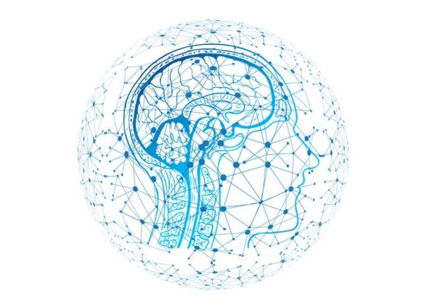
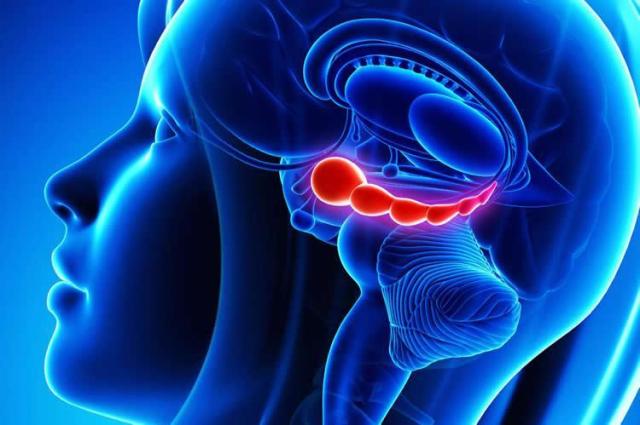
One of the surprises to Drew and his team was finding that brain cells that suppress fear memories hid in the hippocampus. Traditionally, scientists associate fear with another part of the brain, the amygdala. The hippocampus, responsible for many aspects of memory and spatial navigation, seems to play an important role in contextualizing fear, for example, by tying fearful memories to the place where they happened.

The discovery may help explain why one of the leading ways to treat fear-based disorders, exposure therapy, sometimes stops working. Exposure therapy promotes the formation of new memories of safety that can override an original fear memory. For example, if someone becomes afraid of spiders after being bitten by one, he might undertake exposure therapy by letting a harmless spider crawl on him. The safe memories are called “extinction memories.”

“Extinction does not erase the original fear memory but instead creates a new memory that inhibits or competes with the original fear,” Drew said. “Our paper demonstrates that the hippocampus generates memory traces of both fear and extinction, and competition between these hippocampal traces determines whether fear is expressed or suppressed.”

Given this, recommended practices around the frequency and timing of exposure therapy may need revisiting, and new pathways for drug development may be explored.

In experiments, Drew and his team placed mice in a distinctive box and induced fear with a harmless shock. After that, when one of the mice was in the box, it would display fear behavior until, with repeated exposure to the box without a shock, the extinction memories formed, and the mouse was not afraid.

Scientists were able to artificially activate the fear and suppress the extinction trace memories by using a tool called optogenetics to turn the extinction neurons on and off again.

“Artificially suppressing these so-called extinction neurons causes fear to relapse, whereas stimulating them prevents fear relapse,” Drew said. “These experiments reveal potential avenues for suppressing maladaptive fear and preventing relapse.”

The studies were led by graduate student Anthony Lacagnina of The University of Texas at Austin, with contributions from Emma Brockway, Chelsea Crovetti, Francis Shue, Meredith McCarty and Kevin Sattler of The University of Texas; and Sean Lim, Sofia Leal Santos and Christine Denny of Columbia University.

How the brain fights off fears that return to haunt us

A Dozen Dolphins Have Beached Themselves, Showing The Deadly Hallmark of Alzheimer’s disease

by Carly Cassella

Over a dozen dolphins, stranded on the beaches of Florida and Massachusetts, have been found with brains full of amyloid plaques, a hallmark of Alzheimer’s disease. The scientists who made the discovery think it may be a warning to us all: alongside the Alzheimer’s-like plaques, the team also found the environmental toxin BMAA.

Produced by blue-green algae blooms, this neurotoxin is easily caught up in the ocean food web, and chronic dietary exposure has long been suspected to be a cause of neurological disease, including Alzheimers, Parkinson’s and Amyotrophic Lateral Sclerosis (ALS).

The presence of both BMAA and amyloid plaques in 13 stranded dolphins now adds even more weight to this hypothesis.

“Dolphins are an excellent sentinel species for toxic exposures in the marine environment,” says neurologist Deborah Mash from the University of Miami.

“With increasing frequency and duration of cyanobacterial blooms in coastal waters, dolphins might provide early warning of toxic exposures that could impact human health.”

They might also be a good animal model for how BMAA could trigger Alzheimer’s disease. In 2017, it was discovered that dolphins are the only known wild animal to show signs of this common human disease.

Meanwhile, dolphins that inhabit Florida coastal waters are also commonly exposed to recurring harmful algae blooms (HABs). This might just be a coincidence, but experiments have shown that chronic BMAA dietary exposure can trigger neurodegenerative changes in both humans and non-human primates.

“Acute and chronic exposures to such toxins can be harmful to both humans and animals resulting in respiratory illnesses, severe dermatitis, mucosal damage, cancer, organ failure and death,” the authors write.

As the world warms at a rapid rate, these HABs are only becoming more frequent, and the authors worry that dolphins will accumulate even more BMAA as a result, “both by exposure to HABs and by the ingestion of prey previously exposed to the cyanotoxin”.

As such, these creatures may very well be our first indication of poor environmental conditions, and while it’s still not clear if these blooms directly lead to Alzheimer’s in dolphins or in humans, the researchers say it’s a risk we shouldn’t be willing to take.

“The $64,000 question is whether these marine mammals experienced cognitive deficits and disorientation that led to their beaching,” says co-author Paul Alan Cox, an ethnobotanist at the Brain Chemistry Labs in Jackson Hole.

“Until further research clarifies this question, people should take simple steps to avoid cyanobacterial exposure.”

This study has been published in PLOS ONE.

https://www.sciencealert.com/beached-dolphins-had-alzheimer-s-like-plaques-and-it-s-a-warning-to-us-all

New neurons for life? Old people can still make fresh brain cells, study finds

By Emily Underwood

One of the thorniest debates in neuroscience is whether people can make new neurons after their brains stop developing in adolescence—a process known as neurogenesis. Now, a new study finds that even people long past middle age can make fresh brain cells, and that past studies that failed to spot these newcomers may have used flawed methods.

The work “provides clear, definitive evidence that neurogenesis persists throughout life,” says Paul Frankland, a neuroscientist at the Hospital for Sick Children in Toronto, Canada. “For me, this puts the issue to bed.”

Researchers have long hoped that neurogenesis could help treat brain disorders like depression and Alzheimer’s disease. But last year, a study in Nature reported that the process peters out by adolescence, contradicting previous work that had found newborn neurons in older people using a variety of methods. The finding was deflating for neuroscientists like Frankland, who studies adult neurogenesis in the rodent hippocampus, a brain region involved in learning and memory. It “raised questions about the relevance of our work,” he says.

But there may have been problems with some of this earlier research. Last year’s Nature study, for example, looked for new neurons in 59 samples of human brain tissue, some of which came from brain banks where samples are often immersed in the fixative paraformaldehyde for months or even years. Over time, paraformaldehyde forms bonds between the components that make up neurons, turning the cells into a gel, says neuroscientist María Llorens-Martín of the Severo Ochoa Molecular Biology Center in Madrid. This makes it difficult for fluorescent antibodies to bind to the doublecortin (DCX) protein, which many scientists consider the “gold standard” marker of immature neurons, she says.

The number of cells that test positive for DCX in brain tissue declines sharply after just 48 hours in a paraformaldehyde bath, Llorens-Martín and her colleagues report today in Nature Medicine. After 6 months, detecting new neurons “is almost impossible,” she says.

When the researchers used a shorter fixation time—24 hours—to preserve donated brain tissue from 13 deceased adults, ranging in age from 43 to 87, they found tens of thousands of DCX-positive cells in the dentate gyrus, a curled sliver of tissue within the hippocampus that encodes memories of events. Under a microscope, the neurons had hallmarks of youth, Llorens-Martín says: smooth and plump, with simple, undeveloped branches.

In the sample from the youngest donor, who died at 43, the team found roughly 42,000 immature neurons per square millimeter of brain tissue. From the youngest to oldest donors, the number of apparent new neurons decreased by 30%—a trend that fits with previous studies in humans showing that adult neurogenesis declines with age. The team also showed that people with Alzheimer’s disease had 30% fewer immature neurons than healthy donors of the same age, and the more advanced the dementia, the fewer such cells.

Some scientists remain skeptical, including the authors of last year’s Nature paper. “While this study contains valuable data, we did not find the evidence for ongoing production of new neurons in the adult human hippocampus convincing,” says Shawn Sorrells, a neuroscientist at the University of Pittsburgh in Pennsylvania who co-authored the 2018 paper. One critique hinges on the DCX stain, which Sorrells says isn’t an adequate measure of young neurons because the DCX protein is also expressed in mature cells. That suggests the “new” neurons the team found were actually present since childhood, he says. The new study also found no evidence of pools of stem cells that could supply fresh neurons, he notes. What’s more, Sorrells says two of the brain samples he and his colleagues looked at were only fixed for 5 hours, yet they still couldn’t find evidence of young neurons in the hippocampus.

Llorens-Martín says her team used multiple other proteins associated with neuronal development to confirm that the DCX-positive cells were actually young, and were “very strict,” in their criteria for identifying young neurons.

Heather Cameron, a neuroscientist at the National Institute of Mental Health in Bethesda, Maryland, remains persuaded by the new work. Based on the “beauty of the data” in the new study, “I think we can all move forward pretty confidently in the knowledge that what we see in animals will be applicable in humans, she says. “Will this settle the debate? I’m not sure. Should it? Yes.”

https://www.sciencemag.org/news/2019/03/new-neurons-life-old-people-can-still-make-fresh-brain-cells-study-finds?utm_campaign=news_daily_2019-03-25&et_rid=17036503&et_cid=2734364

Scientists Identify The Location of ‘Taste’ in Your Head, And It’s Not The Tongue

by David Nield

How exactly do our brains sort between the five taste groups: sweet, sour, salty, bitter and umami? We’ve now got a much better idea, thanks to research that has pinned down where in the brain this taste processing happens.

Step forward: the insular cortex. Already thought to be responsible for everything from motor control to social empathy, we can now add flavour identification to its list of jobs.

It’s an area of the brain scientists have previously suspected could be responsible for sorting tastes, and which has been linked to taste in rodents, but this new study is much more precise in figuring out the role it plays in decoding what our tongues are telling us.

“We have known that tastes activate the human brain for some time, but not where primary taste types such as sweet, sour, salty, and bitter are distinguished,” says one of the team, Adam Anderson from Cornell University in New York.

“By using some new techniques that analyse fine-grained activity patterns, we found a specific portion of the insular cortex – an older cortex in the brain hidden behind the neocortex – represents distinct tastes.”

Anderson and his team used detailed fMRI scans of 20 adults as well as a new statistical model to dig deeper than previous studies into the link between the insular cortex and taste. This helped separate the taste response from other related responses – like the disgust we might feel when eating something sour or bitter.

Part of the problem in pinning down the taste-testing parts of the brain is that multiple regions of neurons get busy whenever we’re eating something. However, this study helps to cut through some of that noise.

In particular, it seems that different tastes don’t necessarily affect different parts of the insular cortex, but rather prompt different patterns of activity. Those patterns help the brain determine what it’s tasting.

For example, one particular section of the insular cortex was found to light up – in terms of neural activity – whenever something sweet was tasted. It’s a literal sweet spot, in other words, but it also showed that different brains have different wiring.

“While we identified a potential sweet spot, its precise location differed across people and this same spot responded to other tastes, but with distinct patterns of activity,” says Anderson.

“To know what people are tasting, we have to take into account not only where in the insula is stimulated, but also how.”

The work follows on from previous research showing just how big a role the brain plays in perceiving taste. It used to be thought that receptors on the tongue did most of the taste testing, but now it seems the brain is largely in charge of the process.

That prior study showed how switching certain brain cells on and off in mice was enough to prevent them from distinguishing between sweet and bitter. The conclusion is that while the tongue does identify certain chemicals, it’s the brain that interprets them.

The new research adds even more insight into what’s going on in the brain in humans when we need to work out what we’re tasting – and shows just how important a job the insular cortex is doing.

“The insular cortex represents experiences from inside our bodies,” says Anderson. “So taste is a bit like perceiving our own bodies, which is very different from other external senses such as sight, touch, hearing or smell.”

The research has been published in Nature Communications.

https://www.sciencealert.com/now-we-know-the-part-of-the-brain-that-tells-us-what-we-re-tasting

Neuroscience proves Nietzsche right: some people are wired to be more spontaneous than others

by Parashkev Nachev

“Why can’t you just relax into it?” is a question many of us have asked in frustration with ourselves or others – be it on the dance floor, the sporting field or in rather more private circumstances. The task typically requires us to respond spontaneously to external events, without any deliberation whatsoever. It ought to be easy – all you have to do is let go – yet it can be infuriatingly difficult.

“Stop thinking about it!” is the standard remedial advice, although cancelling thought with thought is something of a paradox. The retort, “I am trying!”, is equally puzzling, for deliberate intent is precisely what we are here struggling to avoid. So what is this act of choosing not to choose, of consciously relinquishing control over our actions? Our new study, published in Communications Biology, has finally provided insights into how this capacity is expressed in the brain.

Astonishingly, this fundamental human phenomenon has no name. It might have escaped academic recognition entirely had the German philosopher Friedrich Nietzsche not given it a brilliant gloss in his first book The Birth of Tragedy, itself a paradoxical work of philosophy in tacitly encouraging the reader to stop reading and get a drink instead. Whereas other thinkers saw culture on a single continuum, evolving into ever greater refinement, order and rationality, Nietzsche saw it as distributed across two radically different but equally important planes.

Perpendicular to the conventional “Apolline” dimension of culture, he introduced the “Dionysiac”: chaotic, spontaneous, vigorous and careless of the austere demands of rationality. Neither aspect was held to be superior, each may be done badly or well, and both are needed for a civilisation to find its most profound creative expression. Every Batman needs a Joker, he might have said, had he lived in a more comical age.

Of course, Nietzsche was not the first to observe that human beings sometimes behave with wanton abandon. His innovation consisted in realising it is a constitutional characteristic we could and should develop. And as with any behavioural characteristic, the facility to acquire it will vary from one person to another.

Seeing the light

As Dionysus and neuroscientists are mostly strangers, it should come as no surprise that the capacity for “meta-volition” – to give it a name that captures the notion of choosing not to choose one’s actions – has until now escaped experimental study. To find out how our brains allow us to give up control and explain why some of us are better at it than others, my colleagues and I wanted to develop a behavioural test and examine the patterns of brain activity that go with lesser or greater ability.

Most tests in behavioural neuroscience pit conscious, deliberate, complex actions against their opposites, measuring the power to suppress them. A classic example is the anti-saccade task, which purportedly measures “cognitive control”. Participants are instructed not to look towards the light when they see a brief flash in the visual periphery, but instead to the opposite side. That’s hard to do because looking towards the light is the natural inclination. People who are better at this are said to have greater cognitive control.

To measure how good people are at relinquishing control, we cannot simply flip a task around. If people are asked to look into the light, will and instinct are placed in perfect agreement. To put the two in opposition, we must make the automatic task unconscious so that volition could only be a hindrance.


White matter map of the brain (ray traced rendering), with the area correlated with spontaneity in red. Credit: Parashkev Nachev

It turns out that this is easy to do by flashing two lights on opposite sides of the visual periphery nearly simultaneously, and asking the subject to orient as fast as possible to the one they see first. If a flash comes a few dozen milliseconds before the next, people typically obtain an automatic bias to the first flash. You need at least double that amount of time to reach the threshold for consciously detecting which one comes first. Thinking about what came first could only impair your performance because your instinct operates well beneath the threshold at which the conscious gets a foothold.

Amazingly for such a simple task, people vary dramatically in their ability. Some – the Dionysiacs – effortlessly relax into allowing themselves to be guided by the first light, requiring no more than a few milliseconds between the flashes. Others – the Apollines – cannot let go, even when the flashes are many times further apart. Since trying harder does not help, the differences are not a matter of effort but appear to be part of who we are.

We used magnetic resonance imaging to investigate the brains of people performing the task, focusing on white matter – the brain’s wiring. A striking picture emerged. Extensive sections of the wiring of the right prefrontal lobe, a region heavily implicated in complex decision making, was revealed to be stronger in those who were worse at the task: the Apollines. The more developed the neural substrates of volition, it seems, the harder to switch them off.

By contrast, no part of the Dionysiac brain showed evidence of stronger wiring. Suppressing volition appears to depend less on a “meta-volitional centre” that is better developed than on the interplay between spontaneous and deliberate actions. We can think of it as two coalitions of brain cells in competition, with the outcome dependent on the relative strength of the teams, not the qualities of any umpire.

The competitive brain

The results demonstrate how the brain operates by competition at least as much as by cooperation. It may fail at a task not because it does not have the power, but because another, more dominant power stands in opposition. Our decisions reflect the outcomes of battles between warring factions that differ in their characteristics and evolutionary lineage, battles we can do little to influence because we are ourselves their products.

People also differ widely in their qualities, including spontaneity, not because evolution has not yet arrived at an optimum, but because it seeks to diversify the field as far as possible. That’s why it creates individuals tuned to respond to their environment in very different ways. The task of evolution is less to optimise a species for the present than to prepare it for a multiplicity of futures unknown.

That our lives are now dominated by a rational, Apolline order does not mean we shall not one day descend into an instinctual, Dionysiac chaos. Our brains are ready for it – our culture should be too.

https://medicalxpress.com/news/2019-03-neuroscience-nietzsche-people-wired-spontaneous.html

Having great-grandparents or cousins with Alzheimer’s disease substantially increases your risk of developing it also.

Having a parent with Alzheimer’s disease has been known to raise a person’s risk of developing the disease, but new research published in Neurology suggests that having second- and third-degree relatives who have had Alzheimer’s may also increase risk.

“Family history is an important indicator of risk for Alzheimer’s disease, but most research focuses on dementia in immediate family members, so our study sought to look at the bigger family picture,” said Lisa A. Cannon-Albright, PhD, University of Utah School of Medicine, Salt Lake City, Utah. “We found that having a broader view of family history may help better predict risk. These results potentially could lead to better diagnoses and help patients and their families in making health-related decisions.”

For the study, researchers looked at the Utah Population Database, which includes the genealogy of Utah pioneers from the 1800s and their descendants up until modern day. The database is linked to Utah death certificates, which show causes of death, and in a majority of cases, contributing causes of death.

In that database, researchers analysed data from over 270,800 people who had at least 3 generations of genealogy connected to the original Utah pioneers including genealogy data for both parents, all 4 grandparents, and at least 6 of 8 great-grandparents. Of those, 4,436 have a death certificate that indicates Alzheimer’s disease as a cause of death.

Results showed that people with 1 first-degree relative with Alzheimer’s disease (18,494 people) had a 73% increased risk of developing the disease. Of this group of people, 590 developed Alzheimer’s disease; the researchers would have expected this group to have 341 cases.

People with 2 first-degree relatives were 4 times more likely to develop the disease; those with 3 were 2.5 times more likely; and those with 4 were nearly 15 times more likely to develop Alzheimer’s disease.

Of the 21 people in the study with 4 first-degree relatives with Alzheimer’s, 6 had the disease. The researchers would have expected only 0.4 people to develop the disease.

Those with 1 first-degree relative and 1 second-degree relative had a 21 times greater risk. Examples of this would be a parent and one grandparent with the disease, or a parent and one aunt or uncle. There were 25 people in this category in the study; 4 of them had the disease when researchers would have expected 0.2 cases.

Those who had only third-degree relatives, and 3 such relatives, with Alzheimer’s disease had a 43% greater risk of developing the disease. An example of this would be two great-grandparents with the disease, along with one great uncle, but no parents or grandparents with the disease. Of the 5,320 people in this category, 148 people had the disease when researchers would have expected 103.

“More and more, people are increasingly seeking an estimate of their own genetic risk for Alzheimer’s disease,” said Dr. Cannon-Albright. “Our findings indicate the importance of clinicians taking a person’s full family history that extends beyond their immediate family members.”

She noted that among all of the study participants, 3% had a family history that doubled their risk of Alzheimer’s disease, and a little over one-half of a percent had a family history that increased their risk by ≥3 times that of a person without a family history of the disease.

Limitations of the study include that not all individuals dying from Alzheimer’s disease may have had a death certificate listing it as cause of death. Dr. Cannon-Albright said death certificates are known to underestimate the prevalence of the disease.

“There are still many unknowns about why a person develops Alzheimer’s disease,” she said. “A family history of the disease is not the only possible cause. There may be environmental causes, or both. There is still much more research needed before we can give people a more accurate prediction of their risk of the disease.”

Reference:
https://n.neurology.org/content/early/2019/03/13/WNL.0000000000007231

https://dgnews.docguide.com/having-great-grandparents-cousins-alzheimer-s-linked-higher-risk?overlay=2&nl_ref=newsletter&pk_campaign=newsletter&nl_eventid=20119

AI and MRIs at birth can predict cognitive development at age 2


Researchers at the University of North Carolina School of Medicine used MRI brain scans and machine learning techniques at birth to predict cognitive development at age 2 years with 95 percent accuracy.

“This prediction could help identify children at risk for poor cognitive development shortly after birth with high accuracy,” said senior author John H. Gilmore, MD, Thad and Alice Eure Distinguished Professor of psychiatry and director of the UNC Center for Excellence in Community Mental Health. “For these children, an early intervention in the first year or so of life – when cognitive development is happening – could help improve outcomes. For example, in premature infants who are at risk, one could use imaging to see who could have problems.”

The study, which was published online by the journal NeuroImage, used an application of artificial intelligence called machine learning to look at white matter connections in the brain at birth and the ability of these connections to predict cognitive outcomes.

Gilmore said researchers at UNC and elsewhere are working to find imaging biomarkers of risk for poor cognitive outcomes and for risk of neuropsychiatric conditions such as autism and schizophrenia. In this study, the researchers replicated the initial finding in a second sample of children who were born prematurely.

“Our study finds that the white matter network at birth is highly predictive and may be a useful imaging biomarker. The fact that we could replicate the findings in a second set of children provides strong evidence that this may be a real and generalizable finding,” he said.

Jessica B. Girault, PhD, a postdoctoral researcher at the Carolina Institute for Developmental Disabilities, is the study’s lead author. UNC co-authors are Barbara D. Goldman, PhD, of UNC’s Frank Porter Graham Child Development Institute, Juan C. Prieto, PhD, assistant professor, and Martin Styner, PhD, director of the Neuro Image Research and Analysis Laboratory in the department of psychiatry.

AI and MRIs at birth can predict cognitive development at age 2

FDA approves first drug for postpartum depression

By Elizabeth Chuck and Lauren Dunn

The intrusive thoughts started weeks after Stephanie Hathaway gave birth: an overwhelming feeling that her daughter deserved a better mother; that her husband deserved a better wife; that her future was hopeless.

“They just played on repeat in my head,” Hathaway, 33, of South Glastonbury, Connecticut, said. “I was holding my baby one night, and my husband was at a meeting, and I just thought, ‘Oh, my goodness. If I put the baby down, I might hurt myself.’”

Hathaway was diagnosed with postpartum depression — the intense sadness, anxiety or despair that occurs within the first year after giving birth, according to the Centers for Disease Control and Prevention. It affects about one in nine women, although the rate may be as high as one in every five women, the CDC finds.

Hathaway’s doctor put her on antidepressants, which helped some, but it took two weeks for the medication to kick in, and even longer until her doctor found the appropriate dosage for her. As she waited for relief, Hathaway found herself struggling to bond with her newborn, Hadley, who is now 4.

“It’s heartbreaking,” Hathaway, who had never suffered from depression before and is now a mother to two girls, said. “That’s not what I expected to feel.”

Up until this point, new mothers experiencing postpartum depression have been prescribed the same antidepressants used for treating depression in the general population, such as selective serotonin reuptake inhibitors. The drugs can take weeks to take effect, and do not address the hormonal changes that women go through during and after pregnancy.

But on Tuesday, the Food and Drug Administration approved the first drug specifically developed for postpartum depression, called brexanolone, or Zulresso.

Brexanolone is novel because it has a synthetic form of the hormone allopregnanolone, a progesterone derivative, in it. The hormone increases throughout a woman’s pregnancy and then plummets after she gives birth, a possible contributor to postpartum depression.

“This can potentially transform women’s lives and that of their families,” said Dr. Steve Kanes, chief medical officer of Sage Therapeutics, the Cambridge, Mass., biopharmaceutical company that developed brexanolone. “It’s not just the mother who suffers when there’s postpartum depression. It’s the newborn. It’s the other people in their family.”

Brexanolone is not a pill. The drug is delivered intravenously over the course of a 60-hour infusion, meaning it must be administered in a medically supervised setting, such as a skilled facility or a hospital, rather than at patients’ homes.

IMPROVEMENT IN JUST 24 HOURS

Clinical trials for the drug were promising — not just in the number of women it helped, but in the near-instantaneous relief that is provided.

In double-blind, placebo-controlled trials, many women with moderate to severe postpartum depression saw a marked improvement of their symptoms within just 24 hours of receiving the drug. That improvement was still present 30 days after the infusion, the length of the trial.

“This is for postpartum depression, but it is a step in understanding how we treat depression more broadly,” said Dr. Samantha Meltzer-Brody, director of the perinatal psychiatry program at the University of North Carolina at Chapel Hill and the academic principal investigator in the brexanolone trials. “We have had the same treatments for depression for 30 years. There’s an enormous need for new, novel ways to treat depression, and to treat it quickly.”

The drug’s approval comes just weeks after the FDA signed off on esketamine, a fast-acting nasal spray that uses the active ingredients in the club drug ketamine, as a treatment for severe depression.

For patients who are depressed, rapid relief is a priority. Hathaway, the Connecticut mother, was again diagnosed with postpartum depression after she gave birth to her second, a girl named Brenley who is now 2. This time, the antidepressants did not help at all, and Hathaway felt herself slipping deeper and deeper into a state of hopelessness.

She participated in a brexanolone trial, and her response was striking. Between hours 12 and 18 of the 60-hour infusion, she noticed her despair had waned.

“I woke up from a nap, and the thoughts were gone. And they never came back,” Hathaway said. “And then hour after hour, I got my energy back. I got my appetite back. I was eating because I was actually hungry, not because people were making me eat.”

A COMMON CONDITION

Postpartum depression afflicts as many as 400,000 women in the United States each year. It can include disturbances in sleep or eating patterns in addition to feelings of sadness or apathy. Affected women are often confused and guilt-ridden about why they are feeling down during what is supposed to be a happy time, said Dr. Christine C. Greves, an obstetrician-gynecologist at Orlando Health Winnie Palmer Hospital for Women and Babies.

“As women, we feel like we were born to have a child, and there’s a white picket fence, and life will be great,” said Greves, who does not have ties to Sage Therapeutics. “Then regular life comes into play. You have a child and then you top that with extensive fatigue, hormones, expectations that just can’t be met. It’s all fantasy until we actually have the baby. And then you do feel guilty, because we all want to be Super Mom.”

In the past decade, experts say, there has been more awareness about postpartum depression and more efforts among obstetricians and pediatricians to screen mothers for it.

But having a drug specifically aimed at treating postpartum depression will be one of the most significant steps toward removing any stigma still associated with the condition, said Dr. Kimberly Yonkers, professor of psychiatry, epidemiology and obstetrics, gynecology and reproductive sciences at the Yale School of Medicine.

“It does women a service because it really brings attention to a major medical problem and provides legitimacy, and hopefully will encourage people, whether they use this medication or not, to seek and obtain treatment,” said Yonkers, who does not have ties to the drug company. “We’re all thrilled about that.”

SOME SIDE EFFECTS, AND A HEFTY PRICE TAG

The most common side effects during the brexanolone trial were drowsiness and dizziness. The drug is not believed to have any long-term safety concerns. Kanes, Sage Therapeutics’ chief medical officer, said he expects it will be deemed safe for all mothers, including breastfeeding mothers, but the company is waiting for an FDA ruling on breastfeeding.

The drug comes with a hefty price tag: Sage says it is expected to cost somewhere between $20,000 to $35,000 for the infusion. That does not include the price of a stay in whatever facility it is administered in. It is not clear yet how much insurance would cover.

Kanes pointed out that while high, the cost is a one-time price.

“That’s such an important piece as to why this is so novel. We’re talking about a single treatment that has durable effects,” he said. “This really is a one-time intervention that gets people on their way. It’s transformative.”

For Hathaway, the brexanolone infusion enabled her to return home and be the mother to her daughters that she had wanted to be before postpartum depression took over.

“It’s given them their mom back,” she said. “This is what it was supposed to be like.”

https://www.nbcnews.com/health/womens-health/fda-approves-first-drug-postpartum-depression-n984521

Scientists ‘Clear’ Alzheimer’s Plaque From Mice Using Only Light And Sound

Clumps of harmful proteins that interfere with brain functions have been partially cleared in mice using nothing but light and sound.

Research led by MIT has found strobe lights and a low pitched buzz can be used to recreate brain waves lost in the disease, which in turn remove plaque and improve cognitive function in mice engineered to display Alzheimer’s-like behaviour.

It’s a little like using light and sound to trigger their own brain waves to help fight the disease.

This technique hasn’t been clinically trialled in humans as yet, so it’s too soon to get excited – brain waves are known to work differently in humans and mice.

But, if replicated, these early results hint at a possible cheap and drug-free way to treat the common form of dementia.

So how does it work?

Advancing a previous study that showed flashing light 40 times a second into the eyes of engineered mice treated their version of Alzheimer’s disease, researchers added sound of a similar frequency and found it dramatically improved their results.

“When we combine visual and auditory stimulation for a week, we see the engagement of the prefrontal cortex and a very dramatic reduction of amyloid,” says Li-Huei Tsai, one of the researchers from MIT’s Picower Institute for Learning and Memory.

It’s not the first study to investigate the role sound can play in clearing the brain of the tangles and clumps of tau and amyloid proteins at least partially responsible for the disease.

Previous studies showed bursts of ultrasound make blood vessels leaky enough to allow powerful treatments to slip into the brain, while also encouraging the nervous system’s waste-removal experts, microglia, to pick up the pace.

Several years ago, Tsai discovered light flickering at a frequency of about 40 flashes a second had similar benefits in mice engineered to build up amyloid in their brain’s nerve cells.

“The result was so mind-boggling and so robust, it took a while for the idea to sink in, but we knew we needed to work out a way of trying out the same thing in humans,” Tsai told Helen Thomson at Nature at the time.

The only problem was this effect was confined to visual parts of the brain, missing key areas that contribute to the formation and retrieval of memory.

While the method’s practical applications looked a little limited, the results pointed to a way oscillations could help the brain recover from the grip of Alzheimer’s disease.

As our brain’s neurons transmit signals they also generate electromagnetic waves that help keep remote regions in sync – so-called ‘brain waves’.

One such set of oscillations are defined as gamma-frequencies, rippling across the brain at around 30 to 90 waves per second. These brain waves are most active when we’re paying close attention, searching our memories in order to make sense of what’s going on.

Tsai’s previous study had suggested these gamma waves are impeded in individuals with Alzheimer’s, and might play a pivotal role in the pathology itself.

Light was just one way to trick the parts of the brain into humming in the key of gamma. Sounds can also manage this in other areas.

Instead of the high pitched scream of ultrasound, Tsui used a much lower droning noise of just 40 Hertz, a sound only just high enough for humans to hear.

Exposing their mouse subjects to just one hour of this monotonous buzz every day for a week led to a significant drop in the amount of amyloid build up in the auditory regions, while also stimulating those microglial cells and blood vessels.

“What we have demonstrated here is that we can use a totally different sensory modality to induce gamma oscillations in the brain,” says Tsai.

As an added bonus, it also helped clear the nearby hippocampus – an important section associated with memory.

The effects weren’t just evident in the test subjects’ brain chemistry. Functionally, mice exposed to the treatment performed better in a range of cognitive tasks.

Adding the light therapy from the previous study saw an even more dramatic effect, clearing plaques in a number of areas across the brain, including in the prefrontal cortex. Those trash-clearing microglia also went to town.

“These microglia just pile on top of one another around the plaques,” says Tsai.

Discovering new mechanisms in the way nervous systems clear waste and synchronise activity is a huge step forward in the development of treatments for all kinds of neurological disorders.

Translating discoveries like this to human brains will take more work, especially when there are potential contrasts in how gamma waves appear in mice and human Alzheimer’s brains.

So far early testing for safety has shown the process seems to have no clear side effects.

This research was published in Cell.

https://www.sciencealert.com/astonishing-new-study-treats-alzheimer-s-in-mice-with-a-light-and-sound-show

Neuroscientists can read brain activity to predict decisions 11 seconds before people act

Neuroscientists can read brain activity to predict decisions 11 seconds before people actFree will, from a neuroscience perspective, can look like quite quaint. In a study published this week in the journal Scientific Reports, researchers in Australia were able to predict basic choices participants made 11 seconds before they consciously declared their decisions.

In the study, 14 participants—each placed in an fMRI machine—were shown two patterns, one of red horizontal stripes and one of green vertical stripes. They were given a maximum of 20 seconds to choose between them. Once they’d made a decision, they pressed a button and had 10 seconds to visualize the pattern as hard as they could. Finally, they were asked “what did you imagine?” and “how vivid was it?” They answered these questions by pressing buttons.

Using the fMRI to monitor brain activity and machine learning to analyze the neuroimages, the researchers were able to predict which pattern participants would choose up to 11 seconds before they consciously made the decision. And they were able to predict how vividly the participants would be able to envisage it.

Lead author Joel Pearson, cognitive neuroscience professor at the University of South Wales in Australia, said that the study suggests traces of thoughts exist unconsciously before they become conscious. “We believe that when we are faced with the choice between two or more options of what to think about, non-conscious traces of the thoughts are there already, a bit like unconscious hallucinations,” he said in a statement. “As the decision of what to think about is made, executive areas of the brain choose the thought-trace which is stronger. In, other words, if any pre-existing brain activity matches one of your choices, then your brain will be more likely to pick that option as it gets boosted by the pre-existing brain activity.”

The work has implications for how we understand uncomfortable thoughts: Pearson believes the findings explain why thinking about something only leads to more thoughts on the subject, as it creates “a positive feedback loop.” The study also suggests that unwelcome visualizations, such as those experienced with post-traumatic stress disorder, begin as unconscious thoughts.

Though this is just one study, it’s not the first to show that thoughts can be predicted before they are conscious. As the researchers note, similar techniques have been able to predict motor decisions between seven and 10 seconds before they’re conscious, and abstract decisions up to four seconds before they’re conscious. Taken together, these studies show how understanding how the brain complicates our conception of free will.

Neuroscientists have long known that the brain prepares to act before you’re consciously aware, and there are just a few milliseconds between when a thought is conscious and when you enact it. Those milliseconds give us a chance to consciously reject unconscious impulses, seeming to form a foundation of free will.

Freedom, however, can be enacted by both the unconscious and conscious self—and there are neuroscientists who claim that being controlled by our own unconscious brain is hardly an affront to free will. Studies showing that neuroscientists can predict our actions long before we’re aware of them don’t necessarily negate the concept of free will, but they certainly complicate our conception of our own minds.

https://qz.com/1569158/neuroscientists-read-unconscious-brain-activity-to-predict-decisions/?utm_source=google-news