Teenager Amber Yang is developing a better way to track space junk

Nineteen-year-old Amber Yang has her head in the clouds. Actually, it’s beyond the clouds and well into the Earth’s atmosphere. While some teens are focused on cleaning up the land and water on our planet, Yang has made it her mission to clean up the litter clogging space. And she may just save lives and billions of dollars while she’s at it.

Yang was 15 when she first heard about the escalating issue of space debris that is polluting the Earth’s lower atmosphere. After watching the 2013 movie, “Gravity,” Yang imagined a world in which colliding space debris could set off a series of catastrophic events that threaten lives and technology. While the plot of “Gravity” broke some of the major rules of physics, the underlying premise — that a collision of space debris could lead to disaster — rang true and stuck in Yang’s mind.

That year, over her winter break in Florida, she brushed up on astrophysics, computer coding, and the ins and outs of space junk and developed a program called Seer Tracking to provide an accurate location for each piece of junk orbiting the Earth. Currently, there are millions of pieces of space debris, ranging in size from defunct satellites to tiny specks of paint. Traveling at a rate of around 17,500 miles per hour, each item has the potential to cause a catastrophic collision.

The Department of Defense’s Space Surveillance Network currently analyzes Earth’s space debris using tracking and data that could detect a potential collision as far as 10 days in advance, But according to Yang, her Seer Tracking program is able to predict issues weeks ahead. And the scientific community concurs. Her work has earned her the top award at the Intel International Science and Engineering Fair as well as a spot on Forbes 30 under 30 list. Yang has presented her research at the European Organization for Nuclear Research (CERN) in Geneva, Switzerland and was asked to speak at a TEDx Conference about the issues that women face in STEM careers.

Seer Tracking uses artificial neural networks to track space debris. In other words, Yang has developed an algorithim that allows the program to learn from its mistakes, so that its predictions become more accurate over time. As the data accumulates, Seer Tracking increases in its ability to pinpoint debris locations and predict collisions well in advance.

Yang is now finishing up her sophomore year at Stanford University, pursuing a degree in physics while operating Seer Tracking on the side. She’s still working on improving her software and exploring other avenues of “deep learning” in which computers learn from their mistakes.

https://www.mnn.com/family/family-activities/blogs/meet-teen-whos-cleaning-space-junk

Using science to study whether there should be two or only one space between types sentences

In the beginning, the rules of the space bar were simple. Two spaces after each period. Every time. Easy.

That made sense in the age of the typewriter. Letters of uniform width looked cramped without extra space after the period. Typists learned not to do it.

But then, at the end of the 20th century, the typewriter gave way to the word processor, and the computer, and modern variable-width fonts. And the world divided.

Some insisted on keeping the two-space rule. They couldn’t get used to seeing just one space after a period. It simply looked wrong.

Some said this was blasphemy. The designers of modern fonts had built the perfect amount of spacing, they said. Anything more than a single space between sentences was too much.

And so the rules of typography fell into chaos. “Typing two spaces after a period is totally, completely, utterly, and inarguably wrong,” Farhad Manjoo wrote in Slate in 2011. “You can have my double space when you pry it from my cold, dead hands,” Megan McArdle wrote in the Atlantic the same year. (And yes, she double-spaced it.)

This schism has actually existed throughout most of typed history, the writer and type enthusiast James Felici once observed (in a single-spaced essay).

The rules of spacing have been wildly inconsistent going back to the invention of the printing press. The original printing of the U.S. Declaration of Independence used extra long spaces between sentences. John Baskerville’s 1763 Bible used a single space. WhoevenknowswhateffectPietroBembowasgoingforhere.Single spaces. Double spaces. Em spaces. Trends went back and forth between continents and eras for hundreds of years, Felici wrote.It’s not a good look.

And that’s just English. Somewrittenlanguageshavenospacesatall and o thers re quire a space be tween ev e ry syl la ble.

Ob viously, thereneed to be standards. Unless you’re doing avant – garde po e try, or something , you can’tjustspacew ords ho w e v e r y o u want. That would be insanity. Or at least, obnoxious.

Enter three psychology researchers from Skidmore College, who decided it’s time for modern science to sort this out once and for all.

“Professionals and amateurs in a variety of fields have passionately argued for either one or two spaces following this punctuation mark,” they wrote in a paper published last week in the journal Attention, Perception, & Psychophysics.

They cite dozens of theories and previous research, arguing for one space or two. A 2005 study that found two spaces reduced lateral interference in the eye and helped reading. A 2015 study that found the opposite. A 1998 experiment that suggested it didn’t matter.

“However,” they wrote, “to date, there has been no direct empirical evidence in support of these claims, nor in favor of the one-space convention.”

So the researchers, Rebecca L. Johnson, Becky Bui and Lindsay L. Schmitt, rounded up 60 students and some eye tracking equipment, and set out to heal the divide.

First, they put the students in front of computers and dictated a short paragraph, to see how many spaces they naturally used. Turns out, 21 of the 60 were “two-spacers,” and the rest typed with close-spaced sentences that would have horrified the Founding Fathers.

The researchers then clamped each student’s head into place, and used an Eyelink 1000 to record where they looked as they silently read 20 paragraphs. The paragraphs were written in various styles: one-spaced, two-spaced, and strange combinations like two spaces after commas, but only one after periods. And vice versa, too.

And the verdict was: two spaces after the period is better. It makes reading slightly easier.

Reading speed only improved marginally, the paper found, and only for the 21 “two-spacers,” who naturally typed with two spaces between sentences. The majority of one-spacers, on the other hand, read at pretty much the same speed either way. And reading comprehension was unaffected for everyone, regardless of how many spaces followed a period.

The major reason to use two spaces, the researchers wrote, was to make the reading process smoother, not faster. Everyone tended to spend fewer milliseconds staring at periods when a little extra blank space followed it.

(Putting two spaces after a comma, if you’re wondering, slowed down reading speed, so don’t do that.)

The study’s authors concluded that two-spacers in the digital age actually have science on their side, and more research should be done to “investigate why reading is facilitated when periods are followed by two spaces.”

But no sooner did the paper publish than the researchers discovered that science doesn’t necessarily govern matters of the space bar.

Johnson and her co-authors submitted the paper with two spaces after each period — as was proper. And the journal deleted all the    extra spaces anyway.

https://www.washingtonpost.com/news/speaking-of-science/wp/2018/05/04/one-space-between-each-sentence-they-said-science-just-proved-them-wrong-2/?noredirect=on&utm_term=.857b06f6cb87

Disease detectives determine that typhoid killed 12th century Saladin

Saladin may not be well known in the West, but even 800 years after his death, he remains famous in the Middle East. Born in 1137, he rose to become the Sultan of an enormous area that now includes Egypt, Syria, parts of Iraq, Lebanon, Yemen and other regions of North Africa. He successfully led armies against the invading Crusaders and conquered several kingdoms. Historians have described him as the most famous Kurd ever.

Even today, however, Saladin’s death remains a mystery. The illness began in 1193, when he was 56. After two weeks, the Sultan was dead. Some have speculated that fever was a prominent symptom of the illness.

After closely examining a range of evidence about Saladin’s condition, Stephen J. Gluckman, MD, professor of medicine at the University of Pennsylvania School of Medicine, has developed a diagnosis. Dr. Gluckman theorizes that typhoid, a bacterial disease that was very common in the region at the time, is the most likely culprit. Today of course, antibiotics could have greatly helped Saladin. But in the 12th century these medicines did not exist.

Dr. Gluckman delivered his diagnosis at the 25th annual Historical Clinicopathological Conference, held Friday, May 4 at the University of Maryland School of Medicine. The conference is devoted to the diagnosis of disorders that afflicted historical figures; in the past, experts have focused on the diseases of luminaries such as Lenin, Darwin, Eleanor Roosevelt and Lincoln.

Dr. Gluckman, an expert on parasitic disorders, has provided care and taught in many countries around the world. He carefully reviewed what is known about the Sultan’s medical history. “Practicing medicine over the centuries required a great deal of thought and imagination,” he says. “The question of what happened to Saladin is a fascinating puzzle.”

Saladin is known for destroying King Guy’s army at the Horns of Hattin in 1187 and reclaiming Jerusalem for Islam after it had been ruled for nearly a century by Christian crusaders. He is also famed for treating his enemies generously.

Typhoid fever is a potentially deadly disease spread by contaminated food and water. Symptoms of typhoid include high fever, weakness, stomach pain, headache, and loss of appetite. It is common in most parts of the world except in industrialized regions such as the United States, western Europe, Australia, and Japan. About 300 people get typhoid fever in the United States each year, and most of them have recently traveled. Globally, typhoid infects about 22 million people a year, and kills 200,000.

‌Also speaking at the conference will be Thomas Asbridge, PhD, is a reader of medieval history at Queen Mary University of London. Dr. Asbridge is an expert on Saladin and the Crusades.

The conference was founded in 1995 by Philip A. Mackowiak, MD, the Carolyn Frenkil and Selvin Passen History of Medicine Scholar-in-Residence at UMSOM. “This is an intriguing piece of medical detecting,” says Dr. Mackowiak. “If antibiotics had been around in the 12th century, history may have been quite different.”

For more information on the conference, visit:
http://medicalalumni.org/historicalcpc/home/

http://www.medschool.umaryland.edu/news/2018/Expert-Disease-Detective-Unravels-Mysterious-Illness-That-Killed-Famed-12th-Century-Sultan.html

Vasopressin levels in cerebrospinal fluid may be a biomarker for social deficits in patients with autism


Rhesus macaques live in large family groups, but a few animals consistently show less social interaction than others. New research at the California National Primate Research Center at UC Davis and Stanford University shows that these “low social” animals have low levels of the hormone vasopressin in cerebrospinal fluid. A similar result was seen in a small group of children with autism.

One of the characteristics of children with autism spectrum disorder is reduced social ability. It’s difficult to study the possible causes of social impairment in children, but a new study shows that rhesus macaques with low sociability also had low levels of the peptide vasopressin in cerebrospinal fluid, as did children with autism spectrum disorder.

The study, by researchers at the California National Primate Research Center at the University of California, Davis, and Stanford University, is published May 2 in the journal Science Translational Medicine.

“At this point, we consider vasopressin concentrations to be a biomarker for low sociability,” said John Capitanio, professor of psychology at UC Davis and a research scientist at the CNPRC.

Capitanio studies the interplay between social behavior and health. Over several years, his team has assessed rhesus macaque monkeys born at the center for sociability. The center maintains large field corrals where the macaques live in extended large family groups with the same hierarchies and social behavior that they show in the wild.

About 15 percent of the animals are classed as “low social”: They spend less time interacting with others than most macaques. Capitanio has previously studied how this natural variation affects the course of infectious disease.

Professor Karen Parker at the Stanford Department of Psychiatry and Behavioral Sciences, principal investigator on the project, is interested in why children with autism spectrum disorder have deficits in social ability.

The researchers identified “high social” and “low social” monkeys and tested their blood and cerebrospinal fluid for a number of markers previously identified as possibly playing a role in autism. The one that jumped out was vasopressin in cerebrospinal fluid, which was low in “low social” animals. Other potential markers such as oxytocin were not significantly different, they found.

Parker and Capitanio’s team was able to confirm the link between vasopressin and sociability in a second cohort of monkeys and demonstrate in a third group that vasopressin levels in cerebrospinal fluid are stable over time.

In addition, the Stanford team was able to obtain cerebrospinal fluid from seven children with autism spectrum disorder who were undergoing lumbar puncture for other reasons. They found that these children also had low levels of vasopressin compared to seven other children without autism.

There are some important limitations to the study, Capitanio said. Firstly, all the animals and children studied were male. Although autism spectrum disorder is far more common in males, research on females is also needed. Secondly, the study shows a correlation between sociability and vasopressin, but not whether it is a cause or a consequence.

Parker and Capitanio plan to repeat the study with female animals and to treat “low social” animals with vasopressin to see if that changes their behavior.

The monkey studies have particular relevance to humans because they are based on natural behavior, Capitanio said. “This is naturally occurring variation, we aren’t making the monkeys behave in this way,” he said.

Other authors on the paper are: Erna Tarara, Valentina Sclafani, Laura Del Rosso and Katie Chun at UC Davis; Joseph Garner, Ozge Oztan, Sean Berquist, Antonio Hardan and Sonia Partap at Stanford University; Elliott Sherr and Jiang Li at UC San Francisco; and Michael Chez at Sutter Neurosciences Medical Group, Sacramento. The work was supported by grants from the NIH and the Simons Foundation.

Reference: Parker, K. J., Garner, J. P., Oztan, O., Tarara, E. R., Li, J., Sclafani, V., … Capitanio, J. P. (2018). Arginine vasopressin in cerebrospinal fluid is a marker of sociality in nonhuman primates. Science Translational Medicine, 10(439), eaam9100. https://doi.org/10.1126/scitranslmed.aam9100

https://www.ucdavis.edu/news/vasopressin-possible-biomarker-social-deficits-autism/

Using magnetism to regulate neural activity shows a small benefit in patients with mild forms of Alzheimer’s disease

On the heels of one failed drug trial after another, a recent study suggests people with early Alzheimer’s disease could reap modest benefits from a device that uses magnetic fields to produce small electric currents in the brain.

Alzheimer’s is a degenerative brain disorder that afflicts more than 46 million people worldwide. At present there are no treatments that stop or slow its progression, although several approved drugs offer temporary relief from memory loss and other cognitive symptoms by preventing the breakdown of chemical messengers among nerve cells.

The new study tested a regimen that combines computerized cognitive training with a procedure known as repetitive transcranial magnetic stimulation (rTMS). The U.S. Food and Drug Administration has cleared rTMS devices for some migraine sufferers as well as for people with depression who have not responded to antidepressant medications.

Israel-based Neuronix reported results of a phase III clinical trial of its therapy system, known as neuroAD, in Alzheimer’s patients. More than 99 percent of Alzheimer’s drug trials have failed. The last time a phase III trial for a wholly new treatment succeeded (not just a combination of two already approved drugs) was about 15 years ago. The recent study did not test a drug but rather a device, which usually has an easier time gaining FDA clearance. NeuroAD has been approved for use in Europe and the U.K., where six weeks of therapy costs about $6,700. The system is not commercially available in the U.S., but based on the latest results the company submitted an application for FDA clearance last fall.

The neuroAD setup resembles a dental chair fitted with a touch screen and flexible arms, which generate magnetic fields from metal coils positioned near the person’s scalp. The magnetic fields produce electric currents within the brain that influence the activity of neurons. The procedure can reportedly speed up learning by strengthening synaptic connections between neurons while the person performs tasks that engage those particular brain cells. In the cognitive training that accompanies rTMS, when study participants see a picture of a strawberry and touch the screen to identify it as “fruit” or “furniture,” for instance, the system stimulates Wernicke’s area, the brain region responsible for language comprehension.

For its latest rTMS trial, the company enrolled about 130 people with mild to moderate Alzheimer’s at 10 sites—nine in the U.S. and one in Israel. Four out of five participants were already taking symptom-relieving therapies. At the start of the trial, each person took a cognitive battery—a 30-minute paper-and-pencil test commonly used to gauge mental function in Alzheimer’s studies—and was randomly assigned to receive the rTMS-cognitive therapy or a sham treatment for six weeks. The sessions lasted about an hour each day, five days per week.

A week after the six-week regimen, and again five weeks later, participants retook the paper-and-pencil test to see if their cognition improved. Despite the elaborate protocol, study adherence was high. More than 90 percent of participants completed at least 90 percent of their visits, says Babak Tousi, who heads the Clinical Trials Program at Cleveland Clinic Lou Ruvo Center for Brain Health and reported the trial’s results at the Vienna meeting.

Based on past studies of the neuroAD system in smaller groups (none had more than 30 participants), the company expected to see a cognitive benefit after six weeks of treatment. Curiously, though, the recent study revealed no significant difference in test scores between active and sham groups at the seven-week time point. (The sham group sat in the chair and saw pictures on the screen but received no cognitive training or exposure to magnetic fields.) At week 12—six weeks after the therapy ended—the active group did show an 1.8-point test score advantage over the sham group. “That is a pretty small effect,” says Lon Schneider, who directs the State of California Alzheimer’s Disease Center at the University of Southern California in Los Angeles and heard the study results presented in Vienna. By comparison, he says, drugs currently approved to treat Alzheimer’s symptoms have shown a 2.5- to 3-point improvement in six-month clinical trials. And in a study reported last fall, a leading pharmaceutical candidate tested in more than 2,100 people seemed to work about as well (a roughly 1.5-point improvement) but failed to achieve statistical significance.

Plus, the modest effect seen with the new rTMS trial only turned up in participants with mild Alzheimer’s, Tousi reported. People with more advanced cases did not improve on the therapy. “We’ve got that typical problem of a small study that does seem to give outcomes, but the outcomes are either unclear or not fully evaluable,” Schneider says, adding it is unclear, for instance, if the test scores improved because of the cognitive training or resulted from possible mood-enhancing effects of the rTMS, because some Alzheimer’s patients have depression or other psychiatric symptoms.

John-Paul Taylor, a neuropsychiatrist at Newcastle University in England who was not involved with the study and researches TMS’s prospects for treating visual hallucinations in dementia, agrees that it is hard to tell if the cognitive improvement was indeed “a real TMS effect.” He says, however, this technology is “ripe for more investigation.”

Taylor is working with colleagues who are trying to use computational modeling to get a better idea how rTMS works. “That’s where it’s going to get really interesting,” he says. “I suspect you’ll have to tailor the stimulation to individual patients.” Consistent with that idea, earlier this year researchers reported using brain imaging to identify different types of depression—and patients in one of those subgroups responded especially well to rTMS.

With the computational modeling, one could imagine feeding in a person’s brain scan “and the computer would say, you need to be in this position at this stimulation intensity to equal what another person would receive,” Taylor says. “That’s not that far off.” Ultimately, though, “we want a therapeutic that still works across everybody to some degree,” he says. “There’s a hint of that in this trial. I’m cautiously optimistic.”

https://www.scientificamerican.com/article/could-magnetic-brain-stimulation-help-people-with-alzheimer-rsquo-s/

90% of people worldwide breathe polluted air, causing 7 million annual deaths.

Air pollution levels remain dangerously high in many parts of the world. New data from WHO shows that 9 out of 10 people breathe air containing high levels of pollutants. Updated estimations reveal an alarming death toll of 7 million people every year caused by ambient (outdoor) and household air pollution.

“Air pollution threatens us all, but the poorest and most marginalized people bear the brunt of the burden,” says Dr Tedros Adhanom Ghebreyesus, Director-General of WHO. “It is unacceptable that over 3 billion people – most of them women and children – are still breathing deadly smoke every day from using polluting stoves and fuels in their homes. If we don’t take urgent action on air pollution, we will never come close to achieving sustainable development.”

7 million deaths every year

WHO estimates that around 7 million people die every year from exposure to fine particles in polluted air that penetrate deep into the lungs and cardiovascular system, causing diseases including stroke, heart disease, lung cancer, chronic obstructive pulmonary diseases and respiratory infections, including pneumonia.

Ambient air pollution alone caused some 4.2 million deaths in 2016, while household air pollution from cooking with polluting fuels and technologies caused an estimated 3.8 million deaths in the same period.

More than 90% of air pollution-related deaths occur in low- and middle-income countries, mainly in Asia and Africa, followed by low- and middle-income countries of the Eastern Mediterranean region, Europe and the Americas.

Around 3 billion people – more than 40% of the world’s population – still do not have access to clean cooking fuels and technologies in their homes, the main source of household air pollution. WHO has been monitoring household air pollution for more than a decade and,while the rate of access to clean fuels and technologies is increasing everywhere, improvements are not even keeping pace with population growth in many parts of the world, particularly in sub-Saharan Africa.

WHO recognizes that air pollution is a critical risk factor for noncommunicable diseases (NCDs), causing an estimated one-quarter (24%) of all adult deaths from heart disease, 25% from stroke, 43% from chronic obstructive pulmonary disease and 29% from lung cancer.

More countries taking action

More than 4300 cities in 108 countries are now included in WHO’s ambient air quality database, making this the world’s most comprehensive database on ambient air pollution. Since 2016, more than 1000 additional cities have been added to WHO’s database which shows that more countries are measuring and taking action to reduce air pollution than ever before. The database collects annual mean concentrations of fine particulate matter (PM10 and PM2.5). PM2.5 includes pollutants, such as sulfate, nitrates and black carbon, which pose the greatest risks to human health. WHO air quality recommendations call for countries to reduce their air pollution to annual mean values of 20 μg/m3 (for PM10) and 10 μg/m3 (for PM25).
“Many of the world’s megacities exceed WHO’s guideline levels for air quality by more than 5 times, representing a major risk to people’s health,” says Dr Maria Neira, Director of the Department of Public Health, Social and Environmental Determinants of Health, at WHO. “We are seeing an acceleration of political interest in this global public health challenge. The increase in cities recording air pollution data reflects a commitment to air quality assessment and monitoring. Most of this increase has occurred in high-income countries, but we hope to see a similar scale-up of monitoring efforts worldwide.”

While the latest data show ambient air pollution levels are still dangerously high in most parts of the world, they also show some positive progress. Countries are taking measures to tackle and reduce air pollution from particulate matter. For example, in just two years, India’s Pradhan Mantri Ujjwala Yojana Scheme has provided some 37 million women living below the poverty line with free LPG connections to support them to switch to clean household energy use. Mexico City has committed to cleaner vehicle standards, including a move to soot-free buses and a ban on private diesel cars by 2025.

Major sources of air pollution from particulate matter include the inefficient use of energy by households, industry, the agriculture and transport sectors, and coal-fired power plants. In some regions, sand and desert dust, waste burning and deforestation are additional sources of air pollution. Air quality can also be influenced by natural elements such as geographic, meteorological and seasonal factors.

Air pollution does not recognize borders. Improving air quality demands sustained and coordinated government action at all levels. Countries need to work together on solutions for sustainable transport, more efficient and renewable energy production and use and waste management. WHO works with many sectors including transport and energy, urban planning and rural development to support countries to tackle this problem.

Key findings:

WHO estimates that around 90% of people worldwide breathe polluted air. Over the past 6 years, ambient air pollution levels have remained high and approximatively stable, with declining concentrations in some part of Europe and in the Americas.

The highest ambient air pollution levels are in the Eastern Mediterranean Region and in South-East Asia, with annual mean levels often exceeding more than 5 times WHO limits, followed by low and middle-income cities in Africa and the Western Pacific.

Africa and some of the Western Pacific have a serious lack of air pollution data. For Africa, the database now contains PM measurements for more than twice as many cities as previous versions, however data was identified for only 8 of 47 countries in the region.

Europe has the highest number of places reporting data.

In general, ambient air pollution levels are lowest in high-income countries, particularly in Europe, the Americas and the Western Pacific.In cities of high-income countries in Europe, air pollution has been shown to lower average life expectancy by anywhere between 2 and 24 months, depending on pollution levels.

“Political leaders at all levels of government, including city mayors, are now starting to pay attention and take action,” adds Dr Tedros. “The good news is that we are seeing more and more governments increasing commitments to monitor and reduce air pollution as well as more global action from the health sector and other sectors like transport, housing and energy.”

This year WHO will convene the first Global Conference on Air Pollution and Health (30 October – 1 November 2018) to bring governments and partners together in a global effort to improve air quality and combat climate change. http://www.who.int/airpollution/events/conference/en/

http://www.who.int/news-room/detail/02-05-2018-9-out-of-10-people-worldwide-breathe-polluted-air-but-more-countries-are-taking-action

Researchers discover the ‘Achilles Heel’ that makes treatment-resistant prostate cancers self-destruct

By Nicholas Weiler

UC San Francisco researchers have discovered a promising new line of attack against lethal, treatment-resistant prostate cancer. Analysis of hundreds of human prostate tumors revealed that the most aggressive cancers depend on a built-in cellular stress response to put a brake on their own hot-wired physiology. Experiments in mice and with human cells showed that blocking this stress response with an experimental drug — previously shown to enhance cognition and restore memory after brain damage in rodents — causes treatment-resistant cancer cells to self-destruct while leaving normal cells unaffected.

The new study was published online May 2, 2018 in Science Translational Medicine.

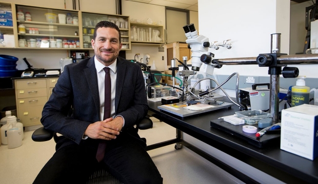
“We have learned that cancer cells become ‘addicted’ to protein synthesis to fuel their need for high-speed growth, but this dependence is also a liability: too much protein synthesis can become toxic,” said senior author Davide Ruggero, PhD, the Helen Diller Family Chair in Basic Cancer Research and a professor of urology and cellular and molecular pharmacology at UCSF. “We have discovered the molecular restraints that let cancer cells keep their addiction under control and showed that if we remove these restraints they quickly burn out under the pressure of their own greed for protein.”

“This is beautiful scientific work that could lead to urgently needed novel treatment strategies for men with very advanced prostate cancer,” added renowned UCSF Health prostate cancer surgeon Peter Carroll, MD, MPH, who is chair of the Department of Urology at UCSF and was a co-author on the new paper.

Prostate cancer is the second leading cause of cancer death for men in the United States: More than one man in ten will be diagnosed in his lifetime, and one in forty-one will die of the disease, according to data from the American Cancer Society. Tumors that recur or fail to respond to surgery or radiation therapy are typically treated with hormonal therapies that target the cancer’s dependence on testosterone. Unfortunately, most cancers eventually develop resistance to hormone therapy, and become even more aggressive, leading to what is known as “castration-resistant” disease, which is nearly always fatal.

As part of a “growth first” strategy, many cancers contain gene mutations that drive them to produce proteins at such a high rate that they risk triggering cells’ built-in self-destruct mechanisms, according to studies previously conducted by Ruggero and colleagues. But aggressive, treatment-resistant prostate cancers typically contain multiple such mutations, which led Ruggero and his team at the UCSF Helen Diller Family Comprehensive Cancer Center to wonder how such cancers sustain themselves under the pressure of so much protein production.

Deadliest Cancers Throttle Excess Protein Synthesis

To explore this question, Ruggero’s team genetically engineered mice to develop prostate tumors containing a pair of mutations seen in nearly 50 percent of patients with castration-resistant prostate cancer: one that causes overexpression of the cancer-driving MYC gene, and one that disables the tumor suppressor gene PTEN. They were surprised to discover that the highly aggressive cancers associated with these mutations actually had lower rates of protein synthesis compared to milder cancers with only a single mutation.

“I spent six months trying to understand if this was actually occurring, because it’s not at all what we expected,” said Crystal Conn, PhD, a postdoctoral researcher in the Ruggero lab and one of the paper’s two lead authors. But she saw the same effects again and again in experiments in mouse and human cancer cell lines as well as in 3-dimensional “organoid” models of the prostate that could be studied and manipulated in lab dishes.

Conn’s experiments eventually revealed that the combination of MYC and PTEN mutations trigger part of a cellular quality control system called the unfolded protein response (UPR), which reacts to cellular stress by reducing levels of protein synthesis throughout the cell. Specifically, these mutations alter the activity of a protein called eIF2a (eukaryotic translation initiation factor 2a key regulator of protein synthesis, by turning it into an alternate form, P-eIF2a, which tunes down cellular protein production.

To assess whether levels of P-eIF2a in patient tumors could be used to predict the development of aggressive, treatment-resistant disease, Conn collaborated with Carroll, who holds the Ken and Donna Derr-Chevron Distinguished Professorship in Urology, and Hao Nguyen, MD, PhD, an assistant professor of urology, to examine 422 tumors surgically extracted from UCSF prostate cancer patients. They used a technique called tissue microarray to measure the levels of PTEN, Myc, and P-eIF2a proteins in these tumors, then asked how these biomarkers predicted patient outcomes using 10 years of clinical follow-up data.

They found that P-eIF2a levels were a powerful predictor of worse outcomes in patients with PTEN-mutant tumors: Only 4 percent of such tumors with low P-eIF2a continued to spread following surgery, while 19 percent of patients with high P-eIF2a went on to develop metastases, and many eventually died. In fact, the presence of PTEN mutations and high P-eIF2a levels in prostate tumors outperformed a current standard test (CAPRA-S) used to assess risk of cancer progression following surgery.

ISRIB Selectively Kills Aggressive Prostate Cancers

Next, the researchers examined whether blocking P-eIF2a‘s suppression of protein synthesis might effectively kill aggressive prostate cancers, said Nguyen, who was co–lead author on the new paper: “Once we realized that these cancers are activating part of the UPR to put the brakes on their own protein synthesis, we began to ask what happens to the cancer if you remove the brakes.”

The researchers collaborated with UCSF biochemist Peter Walter, PhD, whose lab recently identified a molecule called ISRIB that reverses the effects of P-eIF2a activity. (Walter and UCSF neuroscientist Susanna Rosi, PhD, have shown that ISRIB can boost cognition and restore memory after severe brain damage in rodents — likely by restoring the production of proteins needed for learning in injured brain cells.)

Conn tested ISRIB on mice with prostate tumors and in human cancer cell lines and discovered that the drug exposed aggressive cancer cells carrying combined PTEN/MYC mutations to their full drive for protein synthesis, causing them to self-destruct. Intriguingly, she found that the drug had little effect on normal tissue or even on less-aggressive cancers lacking the MYC mutation. In mice, PTEN/MYC prostate tumors began to shrink within 3 weeks of ISRIB treatment, and had not regrown after 6 weeks of treatment, while in contrast, PTEN-only tumors had expanded by 40 percent.

To further investigate the potential use of ISRIB against aggressive human prostate cancer, Nguyen implanted samples of human prostate cancer into mice, a research technique called “patient-derived xenografts” (PDX) that has historically been unsuccessful in studies of prostate cancer.

In one experiment, the researchers transplanted different groups of mice with cells from two tumors extracted from the same prostate cancer patient: one set of cells from the patient’s primary prostate tumor and another from a nearby metastatic colony in the patient’s lymph node. They found that mice implanted with cells from the metastatic sample — which exhibited the expected “aggressive” proteomic profile of high MYC, low PTEN, and high P-eIF2a levels — experienced dramatic tumor shrinkage and extended survival when treated with ISRIB, while mice implanted with cells from the less-aggressive primary prostate tumor experienced only a temporary slowing of tumor growth.

The authors used a third PDX model of metastatic prostate cancer to assess whether blocking the UPR could effectively treat advanced castration-resistant disease: they showed that transplanted tumors, which typically spread and kill mice within 10 days, were significantly reduced and the animals’ lives significantly extended under ISRIB treatment.

“Together these experiments show that blocking P-eIF2α signaling with ISRIB both slows down tumor progression and also kills off the cells that have already progressed or metastasized to become more aggressive,” Conn said. “This is very exciting because finding new treatments for castration-resistant prostate cancer is a pressing and unmet clinical need.”

The researchers hope that this discovery will quickly lead to clinical trials for ISRIB or related drugs for patients with advanced, aggressive prostate cancer. “Most molecules that kill cancer also kill normal cells,” Ruggero said. “But with ISRIB we’ve discovered a beautiful therapeutic window: normal cells are unaffected because they aren’t using this aspect of the UPR to control their protein synthesis but aggressive cancer cells are toast without it.”

“The only side effect we’re aware of,” Conn added, “is that this drug might make you smarter.”

Additional authors on the paper include: Yae Kye, Lingru Xue, Craig M Forester, MD, PhD, Janet E Cowan, Andrew C Hsieh, MD, John T Cunningham, PhD, Charles Truillet, PhD, Michael J Evans, PhD, Byron Hann, MD, PhD, and Peter Walter, PhD, of UCSF; Feven Tameire and Constantinos Koumenis, PhD, of the University of Pennsylvania Perelman School of Medicine; and Christopher P Evans, MD, and Joy C Yang, PhD, of the UC Davis School of Medicine.

The research was supported by the US National Institutes of Health (R01-CA140456, R01-CA154916, and P01-CA165997), the US Department of Defense (W81XWH-15-1-0460), the AUA-SUO-Prostate Cancer Foundation (16YOUN14), the Urology Care Foundation (A130596), the American Cancer Society (PF-14-212-01-RMC), an American Association of Cancer Research (AACR)-Bayer Prostate Cancer Research Fellowship (17-40-44-CONN), the Campini Foundation, the Leukemia and Lymphoma Foundation, the American Cancer Society (130635-RSG-17-005-01-CCE), Calico Life Sciences LLC, the Weill Foundation, the Howard Hughes Medical Institute (HHMI), the UCSF Department of Pediatrics (5K12HDO72222-05), and the Goldberg-Benioff Program in Translational Cancer Biology.

https://www.ucsf.edu/news/2018/05/410336/research-finds-achilles-heel-aggressive-prostate-cancer

We Could Find Aliens by Spotting Their Satellites

By Nola Taylor Redd

Alien civilizations with technology on a par with humanity’s could be detectable using today’s instruments. A new study suggests that if geostationary satellites are thick enough around an alien world, they could be spotted with telescopes already hunting for undiscovered planets.

Both governments and private corporations on our own world use geostationary satellites — which orbit such that they hover over the same spot on Earth — for science, communications, espionage and military applications.

If advanced alien civilizations loft enough satellites into their own geostationary belts, these spacecraft could create a dense, ring-like structure visible from Earth, according to the study.

“It’s … a small chance, but the point is that it’s free,” study lead author Hector Socas-Navarro, of Astrophysics Institute of the Canary Islands, told Space.com by email.

Socas-Navarro simulated the presence of belts of geostationary satellites around exoplanets, to see whether they could be detected by instruments like NASA’s Kepler space telescope and the agency’s recently launched Transiting Exoplanet Survey Satellite (TESS). He found that the belt would need to be about 0.01 percent full for such spacecraft to detect it, whether populated by many small satellites or a handful of large, city-size objects.

“We just need to look for the right signature in the data,” he said.

Socas-Navarro calls this hypothetical structure the Clarke exobelt (CEB), after famed sci-fi author Arthur C. Clarke.

Hunting alien satellites
Both Kepler and TESS detect planets using what’s known as the transit method. The spacecraft watch a field of stars for an extended amount of time. If a planet has the right orbit, and the timing is right, that world will pass in front of its host star from the telescope’s perspective, causing a small, potentially detectable dip in brightness.

In addition to working as an astrophysicist, Socas-Navarro hosts a weekly radio show and podcast. That work helped him come up with the Clarke exobelt idea, he said. One day, a listener asked about a geostationary satellite for the sun.

“As I was doing the calculations to answer this question, I had this mental image of a satellite transiting across the solar disk,” Socas-Navarro said. “That led me to ask myself the question of whether satellites around distant exoplanets would be observable during transit.”

Sufficient material orbiting an exoplanet causes a small dip in starlight before and after the body of the world makes its transit. Scientists have used this method to discover rings around planets outside the solar system and even around distant solar system bodies.

Socas-Navarro said the putative alien-satellite signal would have a signature similar to that of rings — both an exobelt and rings are made up of a swarm of objects orbiting a planet — but there are subtle technical differences in how that signature would look. The signal would also reveal the altitude of the orbiting objects, which could provide a significant clue as to whether the objects were natural or alien-made.

A ring system can occur at any number of distances from the surface of the planet. But if the objects orbited at a planet’s geostationary height — about 22,200 miles (35,700 kilometers) — they are “almost certainly artificial,” Socas-Navarro said.

Similarly, a massive space city or a large station close to a space elevator could look like an exomoon. Again, Socas-Navarro said, altitude is key. If the object hovers at geostationary height, it’s likely to be artificial. [10 Exoplanets That Could Host Alien Life]

“It doesn’t seem to matter too much if they are many small or [a] few large [objects],” he said. “As long as they are spread out all over the orbit, they will basically produce the same signature.”

He also found that the ideal conditions to spot such a satellite belt would be around dim red dwarf stars located within 100 light-years of Earth.

The new study was published last month in The Astrophysical Journal. You can read it for free at the online preprint server arXiv.org.

https://www.space.com/40436-search-alien-life-et-satellites.html

Precise targeting technique could regulate gut bacteria in a way that curtails human disease.

Emerging evidence suggests that microbes in the digestive system have a big influence on human health and may play a role in the onset of disease throughout the body. Now, in a study appearing in ACS Chemical Biology, scientists report that they have potentially found a way to use chemical compounds to target and inhibit the growth of specific microbes in the gut associated with diseases without causing harm to other beneficial organisms.

The digestive system is crammed with trillions of bacteria, fungi, and other microbes that help process food. Recent studies suggest that the changes in these gut flora, or microbiome, may play a role in the onset of a host of diseases and conditions including obesity, diabetes, cancer, allergies, asthma, autism and multiple sclerosis. Antibiotics can help regulate the microbiome, but bacterial resistance is on the rise. In addition, antibiotics can wipe out some of the organisms that contribute to a healthy microbiome, and the microbes that take their place can sometimes cause more harm than good. Researchers have also investigated using probiotics and fecal transplants to resolve some of these problems. But to date, few have really looked at using non-microbicidal small molecules to alter the microbiome in a targeted way to improve health. To help fill this gap, Daniel Whitehead, Kristi Whitehead and colleagues sought to use a chemical compound to precisely target and disrupt the metabolic processes of members of the Bacteroides genus, a group of bacteria commonly found in the gut that appear to be associated with the onset of type I diabetes in genetically susceptible individuals.

In laboratory studies, the researchers found that small concentrations of acarbose, a drug used to treat diabetes, significantly disrupted the activity of a group of proteins involved in the Starch Utilization System (Sus). The model bacteria called Bacteroides thetaiotaomicron (Bt), as well as other Bacteroides members, have this system. With Sus inhibited, Bt couldn’t metabolize a pair of complex carbohydrates that are not digested by humans once they reach the colon, but that are vital to the survival of the microbes. As a result, the bacteria cannot grow. The team found that acarbose was specific, having similar effects on another Bacteroides bacteria, but little or no effect on other types of gut microbes. The researchers conclude that with further study it may be possible to develop drugs that target gut bacteria with pinpoint accuracy to permanently alter the composition of the microbiome and, in turn, prevent or treat disease.

Reference
Nonmicrobicidal Small Molecule Inhibition of Polysaccharide Metabolism in Human Gut Microbes: A Potential Therapeutic Avenue. Anthony D. Santilli, Elizabeth M. Dawson, Kristi J. Whitehead, and Daniel C. Whitehead. CS Chem. Biol., DOI: 10.1021/acschembio.8b00309.

https://www.acs.org/content/acs/en/pressroom/presspacs/2018/acs-presspac-may-2-2018/precise-targeting-technique-could-regulate-gut-bacteria-curtailing-disease.html

Stanford scientists find fear, courage switches in brain


Pinpoint stimulation of a cluster of nerve cells in the brains of mice encouraged timid responses to a perceived threat, whereas stimulation of an adjacent cluster induced boldness and courage.

Researchers at the Stanford University School of Medicine have identified two adjacent clusters of nerve cells in the brains of mice whose activity level upon sighting a visual threat spells the difference between a timid response and a bold or even fierce one.

Located smack-dab in the middle of the brain, these clusters, or nuclei, each send signals to a different area of the brain, igniting opposite behaviors in the face of a visual threat. By selectively altering the activation levels of the two nuclei, the investigators could dispose the mice to freeze or duck into a hiding space, or to aggressively stand their ground, when approached by a simulated predator.

People’s brains probably possess equivalent circuitry, said Andrew Huberman, PhD, associate professor of neurobiology and of ophthalmology. So, finding ways to noninvasively shift the balance between the signaling strengths of the two nuclei in advance of, or in the midst of, situations that people perceive as threatening may help people with excessive anxiety, phobias or post-traumatic stress disorder lead more normal lives.

“This opens the door to future work on how to shift us from paralysis and fear to being able to confront challenges in ways that make our lives better,” said Huberman, the senior author of a paper describing the experimental results. It was published online May 2 in Nature. Graduate student Lindsey Salay is the lead author.

Perilous life of a mouse
There are plenty of real threats in a mouse’s world, and the rodents have evolved to deal with those threats as best they can. For example, they’re innately afraid of aerial predators, such as a hawk or owl swooping down on them. When a mouse in an open field perceives a raptor overhead, it must make a split-second decision to either freeze, making it harder for the predator to detect; duck into a shelter, if one is available; or to run for its life.

To learn how brain activity changes in the face of such a visual threat, Salay simulated a looming predator’s approach using a scenario devised some years ago by neurobiologist Melis Yilmaz Balban, PhD, now a postdoctoral scholar in Huberman’s lab. It involves a chamber about the size of a 20-gallon fish tank, with a video screen covering most of its ceiling. This overhead screen can display an expanding black disc simulating a bird-of-prey’s aerial approach.

Looking for brain regions that were more active in mice exposed to this “looming predator” than in unexposed mice, Salay pinpointed a structure called the ventral midline thalamus, or vMT.

Salay mapped the inputs and outputs of the vMT and found that it receives sensory signals and inputs from regions of the brain that register internal brain states, such as arousal levels. But in contrast to the broad inputs the vMT receives, its output destination points were remarkably selective. The scientists traced these outputs to two main destinations: the basolateral amygdala and the medial prefrontal cortex. Previous work has tied the amygdala to the processing of threat detection and fear, and the medial prefrontal cortex is associated with high-level executive functions and anxiety.

Further inquiry revealed that the nerve tract leading to the basolateral amygdala emanates from a nerve-cell cluster in the vMT called the xiphoid nucleus. The tract that leads to the medial prefrontal cortex, the investigators learned, comes from a cluster called the nucleus reuniens, which snugly envelopes the xiphoid nucleus.

Next, the investigators selectively modified specific sets of nerve cells in mice’s brains so they could stimulate or inhibit signaling in these two nerve tracts. Exclusively stimulating xiphoid activity markedly increased mice’s propensity to freeze in place in the presence of a perceived aerial predator. Exclusively boosting activity in the tract running from the nucleus reuniens to the medial prefrontal cortex in mice exposed to the looming-predator stimulus radically increased a response seldom seen under similar conditions in the wild or in previous open-field experiments: The mice stood their ground, right out in the open, and rattled their tails, an action ordinarily associated with aggression in the species.

Thumping tails

This “courageous” behavior was unmistakable, and loud, Huberman said. “You could hear their tails thumping against the side of the chamber. It’s the mouse equivalent of slapping and beating your chest and saying, ‘OK, let’s fight!’” The mice in which the nucleus reuniens was stimulated also ran around more in the chamber’s open area, as opposed to simply running toward hiding places. But it wasn’t because nucleus reuniens stimulation put ants in their pants; in the absence of a simulated looming predator, the same mice just chilled out.

In another experiment, the researchers showed that stimulating mice’s nucleus reuniens for 30 seconds before displaying the “looming predator” induced the same increase in tail rattling and running around in the unprotected part of the chamber as did vMT stimulation executed concurrently with the display. This suggests, Huberman said, that stimulating nerve cells leading from the nucleus reunions to the prefrontal cortex induces a shift in the brain’s internal state, predisposing mice to act more boldly.

Another experiment pinpointed the likely nature of that internal-state shift: arousal of the autonomic nervous system, which kick-starts the fight, flight or freeze response. Stimulating either the vMT as a whole or just the nucleus reuniens increased the mice’s pupil diameter — a good proxy of autonomic arousal.

On repeated exposures to the looming-predator mockup, the mice became habituated. Their spontaneous vMT firing diminished, as did their behavioral responses. This correlates with lowered autonomic arousal levels.

Human brains harbor a structure equivalent to the vMT, Huberman said. He speculated that in people with phobias, constant anxiety or PTSD, malfunctioning circuitry or traumatic episodes may prevent vMT signaling from dropping off with repeated exposure to a stress-inducing situation. In other experiments, his group is now exploring the efficacy of techniques, such as deep breathing and relaxation of visual fixation, in adjusting the arousal states of people suffering from these problems. The thinking is that reducing vMT signaling in such individuals, or altering the balance of signaling strength from their human equivalents of the xiphoid nucleus and nucleus reuniens may increase their flexibility in coping with stress.

Reference:
Salay, L. D., Ishiko, N., & Huberman, A. D. (2018). A midline thalamic circuit determines reactions to visual threat. Nature. doi:10.1038/s41586-018-0078-2

http://med.stanford.edu/news/all-news/2018/05/scientists-find-fear-courage-switches-in-brain.html