Chinese scientists have put human brain genes in monkeys—and yes, they may be smarter

by Antonio Regalado

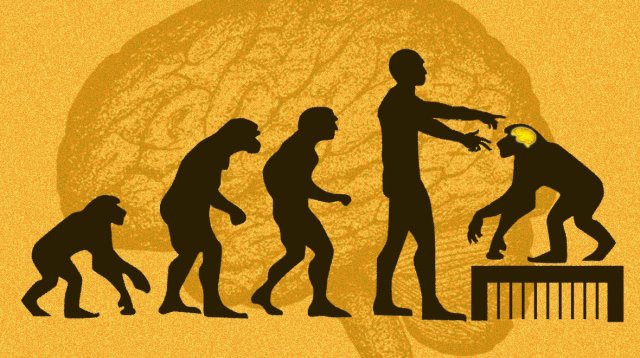
Human intelligence is one of evolution’s most consequential inventions. It is the result of a sprint that started millions of years ago, leading to ever bigger brains and new abilities. Eventually, humans stood upright, took up the plow, and created civilization, while our primate cousins stayed in the trees.

Now scientists in southern China report that they’ve tried to narrow the evolutionary gap, creating several transgenic macaque monkeys with extra copies of a human gene suspected of playing a role in shaping human intelligence.

“This was the first attempt to understand the evolution of human cognition using a transgenic monkey model,” says Bing Su, the geneticist at the Kunming Institute of Zoology who led the effort.

According to their findings, the modified monkeys did better on a memory test involving colors and block pictures, and their brains also took longer to develop—as those of human children do. There wasn’t a difference in brain size.

The experiments, described on March 27 in a Beijing journal, National Science Review, and first reported by Chinese media, remain far from pinpointing the secrets of the human mind or leading to an uprising of brainy primates.

Instead, several Western scientists, including one who collaborated on the effort, called the experiments reckless and said they questioned the ethics of genetically modifying primates, an area where China has seized a technological edge.

“The use of transgenic monkeys to study human genes linked to brain evolution is a very risky road to take,” says James Sikela, a geneticist who carries out comparative studies among primates at the University of Colorado. He is concerned that the experiment shows disregard for the animals and will soon lead to more extreme modifications. “It is a classic slippery slope issue and one that we can expect to recur as this type of research is pursued,” he says.

Research using primates is increasingly difficult in Europe and the US, but China has rushed to apply the latest high-tech DNA tools to the animals. The country was first to create monkeys altered with the gene-editing tool CRISPR, and this January a Chinese institute announced it had produced a half-dozen clones of a monkey with a severe mental disturbance.

“It is troubling that the field is steamrolling along in this manner,” says Sikela.

Evolution story

Su, a researcher at the Kunming Institute of Zoology, specializes in searching for signs of “Darwinian selection”—that is, genes that have been spreading because they’re successful. His quest has spanned such topics as Himalayan yaks’ adaptation to high altitude and the evolution of human skin color in response to cold winters.

The biggest riddle of all, though, is intelligence. What we know is that our humanlike ancestors’ brains rapidly grew in size and power. To find the genes that caused the change, scientists have sought out differences between humans and chimpanzees, whose genes are about 98% similar to ours. The objective, says, Sikela, was to locate “the jewels of our genome”—that is, the DNA that makes us uniquely human.

For instance, one popular candidate gene called FOXP2—the “language gene” in press reports—became famous for its potential link to human speech. (A British family whose members inherited an abnormal version had trouble speaking.) Scientists from Tokyo to Berlin were soon mutating the gene in mice and listening with ultrasonic microphones to see if their squeaks changed.

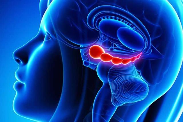
Su was fascinated by a different gene, MCPH1, or microcephalin. Not only did the gene’s sequence differ between humans and apes, but babies with damage to microcephalin are born with tiny heads, providing a link to brain size. With his students, Su once used calipers and head spanners to the measure the heads of 867 Chinese men and women to see if the results could be explained by differences in the gene.

By 2010, though, Su saw a chance to carry out a potentially more definitive experiment—adding the human microcephalin gene to a monkey. China by then had begun pairing its sizeable breeding facilities for monkeys (the country exports more than 30,000 a year) with the newest genetic tools, an effort that has turned it into a mecca for foreign scientists who need monkeys to experiment on.

To create the animals, Su and collaborators at the Yunnan Key Laboratory of Primate Biomedical Research exposed monkey embryos to a virus carrying the human version of microcephalin. They generated 11 monkeys, five of which survived to take part in a battery of brain measurements. Those monkeys each have between two and nine copies of the human gene in their bodies.

Su’s monkeys raise some unusual questions about animal rights. In 2010, Sikela and three colleagues wrote a paper called “The ethics of using transgenic non-human primates to study what makes us human,” in which they concluded that human brain genes should never be added to apes, such as chimpanzees, because they are too similar to us. “You just go to the Planet of the Apes immediately in the popular imagination,” says Jacqueline Glover, a University of Colorado bioethicist who was one of the authors. “To humanize them is to cause harm. Where would they live and what would they do? Do not create a being that can’t have a meaningful life in any context.”

In an e-mail, Su says he agrees that apes are so close to humans that their brains shouldn’t be changed. But monkeys and humans last shared an ancestor 25 million years ago. To Su, that alleviates the ethical concerns. “Although their genome is close to ours, there are also tens of millions of differences,” he says. He doesn’t think the monkeys will become anything more than monkeys. “Impossible by introducing only a few human genes,” he says.

Smart monkey?

Judging by their experiments, the Chinese team did expect that their transgenic monkeys could end up with increased intelligence and brain size. That is why they put the creatures inside MRI machines to measure their white matter and gave them computerized memory tests. According to their report, the transgenic monkeys didn’t have larger brains, but they did better on a short-term memory quiz, a finding the team considers remarkable.

Several scientists think the Chinese experiment didn’t yield much new information. One of them is Martin Styner, a University of North Carolina computer scientist and specialist in MRI who is listed among the coauthors of the Chinese report. Styner says his role was limited to training Chinese students to extract brain volume data from MRI images, and that he considered removing his name from the paper, which he says was not able to find a publisher in the West.

“There are a bunch of aspects of this study that you could not do in the US,” says Styner. “It raised issues about the type of research and whether the animals were properly cared for.”

After what he’s seen, Styner says he’s not looking forward to more evolution research on transgenic monkeys. “I don’t think that is a good direction,” he says. “Now we have created this animal which is different than it is supposed to be. When we do experiments, we have to have a good understanding of what we are trying to learn, to help society, and that is not the case here.” One issue is that genetically modified monkeys are expensive to create and care for. With just five modified monkeys, it’s hard to reach firm conclusions about whether they really differ from normal monkeys in terms of brain size or memory skills. “They are trying to understand brain development. And I don’t think they are getting there,” says Styner.

In an e-mail, Su agreed that the small number of animals was a limitation. He says he has a solution, though. He is making more of the monkeys and is also testing new brain evolution genes. One that he has his eye on is SRGAP2C, a DNA variant that arose about two million years ago, just when Australopithecus was ceding the African savannah to early humans. That gene has been dubbed the “humanity switch” and the “missing genetic link” for its likely role in the emergence of human intelligence.

Su says he’s been adding it to monkeys, but that it’s too soon to say what the results are.

https://www.technologyreview.com/s/613277/chinese-scientists-have-put-human-brain-genes-in-monkeysand-yes-they-may-be-smarter/

Fecal Transplant Therapy in Kids Has Reduced Their Autism Severity by 47%

by MIKE MCRAE

Transforming the microbial environment in the guts of children diagnosed with autism could significantly ease the severity of their condition’s signature traits, according to newly published research.

A study on the effects of a form of faecal transplant therapy in children on the autism spectrum found participants not only experienced fewer gut problems, but continued to show ongoing improvements in autism symptoms two years after the procedure.

Arizona State University researchers had already discovered a dose of healthy gut microflora caused characteristics associated with autism spectrum disorder (ASD) to ease or vanish for at least a couple of months after treatment ended.

But to be taken seriously as a potential therapy, there needed to be long term improvements. So a return to the original group of volunteers for another check-up was in order.

It turned out those new microbes were settling in nicely.

“In our original paper in 2017, we reported an increase in gut diversity together with beneficial bacteria after microbiota transfer therapy (MTT), and after two years, we observed diversity was even higher and the presence of beneficial microbes remained,” says biotechnologist Dae-Wook Kang.

The gut might seem like an odd place to start in developing therapies that assist individuals with a neurological condition such as autism.

But in addition to its defining characteristics of impaired social and communication skills, sensory challenges, and reduced core strength and motor control, for up to half of those with ASD the condition can come with a bunch of gut problems.

“Many kids with autism have gastrointestinal problems, and some studies, including ours, have found that those children also have worse autism-related symptoms,” says environmental engineer Rosa Krajmalnik-Brown.

Previous studies have repeatedly pointed to the potential benefits of swapping out a ‘bad’ microbial communities for a better one, either through using probiotics or courses of antibiotics.

Most showed promising short-term effects, suggesting there was more to be explored when it comes to gut-based therapies.

“In many cases, when you are able to treat those gastrointestinal problems, their behaviour improves,” says Krajmalnik-Brown.

In an attempt to elicit a more long-term result, the researchers pulled out the big guns. Forget dropping in a few microbial tourists or killing off a handful of trouble-makers – they went for a whole mass migration.

Using a customised process of gut microflora transplantation called microbiota transfer therapy, the researchers gave 18 kids aged between 7 and 16 a belly full of new microorganisms.

All of the volunteers had both an autism diagnosis and moderate to severe gastrointestinal problems. This group was compared with 20 equivalent control subjects who had neither gut problems nor an ASD diagnosis.

Both were treated for 10 weeks and then had follow-up test sessions for a further 8 weeks.

Admittedly, the experiment wasn’t blinded, so we do need to be cautious in how we read into the results. Placebo effects can’t be ruled out in cases like this.

But saying they were ‘promising’ isn’t too strong a claim to make. The children not only experienced an 80 percent reduction in gastrointestinal symptoms, they showed significant improvements when tested with common ASD diagnostic tools.

Two years later, those same tests indicate the conditions have only improved.

“The team’s new publication reports that the study demonstrated that two years after treatment stopped the participants still had an average of a 58 percent reduction in GI symptoms compared to baseline,” says Krajmalnik-Brown.

“In addition, the parents of most participants reported a slow but steady improvement in core ASD symptoms.”

An external evaluation using a standard ASD diagnostic tool concluded 83 percent of the initial test group could be considered as severe on the autistic spectrum. Two years later, this dropped to just 17 percent.

Amazingly, 44 percent no longer made the cut-off for being on the mild end of the spectrum at all.

Overall, the evaluator determined the severity of ASD traits was reduced by 47 percent compared with their baseline.

For a therapy that has barely any side-effects, and such remarkable improvements in challenges many with ASD struggle with, it’s surely a treatment that will continue to attract attention for further research.

Faecal transplants might sound a little gross, but you might as well get used to them. We’re bound to be seeing them used for a variety of things in the future, from treating superbugs to winning sports.

Now that we’re learning our neurological health is intimately connected with our digestive system, transplanting microbial communities from a healthy gut is seen as the next big thing in treating brain disorders.

This isn’t to say microflora cause autism. It’s a complex condition that has its roots in a diverse range of genes and environmental influences that nudge the brain’s development early in life.

But if we can swap out even a few of those influences, we just might be able to make life a little easier for those who need it.

This research was published in Scientific Reports.

https://www.sciencealert.com/autism-severity-cut-in-half-in-kids-who-underwent-radical-faecal-transplant-therapy

New type of ‘extinction neuron’ in the brain may fight off fearful memories

Summary: Newly identified ‘extinction neurons’ in the hippocampus suppress fearful memories when activated, and allow the memories to return when deactivated. The findings may provide new treatment avenues for PTSD, phobias, and anxiety.

Neuroscientists at The University of Texas at Austin have discovered a group of cells in the brain that are responsible when a frightening memory re-emerges unexpectedly, like Michael Myers in every “Halloween” movie. The finding could lead to new recommendations about when and how often certain therapies are deployed for the treatment of anxiety, phobias and post-traumatic stress disorder (PTSD).

In the new paper, out today in the journal Nature Neuroscience, researchers describe identifying “extinction neurons,” which suppress fearful memories when they are activated or allow fearful memories to return when they are not.

Since the time of Pavlov and his dogs, scientists have known that memories we thought we had put behind us can pop up at inconvenient times, triggering what is known as spontaneous recovery, a form of relapse. What they didn’t know was why it happened.

“There is frequently a relapse of the original fear, but we knew very little about the mechanisms,” said Michael Drew, associate professor of neuroscience and the senior author of the study. “These kinds of studies can help us understand the potential cause of disorders, like anxiety and PTSD, and they can also help us understand potential treatments.”

One of the surprises to Drew and his team was finding that brain cells that suppress fear memories hid in the hippocampus. Traditionally, scientists associate fear with another part of the brain, the amygdala. The hippocampus, responsible for many aspects of memory and spatial navigation, seems to play an important role in contextualizing fear, for example, by tying fearful memories to the place where they happened.

The discovery may help explain why one of the leading ways to treat fear-based disorders, exposure therapy, sometimes stops working. Exposure therapy promotes the formation of new memories of safety that can override an original fear memory. For example, if someone becomes afraid of spiders after being bitten by one, he might undertake exposure therapy by letting a harmless spider crawl on him. The safe memories are called “extinction memories.”

“Extinction does not erase the original fear memory but instead creates a new memory that inhibits or competes with the original fear,” Drew said. “Our paper demonstrates that the hippocampus generates memory traces of both fear and extinction, and competition between these hippocampal traces determines whether fear is expressed or suppressed.”

Given this, recommended practices around the frequency and timing of exposure therapy may need revisiting, and new pathways for drug development may be explored.

In experiments, Drew and his team placed mice in a distinctive box and induced fear with a harmless shock. After that, when one of the mice was in the box, it would display fear behavior until, with repeated exposure to the box without a shock, the extinction memories formed, and the mouse was not afraid.

Scientists were able to artificially activate the fear and suppress the extinction trace memories by using a tool called optogenetics to turn the extinction neurons on and off again.

“Artificially suppressing these so-called extinction neurons causes fear to relapse, whereas stimulating them prevents fear relapse,” Drew said. “These experiments reveal potential avenues for suppressing maladaptive fear and preventing relapse.”

The studies were led by graduate student Anthony Lacagnina of The University of Texas at Austin, with contributions from Emma Brockway, Chelsea Crovetti, Francis Shue, Meredith McCarty and Kevin Sattler of The University of Texas; and Sean Lim, Sofia Leal Santos and Christine Denny of Columbia University.

How the brain fights off fears that return to haunt us

Forever young: study uncovers protein that keeps skin youthful.

Beauty might only be skin deep, but for those wondering how to keep that skin young, scientists may have found an answer in the form of a protein that encourages cell competition.

The prosaically named COL17A1 might not sound like a fountain of youth, but the new study suggests it does the heavy lifting when it comes to keeping skin intact and unimpaired.

The protein works by encouraging cell competition, a key process to maintain tissue fitness. That effectively “drives out” weaker cells while encouraging replication of stronger ones.

“Damaged or stressed stem cells can be selectively eliminated by intact stem cells every day in our skin,” said Emi Nishimura, a professor at the Tokyo Medical and Dental University’s Stem Cell Biology department, who led the research.

But ageing results in a depletion of COL17A1, as do familiar enemies of youthful skin, like UV radiation and other stress factors.

And when that happens, weaker cells replicate, leaving the skin thinner, more prone to damage and slower to heal.

The research published Thursday in the journal Nature is based on investigations using mice tails, which share many of the same characteristics as human skin.

After confirming the importance of COL17A1, the team decided to investigate whether they could stimulate the protein once it was depleted—effectively looking for compounds that could kickstart the anti-ageing process in skin.

They isolated two chemical compounds—Y27632 and apocynin—and tested both on skin cells, with positive results.

“Application of these drugs to full-thickness skin wounds significantly promoted wound repair,” the study said.

The two compounds point to ways of “facilitating skin regeneration and reducing skin ageing,” the study added.

In a review of the study commissioned by Nature, two professors from the University of Colorado said cell competition had previously only been studied extensively in fruit flies.

The research “provides evidence that healthy cells in mammals can also efficiently repopulate adult tissues, replacing unfit or damaged cells,” wrote professors Ganna Bilousova and James DeGregori.

And they said the research offered “proof-of-principle” that the two chemical compounds could combat ageing.

“Future studies are needed to determine the mechanisms of cell competition in other tissues, and to identify compounds capable of reversing ageing in other organs,” they said.

Nishimura told AFP that the work could eventually lead to products like creams or tablets that could stop skin deterioration and promote repair.

“We are going to collaborate with pharmaceutical or cosmetic companies for the clinical use of the chemicals,” she said.

She said additional research would investigate whether the same process might also be at work in other parts of the body that have so-called epithelial cells like skin does.

“We are working on other epithelial organs as well to find out (whether) similar competition may underlie long-term tissue maintenance as well as organ ageing,” she said.

https://medicalxpress.com/news/2019-04-young-uncovers-protein-skin-youthful.html

A Dozen Dolphins Have Beached Themselves, Showing The Deadly Hallmark of Alzheimer’s disease

by Carly Cassella

Over a dozen dolphins, stranded on the beaches of Florida and Massachusetts, have been found with brains full of amyloid plaques, a hallmark of Alzheimer’s disease. The scientists who made the discovery think it may be a warning to us all: alongside the Alzheimer’s-like plaques, the team also found the environmental toxin BMAA.

Produced by blue-green algae blooms, this neurotoxin is easily caught up in the ocean food web, and chronic dietary exposure has long been suspected to be a cause of neurological disease, including Alzheimers, Parkinson’s and Amyotrophic Lateral Sclerosis (ALS).

The presence of both BMAA and amyloid plaques in 13 stranded dolphins now adds even more weight to this hypothesis.

“Dolphins are an excellent sentinel species for toxic exposures in the marine environment,” says neurologist Deborah Mash from the University of Miami.

“With increasing frequency and duration of cyanobacterial blooms in coastal waters, dolphins might provide early warning of toxic exposures that could impact human health.”

They might also be a good animal model for how BMAA could trigger Alzheimer’s disease. In 2017, it was discovered that dolphins are the only known wild animal to show signs of this common human disease.

Meanwhile, dolphins that inhabit Florida coastal waters are also commonly exposed to recurring harmful algae blooms (HABs). This might just be a coincidence, but experiments have shown that chronic BMAA dietary exposure can trigger neurodegenerative changes in both humans and non-human primates.

“Acute and chronic exposures to such toxins can be harmful to both humans and animals resulting in respiratory illnesses, severe dermatitis, mucosal damage, cancer, organ failure and death,” the authors write.

As the world warms at a rapid rate, these HABs are only becoming more frequent, and the authors worry that dolphins will accumulate even more BMAA as a result, “both by exposure to HABs and by the ingestion of prey previously exposed to the cyanotoxin”.

As such, these creatures may very well be our first indication of poor environmental conditions, and while it’s still not clear if these blooms directly lead to Alzheimer’s in dolphins or in humans, the researchers say it’s a risk we shouldn’t be willing to take.

“The $64,000 question is whether these marine mammals experienced cognitive deficits and disorientation that led to their beaching,” says co-author Paul Alan Cox, an ethnobotanist at the Brain Chemistry Labs in Jackson Hole.

“Until further research clarifies this question, people should take simple steps to avoid cyanobacterial exposure.”

This study has been published in PLOS ONE.

https://www.sciencealert.com/beached-dolphins-had-alzheimer-s-like-plaques-and-it-s-a-warning-to-us-all

Small stickers on the ground trick Tesla autopilot into steering into opposing traffic lane

Researchers from Tencent Keen Security Lab have published a report detailing their successful attacks on Tesla firmware, including remote control over the steering, and an adversarial example attack on the autopilot that confuses the car into driving into the oncoming traffic lane.

The researchers used an attack chain that they disclosed to Tesla, and which Tesla now claims has been eliminated with recent patches.

To effect the remote steering attack, the researchers had to bypass several redundant layers of protection, but having done this, they were able to write an app that would let them connect a video-game controller to a mobile device and then steer a target vehicle, overriding the actual steering wheel in the car as well as the autopilot systems. This attack has some limitations: while a car in Park or traveling at high speed on Cruise Control can be taken over completely, a car that has recently shifted from R to D can only be remote controlled at speeds up to 8km/h.

Tesla vehicles use a variety of neural networks for autopilot and other functions (such as detecting rain on the windscreen and switching on the wipers); the researchers were able to use adversarial examples (small, mostly human-imperceptible changes that cause machine learning systems to make gross, out-of-proportion errors) to attack these.

Most dramatically, the researchers attacked the autopilot’s lane-detection systems. By adding noise to lane-markings, they were able to fool the autopilot into losing the lanes altogether, however, the patches they had to apply to the lane-markings would not be hard for humans to spot.

Much more seriously, they were able to use “small stickers” on the ground to effect a “fake lane attack” that fooled the autopilot into steering into the opposite lanes where oncoming traffic would be moving. This worked even when the targeted vehicle was operating in daylight without snow, dust or other interference.

Misleading the autopilot vehicle to the wrong direction with some patches made by a malicious attacker, in sometimes, is more dangerous than making it fail to recognize the lane. We paint three inconspicuous tiny square in the picture took from camera, and the vision module would recognize it as a lane with a high degree of confidence as below shows…

After that we tried to build such a scene in physical: we pasted some small stickers as interference patches on the ground in an intersection. We hope to use these patches toguide the Tesla vehicle in the Autosteer mode driving to the reverse lane. The test scenario like Fig 34 shows, red dashes are the stickers, the vehicle would regard them as the continuation of its right lane, and ignore the real left lane opposite the intersection. When it travels to the middle of the intersection, it would take the real left lane as its right lane and drive into the reverse lane.

Tesla autopilot module’s lane recognition function has a good robustness in an ordinary external environment (no strong light, rain, snow, sand and dust interference), but it still doesn’t handle the situation correctly in our test scenario. This kind of attack is simple to deploy, and the materials are easy to obtain. As we talked in the previous introduction of Tesla’s lane recognition function, Tesla uses a pure computer vision solution for lane recognition, and we found in this attack experiment that the vehicle driving decision is only based on computer vision lane recognition results. Our experiments proved that this architecture has security risks and reverse lane recognition is one of the necessary functions for autonomous driving in non-closed roads. In the scene we build, if the vehicle knows that the fake lane is pointing to the reverse lane, it should ignore this fake lane and then it could avoid a traffic accident.

Security Research of Tesla Autopilot

Small stickers on the ground trick Tesla autopilot into steering into opposing traffic lane

Dead people and pets are being forged into sparkling blue diamonds — here’s how the process works

by Dave Mosher

When a person dies, cremation is an increasingly popular option. The practice eclipsed burials in the US in 2015 and is expected to make up more than half of all body disposals by 2020, according to the Cremation Association of North America.

But instead of storing a loved one’s cremains in an urn or sprinkling them outside, a growing number of bereaved consumers are doing something more adventurous: forging the ashes into diamonds.

This is possible because carbon is the second-most abundant atomic element in the human body, and diamonds are made of crystallised carbon. Researchers have also improved ways to grow diamonds in the lab in recent years.

While at least five companies offer a “memorial diamond” service, Algordanza in Switzerland is one of the industry leaders — its services are available in 33 countries, and the company told Business Insider it sold nearly 1,000 corporeal gems in 2016. Algordanza also claims to be the only company of its kind that operates its own diamond-growing lab for cremains — one of two in the world. (The other is in Russia.)

“It allows someone to keep their loved one with them forever,” Christina Martoia, a spokesperson for Algordanza US, told Business Insider. “We’re bringing joy out of something that is, for a lot of people, a lot of pain.”

Here’s how the company uses extreme heat and pressure to turn dead people — and sometimes animals — into sparkling gems of all sizes, cuts, and colours.

Read more at https://www.businessinsider.com.au/turn-human-ashes-diamonds-carbon-algordanza-2017-7#14TJLUlcEiVFwIPR.99

FDA Clears Unique Medical Device for Treatment of Anxiety, Depression, and Insomnia

The Food and Drug Administration (FDA) has approved a new cranial electrotherapy stimulator (CES) device for the treatment of anxiety, depression, and insomnia.

The Cervella Cranial Electrotherapy Stimulator by Innovative Neurological Devices is operated using noise-cancelling, Bluetooth-enabled headphones and an app. The device delivers a low-level, constant current to the patient’s cranium via a pair of conductive electrodes incorporated into ear pads of the headphones.

Patients will need a prescription from a licensed healthcare provider in order to purchase the device, which will cost $695, and is due to launch at the end of March (2019).

“We hope that by incorporating treatment electrodes into a noise-cancelling headset, patient compliance will significantly increase and, consequently, treatment outcomes will improve,” said Bart Waclawik, President and CEO of Innovative Neurological Devices. “Also, by making the Cervella device appear indistinguishable from ordinary over-ear headphones, patients will have the freedom to use the device in anxiety-inducing situations without curious looks from onlookers.”

Related Articles
Stimulation Device for MDD Cleared for Much Shorter Treatment Session
OTC Wearable Device for Chronic Pain Cleared by FDA
Investigational Agent for Major Depressive Disorder Gets FDA’s Breakthrough Designation
Waclawik added that patients will be able to share treatment data with providers through the app, which provides automated data recordings and treatment reminders.

For more information visit Cervella.us.

New study shows measures people take to protect their good mood

What does it take to stay in a good mood?

In short: Once happy, steer clear of choices that could invite in negative feelings.

According to new research from Case Western Reserve University, people become protective of their good moods—and avoid options and behaviors that could potentially sully their positive feelings.

“Our study suggests people who are feeling positively are less likely to jeopardize their emotional state, and that affects their choices in life,” said Heath Demaree, professor and chair of the Department of Psychological Sciences in the College of Arts and Sciences at Case Western Reserve.

“They wonder: How much better does it get? If you’re already happy, why risk making your mood worse?” said Demaree, co-author of the study, published in the journal Frontiers of Psychology. “Essentially, it’s quitting while you’re ahead.”

The findings differ from some previous research, which has shown that a person’s risk-taking increases after they’ve become successful—known as the “house money” theory. Still, other studies suggest that people risk more when they’re losing, hoping to break even.

The study

To tap into the emotion of decision-making, Demaree designed an experiment using a modified slot machine that produced winnings half of the time. Each participant was given $50 to begin, and was allowed to take home a portion of their winnings at the end of the experiment. Players were asked about their emotions after each pull of the slot machine in order to decide how much of their decision-making was due to their finances and how much was due to their emotional state.

After winning a trial, and controlling for other variables (e.g., the amount of money just won), researchers saw that people risked the least when they were in the most positive emotional state.

Making sense of emotion and choice

While risk-taking behavior has been studied for more than a century, significant discrepancies remain between actual real-world decisions involving risk and those predicted by research.

“We see that choices are increasingly explained with a better appreciation of the role of emotion,” said Demaree. “While these results differ from some previous studies, they do further confirm that life outcomes affect mood and then mood influences subsequent behavior.”

The results can inform further research into how people can control their emotions (anger, happiness and fear, for example) that influence their decisions, as well as their bodies’ natural responses to stress, such as elevated heart rate, blood pressure, perspiration and the release of cortisol.

“Increasing positive feelings may help reduce risky behavior,” said Demaree. “These results suggest that people could consider the positive and stable aspects of their lives—thinking of their family, job, spouse—to see their risk-taking behaviors in a more positive context.”

Co-authors of the study were former PhD students of Demaree’s: James Juergensen, Joe Weaver and Christine Moran.

Study: To protect a good mood, people play it safe

New neurons for life? Old people can still make fresh brain cells, study finds

By Emily Underwood

One of the thorniest debates in neuroscience is whether people can make new neurons after their brains stop developing in adolescence—a process known as neurogenesis. Now, a new study finds that even people long past middle age can make fresh brain cells, and that past studies that failed to spot these newcomers may have used flawed methods.

The work “provides clear, definitive evidence that neurogenesis persists throughout life,” says Paul Frankland, a neuroscientist at the Hospital for Sick Children in Toronto, Canada. “For me, this puts the issue to bed.”

Researchers have long hoped that neurogenesis could help treat brain disorders like depression and Alzheimer’s disease. But last year, a study in Nature reported that the process peters out by adolescence, contradicting previous work that had found newborn neurons in older people using a variety of methods. The finding was deflating for neuroscientists like Frankland, who studies adult neurogenesis in the rodent hippocampus, a brain region involved in learning and memory. It “raised questions about the relevance of our work,” he says.

But there may have been problems with some of this earlier research. Last year’s Nature study, for example, looked for new neurons in 59 samples of human brain tissue, some of which came from brain banks where samples are often immersed in the fixative paraformaldehyde for months or even years. Over time, paraformaldehyde forms bonds between the components that make up neurons, turning the cells into a gel, says neuroscientist María Llorens-Martín of the Severo Ochoa Molecular Biology Center in Madrid. This makes it difficult for fluorescent antibodies to bind to the doublecortin (DCX) protein, which many scientists consider the “gold standard” marker of immature neurons, she says.

The number of cells that test positive for DCX in brain tissue declines sharply after just 48 hours in a paraformaldehyde bath, Llorens-Martín and her colleagues report today in Nature Medicine. After 6 months, detecting new neurons “is almost impossible,” she says.

When the researchers used a shorter fixation time—24 hours—to preserve donated brain tissue from 13 deceased adults, ranging in age from 43 to 87, they found tens of thousands of DCX-positive cells in the dentate gyrus, a curled sliver of tissue within the hippocampus that encodes memories of events. Under a microscope, the neurons had hallmarks of youth, Llorens-Martín says: smooth and plump, with simple, undeveloped branches.

In the sample from the youngest donor, who died at 43, the team found roughly 42,000 immature neurons per square millimeter of brain tissue. From the youngest to oldest donors, the number of apparent new neurons decreased by 30%—a trend that fits with previous studies in humans showing that adult neurogenesis declines with age. The team also showed that people with Alzheimer’s disease had 30% fewer immature neurons than healthy donors of the same age, and the more advanced the dementia, the fewer such cells.

Some scientists remain skeptical, including the authors of last year’s Nature paper. “While this study contains valuable data, we did not find the evidence for ongoing production of new neurons in the adult human hippocampus convincing,” says Shawn Sorrells, a neuroscientist at the University of Pittsburgh in Pennsylvania who co-authored the 2018 paper. One critique hinges on the DCX stain, which Sorrells says isn’t an adequate measure of young neurons because the DCX protein is also expressed in mature cells. That suggests the “new” neurons the team found were actually present since childhood, he says. The new study also found no evidence of pools of stem cells that could supply fresh neurons, he notes. What’s more, Sorrells says two of the brain samples he and his colleagues looked at were only fixed for 5 hours, yet they still couldn’t find evidence of young neurons in the hippocampus.

Llorens-Martín says her team used multiple other proteins associated with neuronal development to confirm that the DCX-positive cells were actually young, and were “very strict,” in their criteria for identifying young neurons.

Heather Cameron, a neuroscientist at the National Institute of Mental Health in Bethesda, Maryland, remains persuaded by the new work. Based on the “beauty of the data” in the new study, “I think we can all move forward pretty confidently in the knowledge that what we see in animals will be applicable in humans, she says. “Will this settle the debate? I’m not sure. Should it? Yes.”

https://www.sciencemag.org/news/2019/03/new-neurons-life-old-people-can-still-make-fresh-brain-cells-study-finds?utm_campaign=news_daily_2019-03-25&et_rid=17036503&et_cid=2734364