Forest Fungi Ride Out Wildfires by Hiding Inside Plants

by Annie Greene

After the Chimney Tops 2 Wildfire charred 11,000 acres of the Great Smoky Mountains National Park along the North Carolina–Tennessee state line in November 2016, rangers closed affected trails to visitors. Mycologists Andy N. Miller and Karen Hughes and their teams were an exception. Toting hard hats and sample collection kits, these scientists jumped at the opportunity to track down their research subjects: pyrophilous (“fire-loving”) fungi, which produce mushrooms prolifically after forest fires and then disappear as the forest recovers.

The severely burned areas of the Smokies were almost completely lifeless two months after the blaze, when the group first ventured into the affected zone. “The level of destruction was incredible,” recounts Hughes, a researcher at the University of Tennessee, Knoxville, in an email to The Scientist. “Everything I touched left black carbon on my hands. It was incredibly quiet.” Miller, who is based at the University of Illinois Urbana-Champaign, also noted a surreal lack of activity. “There’s nothing running around, no birds singing,” he says. To him, the site smelled “like a house had burned up.” When the researchers returned to their collection sites a few months later, however, their mushrooms of interest had risen from the ashes. Miller noticed that when these fungi surface, they do so en masse: “They’re less than a millimeter in diameter, but there’s a lot of them, and once you train your eyes, they’re just all over the place.”

The researchers were interested in documenting which species of pyrophilous fungi are present in the Smokies. They also wanted to test a theory about where the fungi go during the long periods between forest fires. Some fire-loving fungi are known to lie dormant in the dirt as spores or other heat-tolerant structures until post-fire soil conditions trigger growth and reproduction. Other species exist between burns in a vegetative state, aiding decomposition of dead organisms or interacting with tree roots. But many fire-loving fungi don’t fit into any of these categories. A new proposal, known as the body snatchers hypothesis, posits that some pyrophilous fungi hide out inside plants or lichens in between fires, nestling among host cells in a so-called endophytic or endolichenic state.

To test the hypothesis, the researchers traveled to the burn site every few months for more than a year to sample the soil, as well as the mosses and lichens that sprang up while the forest recovered. They also gathered specimens from unburned areas of the park for comparison. In May 2018, members of Miller’s lab began analyzing the samples they’d collected. DNA sequencing results identified a total of 22 pyrophilous fungal species in the Smokies. Of these, three species were present only in the soil, while the remaining 19 were found inside plant samples from burned and unburned areas, either exclusively or in addition to being found in the soil. In line with the body snatchers hypothesis, “almost all of our pyrophilous fungi were found as endophytes,” Miller says.

Mosses and lichens often live in difficult-to-reach places such as rock crevices and may be hardy enough to withstand minor flames, so fungi living inside these hosts could theoretically survive a low-intensity wildfire. But it’s still unclear how all of these organisms might persist through a moderate or severe burn, and how a fire-loving fungus would escape its host to recolonize a charred forest. Hughes has a hypothesis based on her observations at the burn site: “After the fire, I saw numerous tiny lichen fragments on the burned soil, as if they had been lofted into the air while trees were burning and settled on the ground after the fire,” she says. These burned plant fragments may inoculate the soil with the fungi they harbor, giving the fire-loving fungi a way into the dirt.

This is a feasible way for both a pyrophilous fungus and its host to rebound after a fire and maintain their relationship, according to Keith Clay, who studies plant-fungus interactions at Tulane University and was not involved with this study. “If [a moss fragment] lands in a good place, it can regenerate the whole plant,” says Clay. “If the endophyte is in that fragment, presumably it can just colonize these newly grown plants as well from the get-go.” Post-fire fungi may also acquire new hosts after a burn, Miller notes. One mushroom can produce millions of airborne spores that likely land on nearby mosses and lichens, germinate, and invade the tissues of these new hosts, he says.

To check whether their findings might apply to other forests, Miller and Hughes analyzed moss and lichen samples from other sites around the US. A handful of the fire-loving fungi identified in the Smokies were also present as endophytes in Indiana and Alaska. That result was surprising because “there was really no evidence that a fire had occurred in the last few years in those areas,” Miller says. “What are they doing there if they’re not waiting for a fire to come along?”

One possibility is that, while body snatching between fires, pyrophilous fungi use their plant hosts as nutrient sources, says Clay. He notes that many plants and fungi have mutualistic endophytic relationships, where the plant typically provides the fungus with “a home where they can live and sugars, carbon, from photosynthesis.” In return, the fungus often produces alkaloids that benefit the host. Yet for the pyrophilous fungi examined in this study, Clay says, “what they offer the plant is not clear.” Sydney Glassman, a microbial ecologist at the University of California, Riverside, who was not involved with the study, notes that in vitro assays using carbon isotopes could help uncover these trade-offs by revealing “nutrient transfer between the plants and the fungus.”

Miller and his team plan to examine the details of fungus-host interactions by recreating body snatching in the lab and conducting long-term field studies, he notes. After all, many forests where pyrophilous fungi live go for decades without fire, he says. “So how is that relationship maintained?”

https://www.the-scientist.com/notebook/forest-fungi-ride-out-wildfires-by-hiding-inside-plants-67326?utm_campaign=TS_DAILY%20NEWSLETTER_2020&utm_source=hs_email&utm_medium=email&utm_content=86856096&_hsenc=p2ANqtz-8BKRYRGs_fo90ZncO_fmihHmxcb7igfgKB79gkfdKckRdyLVHnViIWWELwSyNw7QIkAcI47O7ksk1iFQ0kJDaX39xITA&_hsmi=86856096

Paul Matewele, who died from COVID-19, was known for discovering dangerous microbes on surfaces people touch every day.

by Emma Yasinski

Paul Matewele, a microbiologist who identified pathogenic bacteria on surfaces that humans contact everyday, died as a result of COVID-19 on April 7 at the age of 62.

Matewele was a senior lecturer at London Metropolitan University for 30 years and is best known for his work characterizing potentially pathogenic microbes that people are likely to come in contact with in their homes and public places, according to Úna Fairbrother, an interim head of the School of Human Sciences at London Metropolitan University.

Matewele was born in Zimbabwe in 1958 and earned a master’s degree in biochemistry from St. Andrews University and a PhD in microbiology from Southampton University.

Partially inspired by the growing crisis of antibiotic resistance, Matewele conducted studies identifying sometimes-deadly microbes living on handbags, McDonald’s touch screens, reusable water bottles, makeup, vehicle air conditioners, drinks served in cinemas, London transport systems, and coins. His work on the London transportation system led to a deep cleaning of 50 stations in the London Underground in June 2017.

But among colleagues, he was best known for the time he spent lecturing and tutoring thousands of students. “Paul was a warm, kind, intelligent and conscientious man. He was dedicated to his students and a brilliant colleague to have,” Fairbrother tells The Scientist in an email. “He was a genuinely happy, open person and will be much missed from our team on a personal and professional level.”

Several of Matewele’s students and colleagues shared thoughts and memories in a tribute on the university’s webpage describing him as a “dedicated teacher,” “a kind soul,” and someone who “never stopped smiling.”

Sean Frost, a former colleague of Matawele who is currently a lecturer at the University of Hull, writes on the university page that Matewele “took on the biggest challenges and was never afraid to fight for what he believed in, McDonalds being particularly memorable. Even up until March he was broadcasting warnings about risk of infection from cash, Paul always took the side of the little guy, be it colleagues, students or society. He was a fine example of what an academic should aspire to become.”

Matawele is survived by his 18-year-old son, William.

https://www.the-scientist.com/news-opinion/microbiologist-who-studied-deadly-bacteria-in-public-places-dies-67452?utm_campaign=TS_DAILY%20NEWSLETTER_2020&utm_source=hs_email&utm_medium=email&utm_content=86856096&_hsenc=p2ANqtz-8BKRYRGs_fo90ZncO_fmihHmxcb7igfgKB79gkfdKckRdyLVHnViIWWELwSyNw7QIkAcI47O7ksk1iFQ0kJDaX39xITA&_hsmi=86856096

Long Work Hours Linked to Greater Risk for Hypothyroidism

by Rose Reeb

Increased weekly working hours are associated with a higher risk for hypothyroidism in workers with no evidence of thyroid autoimmunity, according to study results published in Thyroid.

To determine if long work hours are associated with thyroid function, researchers conducted a cross-sectional study using data from 2160 South Korean adults (69.9% men). Individuals who had provided blood and urine samples, were working ≥36 hours per week and <12 hours per day, were not pregnant, did not have a history of thyroid disease, did not have a positive thyroid peroxidase antibody test, did not have isolated hypothyroidism or hyperthyroidism, and were not missing relevant data were included.

Hypothyroidism was defined as a serum thyrotropin level above the upper reference limit with a normal or low free thyroxine level. Hyperthyroidism was defined as a serum thyrotropin level below the lower reference limit with a normal or high free thyroxine level.

The median age of the included individuals was 42.4 years (interquartile range, 33.0- 52.1). The median weekly number of work hours was 47.1 (interquartile range, 39.8-54.9) and 15.4% of included individuals were shift workers. Overall, hypothyroidism was observed in 2.1% and hyperthyroidism was observed in 2.9% of the cohort.

Rates of both hypothyroidism (3.6%) and hyperthyroidism (3.5%) were highest in the group with the highest work hours (P =.03 for trend). The mean number of work hours per week in euthyroid, hypothyroid, and hyperthyroid individuals were 49.5±0.3, 53.4±1.5, and 51.6±1.6 hours, respectively (P =.012). There was a significant association between hypothyroidism and longer work hours per week, even after adjusting for all biological and lifestyle covariates (adjusted odds ratio [aOR], 1.46; 95% CI, 1.12-1.90) and excluding outliers in number of work hours per week (aOR, 1.99; 95% CI, 1.19-3.31) and individuals with overt thyroid dysfunction (aOR, 1.44; 95% CI, 1.11-1.88). Hyperthyroidism was not significantly associated with the number of hours worked per week.

Moreover, hypothyroidism was approximately 2.6 times more prevalent in individuals who worked 53 to 83 hours per week compared with those who worked 36 to 42 hours per week.

As the study was cross-sectional and observational in nature, causation could not be determined. The researchers suggested that “[f]urther research is needed to clarify the causal relationship and the underlying mechanism” of hypothyroidism in connection to longer working hours.

Reference

Lee YK, Lee D-E, Hwangbo Y, Lee YJ, Kim HC, Lee EK. Long work hours are associated with hypothyroidism: a cross-sectional study with population-representative data [published online March 31, 2020]. Thyroid. doi:10.1089/thy.2019.0709

68 year old Nigerian woman gives birth to twins

By Bukola Adebayo, CNN

A Nigerian woman has given birth to twins, a boy, and a girl at the age of 68.

Margaret Adenuga went through three previous IVF procedures before finally having twins.

Her husband Noah Adenuga, 77 told CNN the couple, who married in 1974 had long desired to have a child of their own.

Adenuga said they never gave up even after the failed attempts.
The retired stock auditor told CNN, “I am a dreamer, and I was convinced this particular dream of ours will come to pass.”

The babies were delivered via caesarian section at 37 weeks last Tuesday at the Lagos University Teaching Hospital (LUTH) but the hospital only recently made the news public to give the first-time mother time to recuperate, it said.

Dr. Adeyemi Okunowo, who delivered the babies, told CNN a specialist team was assembled at the hospital to monitor the pregnancy because of her age.
“As an elderly woman and a first-time mother, it was a high-risk pregnancy and also because she was going to have twins but we were able to manage her pregnancy to term,” Okunowo told CNN.

https://www.cnn.com/2020/04/22/africa/nigerian-woman-68-gives-birth-intl/index.html

Regular Exercise Helps Patients Combat Cancer


OUTRUNNING CANCER: Tumors on the lungs of sedentary mice (left) and animals that ran on wheels (right) after injection with melanoma cells.
L. PEDERSEN ET AL., CELL METAB, 2016

Bente Klarlund Pedersen

Mathilde was diagnosed with breast cancer at the age of 44. Doctors treated her with surgery, chemotherapy, and radiation, and Mathilde’s physician informed her that, among many other side effects of her cancer treatment, she could expect to lose muscle mass. To fight muscle wasting, Mathilde began the intensive physical training program offered to cancer patients at the Rigshospitalet University Hospital of Copenhagen. The program consists of 3.5-hour sessions of combined resistance and aerobic training, four times a week for six weeks. Although the chemotherapy made her tired, Mathilde (a friend of mine, not pictured, who requested I use her first name only) did not miss a single training session.

“In a way it felt counterintuitive to do intensive, hard training, while I was tired and nauseous, but I was convinced that the training was good for my physical and mental health and general wellbeing,” Mathilde told me in Danish. She followed the chemo- and radiotherapy strictly according to the prescribed schedule. She was not hospitalized, acquired no infections, and did not develop lymphedema, a failure of the lymphatic system that commonly occurs following breast cancer surgery and leads to swelling of the limbs.

Physical exercise is increasingly being integrated into the care of cancer patients such as Mathilde, and for good reason. Evidence is accumulating that exercise improves the wellbeing of these patients by combating the physical and mental deterioration that often occur during anticancer treatments. Most remarkably, we are beginning to understand that exercise can directly or indirectly fight the cancer itself.

An increasing amount of epidemiological literature strongly indicates that exercise training may lower the risk of cancer, control disease progression, amplify the effects of anticancer therapy, and improve physical function and psychosocial outcomes. For example, a 2016 study of more than 1.4 million individuals in the US and Europe found that people could reduce their cancer risk with moderate to vigorous leisure-time exercise training. The phenomenon held across several different cancers, including breast, colon, rectum, esophagus, lung, liver, kidney, bladder, and head and neck. And the combined results of approximately 700 unique exercise intervention trials, involving more than 50,000 cancer patients in total, leave little doubt that patients benefit from physical activity, showing improvements such as reduced toxicity of anticancer treatment, decreased disease progression, and enhanced survival. The same studies showed that exercise training improves mood, decreases loss of muscle mass, and helps cancer patients return to work earlier after successful treatment. Some studies show that 150 minutes per week of moderate exercise nearly double the chance of survival compared with breast cancer patients who don’t exercise during treatment.

Hundreds of animal studies, conducted over decades, suggest that the link is likely causal: in mice and rats, exercise leads to a reduction in the incidence, growth rate, and metastatic potential of cancer across a large variety of models of different human and murine tumor types. But how exercise helps fight cancer is a bit of a black box. Exercise may improve the efficacy of anticancer treatment by boosting the immune system and thereby attenuating the toxicity of chemotherapy and immunotherapy. Cancer patients are also likely to benefit from the overall health-promoting properties of physical activity, such as improved metabolism and enhanced cardiovascular function.

Uncovering the mechanisms whereby exercise induces anticancer effects is crucial to fighting the disease. Exercise-related factors that have a direct or indirect anticancer effect could serve as valuable biomarkers for monitoring the amount, intensity, and type of exercise required to best aid cancer treatment. Such research could also potentially highlight novel therapeutic targets.

Each workout matters

Regardless of the nature of the training, the primary setting of exercise’s effect on cancer is the bloodstream. Long-term training has been associated with a reduction in the blood levels of systemic risk factors, such as sex hormones, insulin, and inflammatory molecules. However, this effect is only seen if exercise training is accompanied by weight loss, and researchers have not yet established causal direct links between regular exercise training and the reductions in the basal levels of these risk factors. Alternatively, the anticancer effect of exercise could also be the result of something that occurs within individual sessions of exercise, during which muscles are known to release spikes of various hormones and other factors into the blood.

To learn more about the effects of individual bouts of exercise versus long-term training regimens, Christine Dethlefsen, a graduate student in my laboratory, incubated breast cancer cells with serum obtained from cancer survivors at rest before and after a six-month training intervention that began after patients completed primary surgery, chemotherapy, and radiotherapy. For comparison, she incubated other cells with serum obtained from blood drawn from these patients immediately after a two-hour acute exercise session during their weeks-long course of chemotherapy. Her study revealed that serum obtained following an exercise session reduced the viability of the cultured breast cancer cells, while serum drawn at rest following six months of training had no effect.

These data suggest that cancer-fighting effects are driven by repeated acute exercise, and each bout matters. In Dethlefsen’s study, incubation with serum obtained after a single bout of exercise (consisting of 30 minutes of warm-up, 60 minutes of resistance training, and a 30-minute high-intensity interval spinning session) reduced breast cancer cell viability by only 10 to 15 percent compared with control cells incubated with serum obtained at rest. But a reduction in tumor cell viability by 10 to 15 percent several times a week may add up to clinically significant inhibition of tumor growth. Indeed, in a separate study, my colleagues and I found that daily, voluntary wheel running in mice inhibits tumor progression across a range of tumor models and anatomical locations, typically by more than 50 percent.

Exercise’s molecular messengers

One prime candidate for helping to explain the link between exercise and anticancer effects is a group of peptides known as myokines, which are produced and released by muscle cells. Several myokines are released only during exercise, and some researchers have proposed that these exercise-dependent myokines contribute to the myriad beneficial effects of physical activity for all individuals, not just cancer patients, perhaps by mediating crosstalk between the muscles and other parts of the body, including the liver, bones, fat, and brain.

Exercise’s Anticancer Mechanisms

Researchers are beginning to understand that not only can exercise improve cancer patients’ overall wellbeing during treatment, but it may also fight the cancer itself. Experiments on cultured cells and in mice hint at some of the mechanisms that may be involved in these direct and indirect effects.

1) Exercising muscles release multiple compounds known as myokines. Several of these have been shown to affect cancer cell proliferation in culture, and some, including interleukin-6, slow tumor growth in mice.

2) Exercise stimulates an increase in levels of the stress hormones epinephrine and norepinephrine, which can both act directly on tumors and stimulate immune cells to enter the bloodstream.

3) Epinephrine also stimulates natural killer cells to enter circulation.

4) In mice, interleukin-6 appears to direct natural killer cells to home in on tumors.

5) In lab-grown cells and in mice, epinephrine, norepinephrine, and some myokines hinder tumor growth and metastasis.

The best-characterized myokine is interleukin-6, levels of which increase exponentially during exercise in humans. At least in mice, interleukin-6 is involved in directing natural killer (NK) cells to tumor sites. But there are approximately 20 known exercise-induced myokines, and the list continues to grow. Preliminary studies show that myokines can reduce cancer growth in cell culture and in mice. For example, when treated with irisin, a myokine best known for its ability to convert white fat into brown fat, cultured breast cancer cells were more likely to lose viability and undergo apoptosis than were control cells. A study I led found that oncostatin M, another myokine that is upregulated in murine muscles after exercise, also inhibits breast cancer proliferation in vitro. And a team led by Toshikazu Yoshikawa of Kyoto Prefectural University determined that in a mouse model of colon cancer, a myokine known as secreted protein acidic and rich in cysteine (SPARC) reduced tumorigenesis in the colon of exercising mice. Overall, skeletal muscle cells may be secreting several hundred myokine types, but of these, only about 5 percent have been investigated for their biological effects. And researchers have tested fewer for whether they regulate cancer cell growth.

Not all of the molecular messengers released in response to exercise come from the muscles. Notably, exercise induces acute increases in epinephrine and norepinephrine, stress hormones released from the adrenal gland that are involved in recruiting NK cells in humans. Murine studies show that NK cells can signal directly to cancer cells. In Dethlefsen’s study, when breast cancer cells incubated with serum obtained after a bout of exercise were then injected into mice, they showed reduced tumor formation. The exercise-induced suppression of breast cancer cell viability and tumor formation were, however, completely blunted when we blockaded β-adrenergic signaling, the pathway through which epinephrine and norepinephrine work. These findings suggested that epinephrine and norepinephrine are responsible for the cancer-inhibiting effects we observed. Epinephrine and norepinephrine, which activate NK cells, have also been shown to act on cancer cells through the Hippo signaling pathway, which is known for regulating cell proliferation and apoptosis. Exercise-induced spikes in these stress hormones activate this pathway, which somehow inhibits the formation of new malignant tumors associated with metastatic processes.

Calling the immune system

In addition to acting directly on tumors, the myokines released during and after exercise are known to mobilize immune cells, particularly NK cells, which appear to be instrumental to the exercise-mediated control of tumor growth in mice.

The late molecular biologist Pernille Højman of the Centre for Physical Activity Research at Rigshospitalet was a leader in discerning this mechanism. In the study described above that compared tumor growth in active and sedentary mice, on which I was also an author, Højman looked more closely at the tumors and found that the running mice had twice as many cytotoxic T cells and five times more NK cells than those animals housed without a wheel.

Højman repeated the experiment on mice that had been engineered to lack cytotoxic T cells. Again, she found that mice with access to running wheels had smaller tumors. When she performed the same test on mice that had intact T cells but lacked NK cells, the tumors of all the mice grew to the same size. This suggested that the NK cells, and not the T cells, were the link between exercise and tumor growth suppression.

Work by other groups had demonstrated that epinephrine has the potential to mobilize NK cells, and Højman and the rest of our team wondered if epinephrine had a role in mediating the anticancer effects of exercise. We injected mice that had malignant melanoma with either epinephrine or saline and found that the hormone indeed reduced the growth of tumors, but to a lesser degree than what was observed in the mice that had access to a wheel. Something else had to be involved.


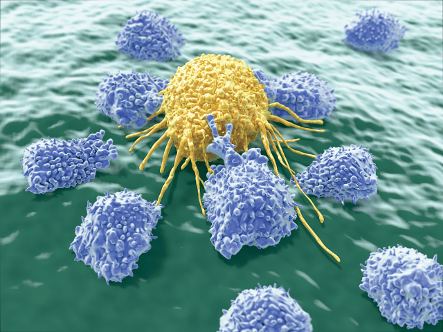
AND STAY OUT: Exercise activates natural killer cells (purple) and helps them home to tumors.

To find out what, our team tested the effects of interleukin-6, which we suspected was the additional exercise factor involved in tumor homing of immune cells. When we exposed inactive mice to both epinephrine and interleukin-6, the rodents’ immune systems attacked the tumors as effectively as if the animals had been running.

While much remains to be learned about how physical exercise influences cancer, evidence shows that exercise training is safe and feasible for patients with the disease and contributes to their physical and psychosocial health. (See “Exercise and Depression” on page 44.) Being active may even delay disease progression and improve survival. A growing number of patients, including Mathilde, are undergoing exercise training to fight physical deterioration during cancer treatment. As they do so, scientists are working hard to understand the pathways by which physical activity results in anticancer activity.

Exercise and Depression

Depression is a severe adverse effect of cancer and cancer therapy. The risk of depression can be as high as 50 percent for some cancer diagnoses, although this number varies a great deal depending on cancer type and stage (J Natl Cancer Inst Monogr, 32:57–71, 2004). In addition to its effects on a patient’s quality of life, depression can hinder compliance with treatment, and it’s a risk factor for mortality in cancer patients (Lancet, 356:1326–27, 2000). In recent years, healthcare providers have increasingly integrated physical exercise into the care of cancer patients with the aim of controlling disease and lessening treatment-related side effects, while researchers have amassed evidence supporting the assertion that such training can lower symptoms of depression in these patients (Acta Oncol, 58:579–87, 2019). The biological mechanisms behind this beneficial effect remain to be determined, although some clues have emerged.

For example, a study in mice found that exercise-dependent changes in metabolism result in reduced accumulation of some neurotoxic products (Cell, 159:33-45, 2014). In cancer patients, systemic levels of kynurenine, a neurotoxic metabolite associated with fatigue and depression, are upregulated (Cancer, 121:2129-36, 2015). In mice, exercise enhances the expression of the enzyme kynurenine aminotransferase, which converts kynurenine into neuroprotective kynurenic acid, thereby reducing depression-like symptoms.

Findings such as these, together with exercise’s well-documented effects in alleviating depression among patients without cancer, suggest that incorporating exercise into cancer treatment may benefit mental as well as physical health.

https://www.the-scientist.com/features/regular-exercise-helps-patients-combat-cancer-67317?utm_campaign=TS_DAILY%20NEWSLETTER_2020&utm_source=hs_email&utm_medium=email&utm_content=86607989&_hsenc=p2ANqtz-8W-OrX7bn_MULo5_Jx-u7E1c2gVfZwwWCD26RHtjZT7CoZ9KWhz0zOuCD53QkfOvre5WKYWWxP0plIm4Lf56uABjYb0A&_hsmi=86607989

ICU Doctor on NYC’s front line Debunks 6 COVID myths

It’s Easter Sunday, just after Passover, just after another exhausting13 hour shift. I can’t watch the news. I’m too busy and too frustrated by all the misinformation. Forgive me, but I need to debunk a few viral myths.

Myth #1: COVID-19 is a disease of the old and sick

This cannot be further from the truth. As a critical care physician, I’m caring for the sickest of the sick. I know the data. What little good data there is shows that 80% of ICU patients are under 65 (in a Wuhan study) or that 40% in ICU were under 60 (in an Italian study). The highest death age group was 60-69. The third highest was 50-59. The most common co-morbid conditions were high blood pressure, diabetes and obesity. These are not weird immune-related illnesses, they’re common, and this hits close to home. I’m 53, I have high blood pressure, diabetes and, like millions of Americans, I’m a little obese. Our stats? 60% of our intubated patients are under 65. Most of my ICU patients have never been sick enough to be hospitalized before this. Sure, many who die are old and have other illnesses, but the popular narrative almost says if you’re not in a nursing home you’re safe. Nothing can be further from the truth. It’s a myth.

Myth number #2: The main concern is a lack of PPE and ventilators

Partially false. Sure, some NYC and UK caregivers have had to use cooking aprons, garbage bags, and other scraps to protect themselves, but many hospitals have all the PPE they need. Luckily, my hospital has been able keep up with all our PPE needs. But many unanticipated shortages go unreported: COVID test swabs, dialysis machines and dialysis fluid needed to keep people alive (COVID causes kidney failure), sedative medications, and we need more oxygen, we’re using so much.

But most of all, we need more amazing people. Especially nurses and respiratory therapists, because many are now sick and some have died. Over 100 doctors have died in Italy. Doctors, therapists, pharmacists, students, and others now have a new career as nursing assistants. No-one is a specialist anymore, we are all COVID care providers. Thank you to the many volunteer doctors and nurses from all across the US that have come to NYC to help. Recovery for patients can take weeks to months, so we’ll need your help and sacrifice for a while yet.

Myth #3: Hydroxychoriquine is a “game changer” and it’s safe.

This potentially false idea was launched on the back of a very small trial from France. I’ve read the paper and it has major flaws. Three larger and more recent trials were negative but they don’t get press. These “game changer” drugs have dangerous side-effects. A recent trial in Brazil was stopped early for fear that high-dose chloroquine was killing people. Other drugs, however, show promise. Watch this space but no “game changers” yet.

Myth #4: Social distancing is our only option and it’s easy to do

This is also untrue. My home, NYC, is one of the most densely populated cities in the world. Many of my patients are poor and immigrated here. They live in small apartments with large families. Social distancing is impossible for many parts of NYC. And in the US more than 10% of the work force is unemployed. Sure, we’re finally flattening the curve, but as a Korean-American, I am proud to say that South Korea did it better and they didn’t shut down their economy. They tested, tested, tested, tracked, and isolated people and provided a mobile app, food, masks, and a thermometer to track their fever. This was done for visitors as well as citizens. The US hasn’t tested widely or efficiently enough. And we need to talk about the painful economic and human impacts of social distancing. Banning all hospital visitors means many terrified patients dying lonely deaths. The loss of human dignity is unimaginable.

Myth #5: We can blame China for the current US pandemic

This is false. Recent research shows that our outbreak in NYC came from Europe. And how helpful are country labels anyway? The 1918 Spanish Flu apparently didn’t originate in Spain, so should we rename it? When it comes to infectious diseases, borders mean nothing in our global economic village, but anti-Asian sentiment has spiked all over the world. Just read the online hate speech about the “KungFlu” and the “WuhanVirus”. As an Asian American, who is doing as much as I can, this is very distressing.
Andrew Yang wrote “We need to step up, help our neighbors, donate … and do everything in our power to accelerate the end of this crisis.” This is what my wife and I and so many others are doing. I work 12-15 hours days alongside residents, doctors, nurses, pharmacists and others. (BTW, many of these heroes are Asian-American.) We’re active in our local church, and my wife has a Facebook group that donates tens of thousands of dollars to food and supplies for front-line workers. Daily, she buys food from struggling restaurants, delivers it to the hospital, and I distribute it in between seeing my patients. This has been our life for months and will be our future for a while.

Does it really matter if the virus is from China, Europe or Mars? Our response would have been the same: to save as many lives as we can.

Myth number #6: This is all overblown, COVID is just like the Flu

I’m just shocked by this one. The infectivity of COVID 19 is three times that of the flu, and it is 40 times more deadly (Dr. Fauci says “10 times”). On Good Friday in NYC, 783 patients died; that’s one death every 2 minutes. In the US, it was one death every 42 seconds. Brace yourself. This is nothing like the flu. If you don’t believe me, just walk into any emergency room in New York, Detroit, Miami, LA or New Orleans.

On a final and personal note, I’m blown away by the response of my residents, my colleagues, the people around me, and all NYC hospital staff. Never have I been more proud to be a health care worker and a residency director. I’m impressed by the sacrifice and commitment of all my residents. I’m in awe of their hard work. These are the finest people on earth. I am humbled by their sacrifice and courage to go above and beyond the call of duty. Oddly, it took a pandemic to bring us this level of mass cooperation. But it’s also frightening. I have practiced critical care medicine for more than 25 years and never have I been so challenged, saddened and emotional. Almost every hour of every shift, someone needs intensive care. I’m very used to comforting patients and their families to prepare for death. I used to do this for someone weekly; , now it’s hourly. Death has become very common: every shift, every ward, and in every emergency room. It feels like a bomb went off somewhere and the whole of New York is slowly suffocating.
The 7pm cheering for health care workers moves me. Previously, at parties, I’d say “I work in an ICU and I ventilate people”. That was a big conversation killer. Now, I feel like a rock star or a military veteran. Who knows? Maybe one day I’ll get to priority board an airplane. But seriously; this experience will lead to future PTSD, pain, scars, and tears, for me and so many residents and health care workers. For now, however, we really need your prayers and support.

I hope this demystifies a few things. Thanks for reading. #columbiamedicine #columbiastrong

With Humans Indoors, Animals Go Wild


Across the globe, wildlife is exploring empty places usually occupied by people.

As humans are remaining indoors in response to the coronavirus pandemic, it appears that wildlife around the world took notice of our absence. There seems to be a never-ending list of animals becoming emboldened during this time to explore areas that are typically heavily populated: Buffalo have taken to the deserted highways in India. Mountain lions have rested in trees in Boulder, Colorado. Wild boar walk the streets of Barcelona while peacocks strut along open streets in Brazil.

Rats in New York City have somehow become even more confident in their quest for food. And a groundhog appeared to stare down two dogs watching through a window while eating a piece of pizza, which probably doesn’t have anything to do with the lockdown, but was a welcome distraction on social media nonetheless.

The Washington Post reports that a tribe of goats overtook the streets of Wales. Video taken by resident Andrew Stuart shows the animals nonchalantly roaming the empty streets and helping themselves to a meal of hedges and flower gardens.

According to SFGate, an employee from Yosemite National Park claims that since the park closed to the public in late March, the sightings of large animals including bears, bobcats, and coyotes have gone up fourfold.

“It’s not like [bears] aren’t usually here,” Yosemite employee Dane Peterson tells SFGate, “it’s that they usually hang back at the edges, or move in the shadows.”

In Mexico, crocodiles that generally stay hidden in lagoons near the beaches in La Ventanilla, Oaxaca, have been coming out in the open since the beaches were closed to the public about two weeks ago, Mexico News Daily reports.

Endangered sea turtles have also taken advantage of empty beaches to nest in Brazil and Florida. It’s too early to tell how lockdown measures will affect sea turtle numbers when it is time for the eggs to hatch. Decreased traffic could create less artificial light to confuse the hatchlings about which direction to go, Shanon Gann, the program manager at Brevard Zoo Sea Turtle Healing Center in Florida, tells weather.com.

A mixed bag for animals that depend on humans

In urban areas where wildlife is, for better or worse, dependent on human activity, the lockdown brings new challenges. The New York Times describes scenes in Thailand, where macaques have come to rely on humans for food. Their populations have become so dense in these areas because of that food supply that people staying home has quickly created a scarcity of resources, leading to aggressive behavior.

The same goes for duck ponds, ecologist Becky Thomas of Royal Holloway in London writes for The Conversation. Although feeding bread to ducks is harmful to their health and the water around them, there will be an adjustment as they compete for healthier resources.

Thomas notes that decreased traffic will lead to less hedgehog roadkill as well as reduced noise pollution that negatively affects the ability of bats, birds, and other animals to communicate.

The lack of human presence hasn’t benefited all animals, as the Times reports, particularly animals in African nature preserves. With fewer tourists around, poachers are killing rhinos with an increased frequency in Botswana and South Africa.

“We’re in a situation of zero income, and our expenses are actually going up all the time just trying to fight off the poachers and protect the reserve,” Lynne MacTavish, operations manager at Mankwe Wildlife Reserve in South Africa, tells the Times. “To say it’s desperate is an understatement. We’re really in crisis here.”

Some of the earliest widely shared reports of wildlife emerging in populated areas turned out to be false, according to National Geographic’s debunking of some of the more common untruths. One such tale says baby elephants in China got drunk on corn wine and passed out in a tea field, which might be very relatable during these times, but never happened. The absence of boats in the canals of Venice brought claims of dolphins appearing for the first time in decades, but the images were from the island of Sardinia, nearly 500 miles away.

There may not be dolphins in Venice, but the waters have gotten astonishingly clear, as the lack of gondolas and other boats on the water haven’t been stirring up sediment, CNBC reports.

Right now, it isn’t clear what the long-term effects of this lockdown will be on nature, primarily because this is occurring when many species in the Northern Hemisphere are mating, giving birth, or coming out of hibernation. Air pollution in some areas has been cut in half since the lockdowns began, Forbes reports, due to the lack of emissions from vehicles and factories. Some cities notorious for smoggy skies, including Los Angeles and Beijing, are enjoying some of the cleanest air they’ve experienced in decades. While the tolls of air pollution on human health are widely known, animals are also at risk, according to the National Wildlife Refuge System.

As many are still sheltering-in-places as we approach the 50th annual Earth Day, this resurgence of wildlife is giving some cause for hope that this evidence will ultimately lead to better policies to protect the environment and create a new normal.

“I am hopeful,” anthropologist Jane Goodall tells the Post. “I am. I lived through World War II. By the time you get to 86, you realize that we can overcome these things. One day we will be better people, more responsible in our attitudes toward nature.”

https://www.the-scientist.com/news-opinion/with-humans-indoors-animals-go-wild-67434?utm_campaign=TS_DAILY%20NEWSLETTER_2020&utm_source=hs_email&utm_medium=email&utm_content=86538478&_hsenc=p2ANqtz-92e5YchE_c5eEZJOR2VWChyXs-TUYFALDBiX0cEwNWRvtMhsuRr4MWSGBf0DCvU1hKkYi4eEAJ3QErLAitWrBijvumwg&_hsmi=86538478

Long space flights can increase the volume of astronauts’ brains

Astronauts’ brains increase in volume after long space flights, causing pressure to build up inside their heads. This may explain why some astronauts experience worsened vision after prolonged periods in space.

“This raises additional concerns for long-duration interplanetary travel, such as the future mission to Mars,” says Larry Kramer at the University of Texas Health Science Center at Houston, who led the study.

Kramer and his colleagues scanned the brains of 11 astronauts before they spent about six months on the International Space Station, and at six points over the year after they returned to Earth. They found that all the astronauts had increased brain volume – including white matter, grey matter and cerebrospinal fluid around the brain – after returning from space.

Under normal gravity, it is thought that fluid in the brain naturally moves downwards when we stand upright. But there is evidence that microgravity prevents this, resulting in accumulation of fluid in the brain and skull.

The astronauts’ brain volume increased by 2 per cent on average and the increases were still present one year after they returned to Earth, which could result in higher intracranial pressure, Kramer says. He suspects this might press on the optic nerve, potentially explaining the vision problems frequently reported by astronauts.

Kramer and his team also observed that part of the brain called the pituitary gland was deformed in six out of the 11 astronauts. These results add to a body of evidence suggesting that brain structure can be altered after space flight.

“This study is important because it provides data, for the first time in NASA astronauts, demonstrating the persistence of structural brain changes even up to one year following return to Earth,” says Donna Roberts at the Medical University of South Carolina.

“We are currently working on methods to counteract the changes we are observing in the brain using artificial gravity,” says Kramer. These methods to pull blood back towards the feet could include a human-sized centrifuge that would spin a person around at high speed, or a vacuum chamber around the lower half of the body.

“Hopefully one of these or other methods will be tested in microgravity and show efficacy,” he says.

Journal reference: Radiology, DOI: 10.1148/radiol.2020191413

Read more: https://www.newscientist.com/article/2240405-long-space-flights-can-increase-the-volume-of-astronauts-brains/#ixzz6Jh5CtujT

Cleveland Ohio company invents ‘Second Breath,’ a ventilator that will save lives during coronavirus pandemic


Second Breath will help save lives during coronavirus pandemic

It’s called “Second Breath,” and this piece of equipment will save lives.

“Without this ventilator there are a lot of folks who might not have a chance at a breath,” said Dr. Mada Helou, from University Hospitals.

Three Cleveland organizations put their innovative skills, knowledge and can-do attitude together to create and manufacturer a breathing pump that will alleviate some of the demand for ventilators, all across the country.

“We like to respond to things, and more importantly the team likes to respond. There were eight engineers that developed this and these guys wanted to do something and they came to me,” said Dan T. Moore, president and CEO, Dan T. Moore Co.

It collaborated with several other organizations and in three weeks, designed “Second Breath.”

“Coronavirus’ main target is the lungs. It effects many organs, but it has a profound effect on our ability to hold oxygen within our blood,” said Dr. Helou.

Experts said an average ventilator costs anywhere from $20,000 and up to $100,000. Second Breath costs about $6,000.

Dan T. Moore Co. has made about 36 ventilators and they’re ready to ship out around the world. Engineers told 19 News they can design about a hundred ventilators a day.

The team tells 19 News they are proud.

“I think great innovation comes under pressure and when the COVID-19 surge showed up folks thought you know what, we need to respond to this quickly,” said Dr. Helou. “Everything about this says Cleveland. It speaks helping people and it speaks collaboration,” she said. “Cleveland, you’ve done this!”

https://www.fox19.com/2020/04/16/ohio-company-invents-second-breath-ventilator-that-will-save-lives-during-coronavirus-pandemic/