Key takeaways:
- A non-deceptive placebo injection reduced chronic back pain with effect size similar to typical treatments.
- Secondary outcome benefits and brain changes lasted up to 1 year.
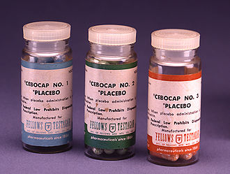
A single saline injection, openly prescribed as a placebo, yielded approximately 1 month of chronic back pain improvement, along with longer-term benefits in depression and sleep, according to data published in JAMA Network Open.
“We have known that placebos can be powerful pain relievers, but it has been unclear how to use them ethically, without patient deception,” Yoni K. Ashar, PhD, assistant professor at the University of Colorado Anschutz Medical Campus, told Healio. “This spurred the development of the ‘open label,’ non-deceptive placebo treatment, which we studied here.”
To investigate the long-term efficacy of open label placebo in chronic back pain, Ashar and colleagues recruited 101 adults (mean age, 40.4 years) with moderate chronic back pain from the Boulder, Colorado, area between November 2017 and August 2018, with a follow-up at 1 year.
Trial participants were randomly assigned to either continue their usual care alone or to also receive a single, open label lumbar saline injection, along with information about how the placebo effect can lead to pain relief. The primary outcome was average pain over the last week 1 month after treatment, measured using a scale of 0 to 10. Secondary outcomes also assessed pain interference, depression, anxiety, anger and sleep quality.
At 1 month, those who received placebo injections reported greater reductions in chronic back pain than the usual care group (relative reduction, 0.61; Hedges g = 0.45; 95% CI, –0.89 to 0.04), according to the researchers.
By 1 year post-treatment, the between-group difference in pain relief was reduced to insignificance. However, after 1 month, other significant benefits were seen in depression, anger, anxiety and sleep disruption, with “medium sized” effect sizes ranging from 0.3 to 0.5 (P < .03 for all).
The researchers also compared neuroimaging between the groups. Functional MRI scans were taken as participants performed both an “evoked” back pain procedure, which used an inflating balloon to cause back distention and pain, and a “spontaneous” pain procedure, where patients rated their pain once per minute over the course of an 8-minute scan.
Overall, the neuroimaging showed “altered brain responses to evoked back pain and altered functional connectivity during spontaneous pain consistent with engagement of descending modulatory pain pathways,” Ashar and colleagues wrote.
The researchers described the placebo injection’s pain relief benefit as “modest in magnitude” but clinically significant and comparable with the effect sizes of typical treatments such as NSAIDs, but with fewer adverse events.
“These findings speak to the power of healing rituals, even when we know they are healing rituals,” Ashar said. “Although we view this study as more mechanistic and conceptually provocative than as clinically applicable, it suggests that providers may be able to ethically prescribe a placebo for their patients one day, without deception. In addition, the duration of benefits on secondary outcomes and the observed brain changes were surprising, considering how brief and minimalist the intervention was.”